Ablation, a great resolution for certain arrhythmias.

afib-with-rvr-7  afib-with-rvr-6afib

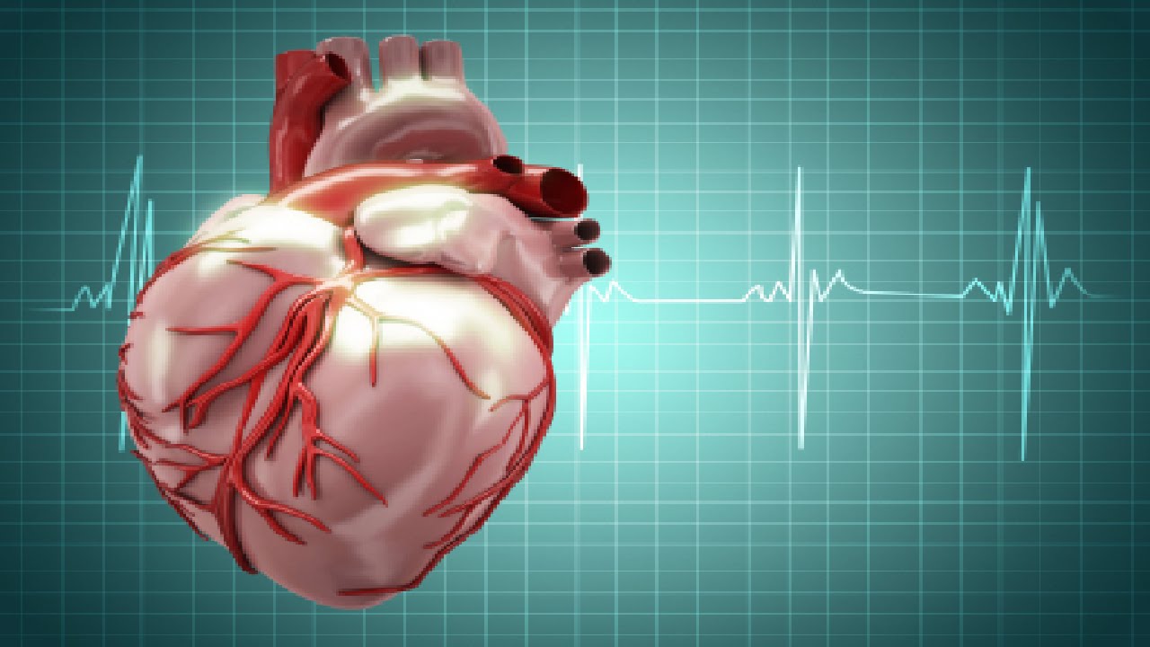
Ablation is used to treat abnormal heart rhythms, or arrhythmias. The type of arrhythmia and the presence of other heart disease will determine whether ablation can be performed surgically or non-surgically.

Ablation therapy using radio frequency waves on the heart is used to cure a variety of cardiac arrhythmiae such as supraventricular tachycardia, Wolff–Parkinson–White syndrome (WPW), ventricular tachycardia, and more recently as management of atrial fibrillation (especially when its newly diagnosed when medical management can’t change it back to normal sinus rhythm, which is the normal cardiac rhythm seen on a telemetry monitor or of an EKG taken on a patient).

An arrhythmia is a change in the heart’s normal rate or rhythm, normally between 60 and 100 beats per minute. Arrhythmias are classified by their location in the heart and by their speed or rhythm. An atrial arrhythmia is an abnormality that occurs in one of the two upper chambers of the heart, the left or right atrium. Arrhythmias are associated with aging and typically happen more frequently during middle age. At least 10 to 15 percent of people older than 70 years experience arrhythmias.  We have what we call our human pacemaker of the heart that naturally sends conduction for the heart to pump, which is called the sinus node.  This is in the upper left corner of the right chamber of the heart.  That is where the name sinus rhythm derives from (the sinus node) which is the best rhythm a human can be in as long as the pulse rate stays above 60 and stays under 100. Now if that sinus node for some reason breaks down and no longer works; so than the pace site starts somewhere in the right atrium below the sinus node (the heart is compensating for whatever is the reason the sinus node is not working).  So now the rhythms are called atrial rhythms because of where the new natural pacemaker site is in the heart.  This is where ablation comes into play if the type of atrial rhythm they have is detrimental to the patient; including if that patient is a candidate for this procedure.  Between our heart chambers on the top (called atriums) and below (called the ventricles) is a AV (meaning atrioventricular valve).  Rhythms above the ventricles are also grouped as supraventricular rhythms.  Which is what ablation is used for.

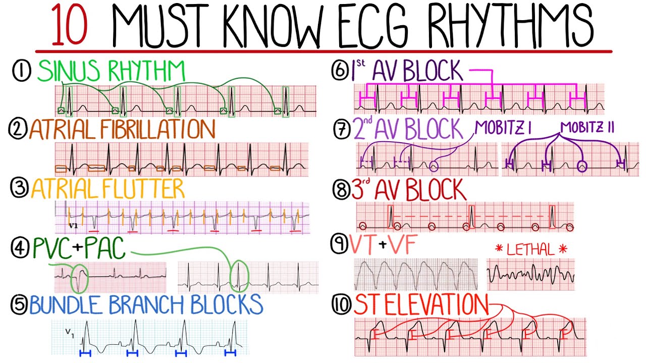
Types of rhythms a patient would be considered for ablation as a possible treatment:

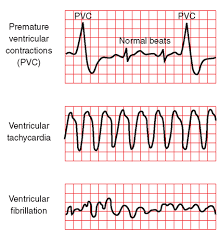
Atrial fibrillation. The electrical signal that circles uncoordinated through the muscles of the atria (the upper chambers of the heart), causing them to quiver (sometimes more than 400 times per minute) without contracting. The ventricles (the lower chambers of the heart) do not receive regular impulses and contract out of rhythm, and the heartbeat becomes uncontrolled and irregular. It is the most common atrial arrhythmia, and 85 percent of people who experience it are older than 65 years.

Atrial fibrillation can cause a blood clot to form, which can enter the bloodstream and trigger a stroke. Underlying heart disease or hypertension increases the risk of stroke from atrial fibrillation as does age even without heart disease or hypertension.

Premature atrial contraction (PAC or premature atrial impulses). A common and benign arrhythmia, a PAC is a heartbeat that originates away from the sinus node, which sends electrical signals through the upper chamber. It typically occurs after the sinus node has initiated one heartbeat and before the next regular sinus discharge. A PAC can cause a feeling of a skipped heartbeat. Use of caffeine, tobacco, and/or alcohol, or stress can bring on PACs or increase their frequency.

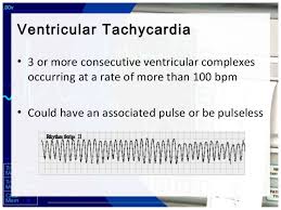
Supraventricular tachycardia (SVT). Characterized by a rapid heart rate that ranges between 100 and 240 beats per minute, SVT usually begins and ends suddenly. SVT occurs when an electrical impulse ‘re-enters’ the atrial muscles. A disorder that a person may have at birth, SVT is commonly caused by a variation in the electrical system of the heart. SVT often begins in childhood or adolescence and can be triggered by exercise, alcohol, or caffeine. SVT is rarely dangerous, but can cause a drop in blood pressure, causing lightheadedness or near-fainting episodes, and, rarely, fainting episodes.

Atrial flutter. Differentiated from atrial fibrillation by its coordinated, regular pattern, atrial flutter is a coordinated rapid beating of the atria. Most who experience atrial flutter are 60 years and older and have some heart disorder, such as heart valve problems or a thickening of the heart muscle. Atrial flutter is classified into two types, according to the pathways responsible for it. Type I normally causes the heart rate to increase to and remain at 150 beats per minute. Rarely, the rate may reach 300 beats per minute; sometimes it decreases to 75 beats per minute. Type II increases the atrial rate faster, so the ventricular rate may be 160 to 170 beats per minute. As with atrial fibrillation, atrial flutter increases the risk of stroke.

Sick sinus syndrome (SSS). Common among older people, SSS is an improper firing of electrical impulses caused by disease or scarring in the sinus or Sinoatrial node (SA node). SSS normally causes the heart rate to slow, but sometimes it alternates between abnormally slow and fast. A progressive condition, with episodes increasing in frequency and duration, SSS can be caused by:

  • Degeneration of the heart’s electrical system; or
  • Diseases of the atrial muscle.                                                                                                                                                                                                                                                                                                                                                    Sinus tachycardia. The sinus node emits abnormally fast electrical signals, which increases the heart rate to between 100 beats per minute to 140 beats per minute at rest, and 200 beats per minute during exercise. A normal response to exercise or stress, it can also be caused by:
  • Adrenaline;
  • Consumption of caffeine, nicotine, or alcohol; and
  • Heart conditions.                                                                                                                                                                                                                                                                                                                                                                                   Wolff-Parkinson-White syndrome (WPW). WPW syndrome occurs when electrical signals fail to pause in the atrioventricular node because an extra pathway allows the impulse to “bypass” the normal pathway; and the syndrome is sometimes called bypass tract. WPW syndrome causes heart rates approaching 240 beats per minute.
  • Occasionally, impulses can go down one extra pathway and up another, creating a “loop” or “short circuit,” (called SVT because of WPW). Patients with WPW syndrome may develop atrial fibrillation and are at increased risk for developing a dangerous ventricular arrhythmia when this occurs. *
  • Sinus bradycardia. Associated with impaired impulse generation in the SA node, it causes the heart rate to decrease to fewer than 60 beats per minute. Commonly caused by SSS, drugs like beta-blockers and calcium-channel blockers can also cause sinus bradycardia. Occasionally sinus bradycardia can be caused by impaired conduction of impulses to the atrial muscles.

QUOTE FOR THURSDAY:

“Heart block is a problem with your heartbeat signal moving from the upper to lower part of your heart. The signal can only get through sometimes, or not at all. This makes your heart beat slowly or skip beats. People with second-degree or third-degree heart block may experience fainting, tiredness and shortness of breath.

Heart block is an issue with a heartbeat signal traveling from the top chambers of your heart to the bottom chambers of your heart. Normally, electrical signals (impulses) travel from your heart’s upper chambers (atria) to your lower chambers (ventricles). The signal moves through your AV node, a cluster of cells that link the electrical activity from your top to bottom chambers. If you have heart block, the signal only makes it to your ventricles some of the time, if at all.

The result is a heart that may not work well. Your heart may beat slowly or skip beats. In severe cases, heart block can affect your heart’s ability to pump blood, causing low blood flow to your entire body.

Other names for heart block are atrioventricular (AV) block or a conduction disorder.  The heart block is between the upper chambers (atriums) and the lower chamber (ventricles) with the electrical activity responsible to make your heart beat properly.”

Cleveland Clinc (Heart Block: Types, Symptoms & Causes)


The Heart Blocks with the heart: Type I HB, Type II HB= Mobitz I and Mobitz II, and lastly Type III HB. Learn the severity of the 4 A-V HBs and the Rxs.

HeartBlocks2

Than when AV nodes which can be blocked=atrio-ventricular block (AV heart block)=HB is a type of heart block in which the conduction between the atria and ventricles of the heart is impaired. Under normal conditions, the sinoatrial node (SA node) in the atria sets the intial pace for the heart, and these impulses travel down to the atriums than passing the AV valves into the ventricles and up the purkinje fibers . In an AV block, this message does not reach the ventricles or is impaired along the way. The ventricles of the heart have their own pacing mechanisms, which can maintain a lowered heart rate in the absence of SA stimulation.

The causes of pathological AV block are varied and include ischemia, infarction, fibrosis or drugs, and the blocks may be complete or may only impair the signaling between the SA and AV nodes.

Types of AV blocks: 1st degree HB which is NSR with a prolonged PR interval and can live a totally normal life with it. PR interval is the time from beginning of the p wave to the beginning of the qrs wave.  In all degrees of HB the PRI interval is prolonged.   You can live a normal life with 1st degree HB.

Mobitz type I is benign, and most people do not experience symptoms. A doctor may only discover the condition during a routine exam or when checking for other conditions using an ECG. Those who do not experience symptoms may require monitoring on an outpatient basis.  If people do experience symptoms, they may require treatment.

2nd degree HB-Mobitz I called Wenkebach is a NSR with a progressing increasing PR interval longer and longer till it actually drops a QRS and you see just the p wave without a QRS once and the rhythm restarts the pattern all over again.

Than 2nd degree HB-Mobitz II and this is a NSR with a prolonged PRI that measures the same amount each time but even though its not progressively getting longer its worse than Mobitz I always.  This is because with the same measurement prolonged PRI interval you only see 2 or 3 more  p waves without the QRS and these need immediate external subcutaneous pacing or just as effective if its not available (external subcutaneous pacing) than a dopamine IV drip 5-20 mcg/kg/min or epinephrine IV drip 2 to 10 mcg/min or epinephrine IV drip 2-10 mcg/min (not kg based).

Than there is complete HB 3rd degree heart block where no conduction from atriums to ventricles is going through so the atriums are contracting the way they want and the ventricles contracting the way they want.  The problem is there is a total disassociation between the upper and lower chambers. Treatment for most patients with acquired complete heart block will require a permanent pacemaker or an implantable cardioverter defibrillator (ICD).  Temporarily till OR the pt may have subcutaneous pacing but may not work at in some cases.

QUOTE FOR WEDNESDAY:

“Cardiac arrest may be caused by almost any known heart condition.

Most cardiac arrests occur when a diseased heart’s electrical system malfunctions. This malfunction causes an abnormal heart rhythm such as ventricular tachycardia or ventricular fibrillation. Some cardiac arrests are also caused by extreme slowing of the heart’s rate (bradycardia).

Irregular heartbeats such as these are life-threatening.

Other causes of cardiac arrest include:

Scarring of the heart tissue – It may be the result of a prior heart attack or another cause. A heart that’s scarred or enlarged from any cause is prone to develop life-threatening ventricular arrhythmias. The first six months after a heart attack is a high-risk period for sudden cardiac arrest in patients with atherosclerotic heart disease (the buildup of fatty deposits, or plaque, in the arteries).
Thickened heart muscle (cardiomyopathy) – Damage to the heart muscle can be the result of high blood pressure, heart valve disease or other causes. A diseased heart muscle can make you more prone to sudden cardiac arrest, especially if you also have heart failure. Learn more about cardiomyopathy.
Heart medications – Under certain conditions, some heart medications can set the stage for arrhythmias that cause sudden cardiac arrest. (Oddly, antiarrhythmic drugs that treat arrhythmias can sometimes produce ventricular arrhythmias even at normal doses. This is called a “proarrhythmic” effect.) Significant changes in blood levels of potassium and magnesium (from using diuretics, for example) can also cause life-threatening arrhythmias and cardiac arrest.
Electrical abnormalities – These, including Wolff-Parkinson-White syndrome and long QT syndrome, may cause sudden cardiac arrest in children and young people.
Blood vessel abnormalities – These rare cases occur particularly in the coronary arteries and aorta. Adrenaline released during intense physical activity can trigger sudden cardiac arrest when these abnormalities are present.
Recreational drug use – This can be associated with cardiac arrest in otherwise healthy people.
Commotio cordis – Commotio cordis occurs as a result of a blow to the left side of the chest during a narrow window in the heart rhythm.”

American Heart Association (Causes of Cardiac Arrest | American Heart Association)

Certain cardiac rhythms can lead to a cardiac arrest or other things & how to prevent it!

HeartBlocks1

The rhythms above are heart blocks (HB) that occur in the bottom of the upper chambers which can occur in some people. There is 1st degree HB where you can live a completely normal life with but 2nd and 3rd degree HB needs treatment (usually a pacemaker) by cardiologist surgeon.  After treatment with 2nd and 3rd degree HB you can live a completely normal life with follow up with your cardiologist and yearly pacemaker checks.

In this rhythm below the Ventricular Tachycardia is with a point on the top but than flips upside down (commonly called Torsedes Pointes).  This is commonly due to Magnesium Level low and IV Magnesium in the hospital is given 1 to 2 gm.

ventrhy4

This  rhythm above with a pulse=also a rhythm pulsating in different areas of the heart in the ventricles only causing the rhythm not to look identical throughout the tele strip above = Polymorphic V- Tac- meaning the stimulus in the ventricles to make the heart beat is coming from different areas of the ventricles for each beat.  Each jagged tooth is a beat that makes up the whole strip shown above for Ventricular Tachycardia.

Than when the atriums aren’t working as the natural pacemaker that took over for the sinus node but now they don’t work so now the ventricles take over and the rhythms of all ventricle rhythms are with NO p waves since the atriums are not working so no p wave is involved but we have QRS waves but their wide in measurement because the rhythm starts in the ventricles. The rhythms are PVC (Premature Ventricular Contractions), Idioventricular Rhythm, Ventricular tachycardia (Monomorphic and Polymorphic-rhythm getting more irregular. When regular and monomorphic=looking identical with every ventricular beat or contraction as opposed to polymorphic=not looking identical each contraction but each one is a ventricular contraction), Torsades De Pointes Ventricular Tachycardia (the rhythm starts upright but turns upside down but each contraction without a p wave and a wide contraction meaning a ventricular contraction), and Ventricular Fibrillation, to asystole.

Here’s what they look like:

 Accelerated Idioventricular Rhythm

Accelerated idioventricular rhythm occurs when three or more ventricular escape beats appear in a sequence. Heart rate will be 50-100 bpm. The QRS complex will be wide (0.12 sec. or more).

A regular QRS measures less than 0.12 which is with all atriums rhythms.

 Asystole

Asystole is the state of no cardiac electrical activity and no cardiac output. Immediate action is required.

Idioventricular Rhythm

Idioventricular rhythm is a slow rhythm of under 50 bpm. It indicates that then ventricules are producing escape beats.

Premature Ventricular Complex-PVC (above 1st strip in #4)

Premature ventricular complexes (PVCs) occur when a ventricular site generates an impulse. This happens before the next regular sinus beat. Look for a wide QRS complex, equal or greater than 0.12 sec. The QRS complex shape can be bizarre. The P wave will be absent.

Premature Ventricular Complex – Bigeminy a QRS after every 2 regular beats

Premature Ventricular Complex – Trigeminy a QRS after every 3 regular beats

Premature Ventricular Complex – Quadrigeminy a QRS after every 4 regular beats

The more PVC’s especially right next to each other can lead to Ventricular Tachycardia to Ventricular Fibrillation if not treated in time.

 Ventricular Fibrillation (in above strip-3rd one)

Ventricular fibrillation originates in the ventricules and it chaotic. No normal EKG waves are present. No heart rate can be observed. Ventricular fibrillation is an emergency condition requiring immediate action.

Ventricular Tachycardia  (in above strip-2nd one)

A sequence of three PVCs in a row is ventricular tachycardia. The rate will be 120-200 bpm. Ventricular Tachycardia has two variations, monomorphic and polymorphic. These variations are discussed separately.

Ventricular Tachycardia Monomorphic

Monomorphic ventricular tachycardia occurs when the electrical impulse originates in one of the ventricules. The QRS complex is wide. Rate is above 100 bpm.  Each V tac beat looks identical like in the strip above.

Ventricular Tachycardia Polymorphic

Polymorphic ventricular tachycardia has QRS complexes that very in shape and size. If a polymorphic ventricular tachycardia has a long QT Interval, it could be Torsade de Pointes.  The strip shows the pulses are not identical=polymorphic since the pulse beats are coming from all different areas of the ventricles.

Torsade de Pointes  (the rhythm strip at the top under Heart Blocks)

Torsade de Pointes is a special form of ventricular tachycardia. The QRS complexes vary in shape and amplitude and appear to wind around the baseline.  This is an example or polymorphic ventricular tachycardia.

Ventricular ending line needs to be treated stat to be switched back to atrial rhythm since the heart is missing ½ of the conduction it’s to normally receive from the atriums and if not reversed the heart will go into failure to heart attack or to asystole flat line and go into a cardiac arrest.

With PVCs=Premature Ventricle Contractions asymptomatic we just closely monitor the pt and telemetry the pt is on. Now a pt with PVCs and symtomatic usually meds with 0xygen (sometimes 02 alone resolves it but other times with meds) but if it gets worse into V Tachycardia the treatment is below.

Idioventricular Rhythm (IVR)is usually with a slow brady pulse and needs meds.   Accelerated IVR (AIVR) is usually hemodynamically tolerated and self-limited; thus, it rarely requires treatment.

Occasionally, patients may not tolerate AIVR due to (1) loss of atrial-ventricular synchrony, (2) relative rapid ventricular rate, or (3) ventricular tachycardia or ventricular fibrillation degenerated from AIVR (extremely rare). Under these situations, atropine can be used to increase the underlying sinus rate to inhibit AIVR.

Other treatments for AIVR, which include isoproterenol, verapamil, antiarrhythmic drugs such as lidocaine and amiodarone, and atrial overdriving pacing are only occasionally used today.

Patients with AIVR should be treated mainly for its underlying causes, such as digoxin toxicity, myocardial ischemia, and structure heart diseases. Beta-blockers are often used in patients with myocardial ischemia-reperfusion and cardiomyopathy

With Ventricular rhythms with fast pulse over 100 with symptomatic signs for the patient we may use as simple as valsalva pressure on the neck that medical staff only do but when pt is in asymptomatic (no symptoms) Ventricular Tachycardia (V-Tac) to even medications but when symptomatic if in V-Tac start cardioversion with a pulse if no pulse called pulseless V-Tac we use a defibrillator since there is no pulse there is no QRS to pace with in having the shock hit at the R wave, why? NO PULSE.

Treatment for Torsade de Pointes is Magnesium deficiency and Mag. Supplement given IV 2gms. Usually effective but if necessary the same as above as directed for it with a pulse or the other V Tac. (without a pulse)-See above.

Ventricular Fibrillation is when the ventricles are just quivering and the atriums in any ventricular rhythm doing nothing. The pt needs CPR and ASAP a defibrillator in hopes the shock will knock the rhythm back to a normal sinus or some form of a real rhythm.

Asystole which is a straight line, no pulse and this is CPR with epinephrine or Vasopressin 40 for only the replacement of the 1st or 2nd dose of Epinephrine 1mg. This is given 3-5 minutes (epinephrine). No defibrillation since no pulse. A rhythm may come back and if not the MD will call when CPR stops. Asystole is hard to resolve in most cases highier probability of resolution if in a hospital where close monitoring is done and its detected quicker.

The PURPOSE in treating any rhythm abnormal to the human heart is to reach the goal of a optimal or healthiest rhythm (a normal sinus rhythm , the best rhythm the heart can be in) and if not reaching an atrial rhythm.  We the medical field aim to reach a heart rhythm the patient can live with and hopefully reaching the best NSR-Normal Sinus Rhythm.  Normal sinus rhythm that is a rhythm starting from the upper right chamber extending to the left one and continues down on both sides to the bottom of the ventricles.  This rhythm is giving the most effective oxygen perfusion to the heart to allow it to do its function (pumping good oxygenated blood flow out of the left ventricle at the same time pumping highly carbon dioxide blood from the right side of the heart to the lungs to get more oxygen).   Doing this it allows the human body to get good amounts of oxygen to all our tissues=good overall oxygen perfusion to all tissues.  At the same time what happens is red blood cells from all tissues with mostly used up oxygen from the cell and more carbon dioxide in the cell are also being pumped by the heart to return to the right side  to the lungs to go through this whole process again in getting more oxygen in the RBCs which keeps us alive. A human without oxygen or low oxygen to their tissues or any tissue is going to reach cellular starvation which in turn causes starvation to the tissues (in general) or to a tissue area (Ex. Take the diabetic-regarding the foot to lack of 02 to cyanotic purple tissue to necrotic black tissue=dead to amputated since the tissue is dead.  Remember by gravity the foot is the furthest from the heart).

Cardiac Arrest or Heart Attack are more likely to occur in  a irregular rhythm especially making the heart work to hard being RVR afib in the atriums that can lead easily to ventricular tachycardia to ventricular fibrillation and not treated immediately.

Cardiac Arrest or an abnormal heart rhythm is an electrical problem with the conduction of the heart whereas a Heart Attack can be caused by a blockage of blood.  An example could be the coronary arteries-main arteries of oxygenated blood to the heart that can lead to a bad rhythm due to lack of 0xygen that leads to worse rhythms as the heart gets more stressed out.

For direction many experts like Atlantic Endocrinology states the following:

“You feel fine—no chest pain, no shortness of breath, no obvious red flags. But what if heart disease is developing silently?  It does for many where the cardiac condition was there awhile but no symptoms and the symptoms arise when the condition gets worse for many cardiac Dx’s.  Who wants to go to the doctor when they feel fine but there is always the silent symptoms and not just for cardiac issues but for this topic we will keep to just cardiac issues.

Many people assume a cardiologist is only for the elderly or those with symptoms, yet heart issues often start long before warning signs appear. So, when is the right time to schedule that first cardiac appointment? The answer might be earlier than you think.

Seeing a cardiologist isn’t just for the elderly or those with symptoms—heart disease can develop silently. Men over 40 and women after menopause face increased risk, especially with a family cardiac history of heart disease, high blood pressure, cholesterol, or diabetes. Preventive check-ups before age 60–70 improve outcomes.

Warning signs could definitely be such as chest pain or chest discomfort, shortness of breath or difficulty breathing, continuous headaches, palpitations continuous or intermittent, fatigue, dizziness or swelling or edema, require medical attention. Monitoring blood pressure, cholesterol, and managing lifestyle factors like diet, exercise, and stress are crucial. Those with hereditary or pre-existing heart conditions should see a cardiologist early for specialized care and prevention.”

Now even with rhythms that could lead to a heart attack!   Not that the rhythms were the primary cause of the heart attack but in most cases the rhythm is due to the cardiac condition. 

Now the rhythm can be used in helping the doctor as a preventative measure if found earlier enough to stop the heart attack from occurring! 

Now you know why there are many reasons to see a cardiologist at least yearly from 40 and up in men and post-menopausal in women!  Gave some examples or I would be writing a few novels!

QUOTE FOR TUESDAY:

“As the most commonly performed heart test, an electrocardiogram measures and records the electrical activity of your heart. Also known as an ECG or EKG, this key diagnostic tool provides invaluable and insightful information about the rhythm and function of your heart.

Simply put, an EKG is a “heart tracing” that offers a reliable (if not preliminary) snapshot of your cardiovascular health.

An EKG is a painless and noninvasive test that measures your heart’s electrical efficiency as it beats. As one of the fastest informational or diagnostic heart tests available, EKG testing can usually be completed in just five minutes.

To conduct an EKG test, a patient attaches up to 12 small, flat, sticky patches called electrodes at various points on your chest, arms, and legs. The electrodes are connected to a monitor that registers your heart’s electrical activity over the course of the exam.

During standard EKG testing, you lie still on a table as the electrodes detect and transmit the electrical activity of your heart to the monitor. EKG testing may also be used to measure and record your heart’s electrical efficiency under stress and through recovery.

The electrical activity of your heart doesn’t just drive your heartbeat, it also sets the rhythm and rate of that beat. A healthy heart usually has a regular beat that’s powered by steady electrical patterns, while a diseased or dysfunctional heart is more likely to have an irregular beat that’s controlled by fast, slow, or erratic electrical patterns.

Every single heartbeat is driven by an electrical impulse, or wave, that causes your heart to contract; each vital contraction keeps blood flowing seamlessly through your body.

An EKG monitors the strength, timing, and efficiency of this wave as it travels through the upper chambers of your heart to the lower chambers. It also monitors the electrical recovery between waves, or your heart’s momentary return to a resting state between each beat.”

ECCA Cardiologists (What an EKG Test Can Tell Your Doctor About Your Heart)

What cardiac rhythms tells your doctor about your heart!

Heart Beat symbol design element

Why cardiac monitoring can be vital important in quickly telling the doctors and nurses very important messages in what is going on with the patient’s heart and overall condition problem (Example A Myocardial Infarction or even to Cardiac Arrest).

Cardiac monitoring is a great way for doctors to understand a patients’ overall heart health, and can provide enough information to quickly and accurately helping the doctor or nurse as a diagnostic tool based on several details within a heart rhythm. While each arrhythmia monitoring device is a little different, these details are essential in diagnosing any underlying and potentially life-threatening events.

Your heart can have the best rhythm it can be in called Normal Sinus Rhythm which is a rhythm that is produced by the sinus node (SA node) that is the human pacemaker of the heart in out right upper atrium.  It starts a impulse (think of it as a message) that starts from the SA node and goes down the right atrium across to the left atrium (the upper chambers of the heart) with contiuing to send both impulses down to the bottom chambers of the heart which we call Ventricles creating the sound we all know the heart make called “lub dub”. This sound is creating when our heart valves open and close between the heart to allow complete fill up and release for the cardiac filling of our blood from top chambers to bottom and out of the heart to our circulation to send oxygenation out to all our tissues from feet to brain and back to lungs where our red blood cells carry the oxygen to tissues but take carbon dioxide back to the lungs for an exchange of new oxygen we breath in to exchange the carbon dioxide for new oxygen in the red blood cells and is send to the heart sent out back to our circulation to keep our body tissues oxygenated.  Without oxygenation that would be red blood cell starvation resulting into death for the human body.

There are times the SA node does not work for some reasons which causes the heart to start sending impulses from areas lower than where the SA node sits in the heart, in the upper right area of the heart.  Now some rhythms under the SA node can live a normal life with being checked up by a Cardiologist preferably or a Primary Care Doctor but know the Cardiologist will probably pick up before any other MD, if numerous years of experience.

Here is the basics to know about telemetry monitoring and your heart rate (also known as pulse):

1. Arrhythmias: Ambulatory heart monitors can be assigned for short-term use (24 to 72 hours) or for long-term use (up to 30 days or more) depending on what your doctor needs to know. Many cardiac monitoring devices record the ups and downs of your heartbeat to determine the presence of any irregularities in your rhythm that could be associated with an arrhythmia that’s new but possibly easily treatable or even curable to dangerous possibly or any underlying conditions.  There’s a device that we call holter monitor.  This device is what you wear for days and bring back to your doctor with leaving on 24hrs or  couple of days till you take off when the MD tells you too.  Than there is continuous telemetry monitoring in the hospital that records on the unit computer the patient usually is on.  This the MD reviews when you come back to his office with the holter monitor or the MD reviews daily or more when in the hospital.  This helps direct the MD in your care since it is a diagnostic tool for him or her.

2. Heart Rate: Your heart rate is the number of times your heart beats per unit of time, and can vary depending on your activities, sleep, and even what you eat. If it gets too low or too high when performing a specific activity, it’s essential that your doctor knows about it. A normal resting heart rate for adults ranges from 60 to 99 beats a minute.  The lower the better but usually not more than 50 if you have been in the heart rate or pulse of 50’s all your life due to being an athelete (some even in there 40’s) but if you have symptoms like dizzy, weakness, change in mental status, chest pain/discomfort, to indigestion that just won’t go away SEE THE MD; especially if the HR or pulse rate is new in a low rate that you are in.

3. P-wave analysis: On the telemetry monitor what MD’s, nurses and even technicians see are rhythm waves that is represented by names for each aspect of the wave we study.  The first wave if in normal sinus rhythm or some type of sinus rhythm we see what we call a p-wave represents the spread of electrical activity over the atrium, and normally lasts less than 0.11 seconds that derived from the SA node.  This is how sinus rhythms got their names.  An abnormally long p-wave occurs when it takes extra time for the electrical wave to reach the entire atrium.  This is the area right before that bigger wave we call QRS wave.   The prolongation for the PR interval signifies usually some type of AV block.  This occurs down at the valves between the upper chambers (atriums) and lower chambers (ventricles).  Ventricle rhythms means their is no impulse going through the atrium or we would see a atrium rhythm so now the ventricles take over to make a rhythm showing Ventricular Rhythms.  These are dangerous rhythms.

4. Morphology: This refers to the form of cardiac rhythms and how they differ depending on underlying conditions. The morphology of a heart rhythm can be observed as a series of deflections away from the baseline of an ECG, and can vary if you have any type of condition that could affect your heart  (Let’s say heart failure to even drugs like Cocaine which is famous for speeding the heart up commonly know for putting patients in atrial RVR; meaning atrial fibrillation but at a high heart rate in the atriums putting your pulse at a HR of 150 to 250 and can lead to a heart attack.

Cardiac and arrhythmia monitoring solutions means that you can start treatment much sooner. Your heart monitor provides your physician with data necessary for diagnoses for a wide range of populations including geriatric, diabetic and pediatric patients, all age groups.

QUOTE FOR THE WEEKEND:

“Other than skin cancer, prostate cancer is the most common cancer in men in the United States.

Prostate cancer can be a serious disease, but most men diagnosed with prostate cancer do not die from it. In fact, more than 3.5 million men in the United States who have been diagnosed with prostate cancer at some point are still alive today.

The prostate cancer death rate declined by about half from 1993 to 2022, most likely due to earlier detection and advances in treatment. In recent years, the decline in the death rate has slowed, likely reflecting the rise in cancers being found at an advanced stage.”

American Cancer Society (Key Statistics for Prostate Cancer | Prostate Cancer Facts | American Cancer Society)

Prostate Cancer: Key Statistics, How common is this Dx, the risk factors, and deaths from prostate cancer!

Prostate Cancer:

Prostate cancer is the second most common cancer among men, first is skin cancer.

African-American men are at the greatest risk to develop prostate cancer. 

The American Cancer Society recommends men with an average risk of prostate cancer should begin the discussion about screening at age 50, while men with higher risk of prostate cancer should begin earlier.

Key statistics on Prostate Cancer from the American Cancer Society:

“Other than skin cancer, prostate cancer is the most common cancer in men in the United States.

Prostate cancer can be a serious disease, but most men diagnosed with prostate cancer do not die from it. In fact, more than 3.5 million men in the United States who have been diagnosed with prostate cancer at some point are still alive today.

The prostate cancer death rate declined by about half from 1993 to 2022, most likely due to earlier detection and advances in treatment. In recent years, the decline in the death rate has slowed, likely reflecting the rise in cancers being found at an advanced stage.”

Again, its still the 2nd most common cancer for men!”

How common is prostate cancer?

The American Cancer Society’s estimates for prostate cancer in the United States for 2025 are:

  • About 313,780 new cases of prostate cancer
  • About 35,770 deaths from prostate cancer

The number of prostate cancers diagnosed each year declined sharply from 2007 to 2014, coinciding with fewer men being screened because of changes in screening recommendations. Since 2014, however, the incidence rate has increased by 3% per year.

Risk of getting prostate cancer

About 1 in 8 men will be diagnosed with prostate cancer during their lifetime. But each man’s risk of prostate cancer can vary, based on his age, race/ethnicity, and other factors.

For example, prostate cancer is more likely to develop in older men. About 6 in 10 prostate cancers are diagnosed in men who are 65 or older, and it is rare in men under 40. The average age of men when they are first diagnosed is about 67.

Prostate cancer risk is also higher in African American men and in Caribbean men of African ancestry than in men of other races.

What is a risk factor?

A risk factor is anything that raises your chances of getting a disease such as cancer.

Different cancers have different risk factors. Some risk factors, like smoking, can be changed. Others, like a person’s age or family history, can’t be changed.

But having a risk factor, or even several, does not mean that you will get the disease. Many people with one or more risk factors never get cancer, while others who get cancer may have had few or no known risk factors.

Researchers have found some factors that can affect prostate cancer risk.

Deaths from prostate cancer

Prostate cancer is the second-leading cause of cancer death in American men, behind only lung cancer. About 1 in 44 men will die of prostate cancer.

Prostate cancer can be a serious disease, but most men diagnosed with prostate cancer do not die from it. In fact, more than 3.5 million men in the United States who have been diagnosed with prostate cancer at some point are still alive today.

The prostate cancer death rate declined by about half from 1993 to 2022, most likely due to earlier detection and advances in treatment. In recent years, the decline in the death rate has slowed, likely reflecting the rise in cancers being found at an advanced stage.

QUOTE FOR FRIDAY:

“The American Cancer Society’s estimates for prostate cancer in the United States for 2025 are:

  • About 313,780 new cases of prostate cancer
  • About 35,770 deaths from prostate cancer

The number of prostate cancers diagnosed each year declined sharply from 2007 to 2014, coinciding with fewer men being screened because of changes in screening recommendations. Since 2014, however, the incidence rate has increased by 3% per year.

Prostate cancer remains the second-leading cause of cancer death in American men, behind only lung cancer. About 1 in 44 men will die of prostate cancer. 

About 1 in 8 men will be diagnosed with prostate cancer during their lifetime. But each man’s risk of prostate cancer can vary, based on his age, race/ethnicity, and other factors.

Prostate cancer can be a serious disease, but most men diagnosed with prostate cancer do not die from it. In fact, more than 3.5 million men in the United States who have been diagnosed with prostate cancer at some point are still alive today.

The prostate cancer death rate declined by about half from 1993 to 2022, most likely due to earlier detection and advances in treatment. In recent years, the decline in the death rate has slowed, likely reflecting the rise in cancers being found at an advanced stage.”

American Cancer Society (Key Statistics for Prostate Cancer | Prostate Cancer Facts | American Cancer Society)