Archive | February 2021

QUOTE FOR THE WEEKEND:

“In the United States, about 795,000 people suffer a stroke each year. Someone has a stroke every 40 seconds, and every 4 minutes someone dies from stroke. There are more than 140,000 deaths each year from stroke. Statistics show that about 40% of stroke deaths occur in males and 60% in females.  Stroke is also the leading cause of serious long-term disability in the United States. There are over 7 million stroke survivors live in United States and two-thirds of them are currently disabled. Around 25% of people who recover their first stroke will have another within 5 years.”

Stroke Awareness Foundation  (www.strokeinfo.org)

Part II Stroke Awareness – Signs/symptoms and diagnostic testing!

Signs and Symptoms of a stroke happening:

Symptoms of stroke include trouble walking, speaking, and understanding, as well as paralysis or numbness of the face, arm, or leg.

People may experience the following:                                                                      

Muscular: difficulty walking, paralysis with weak muscles, problems with coordination, stiff muscles, overactive reflexes, or paralysis of one side of the body

Visual: blurred vision, double vision, sudden visual loss, or temporary loss of vision in one eye

Whole body: balance disorder, fatigue, or lightheadedness

Speech: difficulty speaking, slurred speech, or speech loss

Sensory: pins and needles or reduced sensation of touch

Facial: muscle weakness or numbness

Limbs: numbness or weakness

Also common: difficulty swallowing, headache, inability to understand, mental confusion, numbness, or rapid involuntary eye movement

What is done for a stroke regarding diagnostic tooling:

To determine the most appropriate treatment for your stroke, your emergency team needs to evaluate the type of stroke you’re having and the areas of your brain affected by the stroke. They also need to rule out other possible causes of your symptoms, such as a brain tumor or a drug reaction. Your doctor may use several tests to determine your risk of stroke, including:

CT scan of brain tissue damaged by stroke

Cerebral angiogram  A cerebral angiogram showing a carotid aneurysm due to a stroke.

Physical examination. Your doctor will ask you or a family member what symptoms you’ve been having, when they started and what you were doing when they began. Your doctor then will evaluate whether these symptoms are still present.

Your doctor will want to know what medications you take and whether you have experienced any head injuries. You’ll be asked about your personal and family history of heart disease, transient ischemic attack or stroke.

Your doctor will check your blood pressure and use a stethoscope to listen to your heart and to listen for a whooshing sound (bruit) over your neck (carotid) arteries, which may indicate atherosclerosis. Your doctor may also use an ophthalmoscope to check for signs of tiny cholesterol crystals or clots in the blood vessels at the back of your eyes.

Blood tests. You may have several blood tests, which tell your care team how fast your blood clots, whether your blood sugar is abnormally high or low, whether critical blood chemicals are out of balance, or whether you may have an infection. Managing your blood’s clotting time and levels of sugar and other key chemicals will be part of your stroke care.

Computerized tomography (CT) scan. A CT scan uses a series of X-rays to create a detailed image of your brain. A CT scan can show a hemorrhage, tumor, stroke and other conditions. Doctors may inject a dye into your bloodstream to view your blood vessels in your neck and brain in greater detail (computerized tomography angiography).

Magnetic resonance imaging (MRI). An MRI uses powerful radio waves and magnets to create a detailed view of your brain. An MRI can detect brain tissue damaged by an ischemic stroke and brain hemorrhages. Your doctor may inject a dye into a blood vessel to view the arteries and veins and highlight blood flow (magnetic resonance angiography, or magnetic resonance venography).

Carotid ultrasound. In this test, sound waves create detailed images of the inside of the carotid arteries in your neck. This test shows buildup of fatty deposits (plaques) and blood flow in your carotid arteries.

Cerebral angiogram. In this test, your doctor inserts a thin, flexible tube (catheter) through a small incision, usually in your groin, and guides it through your major arteries and into your carotid or vertebral artery. Then your doctor injects a dye into your blood vessels to make them visible under X-ray imaging. This procedure gives a detailed view of arteries in your brain and neck.

Echocardiogram. An echocardiogram uses sound waves to create detailed images of your heart. An echocardiogram can find a source of clots in your heart that may have traveled from your heart to your brain and caused your stroke.

You may have a transesophageal echocardiogram. In this test, your doctor inserts a flexible tube with a small device (transducer) attached into your throat and down into the tube that connects the back of your mouth to your stomach (esophagus). Because your esophagus is directly behind your heart, a transesophageal echocardiogram can create clear, detailed ultrasound images of your heart and any blood clots.

In reality going to an ER room if the pt comes suspected of a stroke and has symptoms or not than  nationally in America the hospitals are to do the following:                                                                                 -A neuro assessment should be done in 10 minutes

-A CT SCAN ordered and pt sent off for the test and done within 25 minutes.

-The CT SCAN read and interpreted by the radiologist / neuro doctor.  At this point it tells the MD if the pt has a blockage or a hemmorage in the brain that caused the stroke.  Remember a ischemic stroke and hemmoragic stroke are treated differently.

We’ll get into treatment tomorrow in Part III Treatment of a stroke.

QUOTE FOR FRIDAY:

“If you have been diagnosed with heart disease, it is important to understand the condition can lead to serious health problem; if left untreated.  Hypercholesteremia can happen to multiple areas.  Healthy, regular blood flow to all organs is a necessity for survival and the brain needs to get oxygen and nutrients it needs to function. The association between heart disease and stroke risk is based on the heart’s role in providing good blood flow to the brain.”.
Heidi Moawad is a neurologist and expert in the field of brain health and neurological disorders.  (verywellhealth.com)

QUOTE FOR WEDNESDAY:

“Cholesterol is a waxy substance found in your blood. Your body needs cholesterol to build healthy cells, but high levels of cholesterol can increase your risk of heart disease. With high cholesterol, you can develop fatty deposits in your blood vessels. Eventually, these deposits grow, making it difficult for enough blood to flow through your arteries. Sometimes, those deposits can break suddenly and form a clot that causes a heart attack or stroke. “.

MAYO CLINIC

QUOTE FOR TUESDAY:

“1-According to the Heart Foundation, listening to your loved one is one of the most important things you can do to help their recovery. Hearing their needs and concerns will mean much more than rushing to give advice.  2-There are many ways to offer support to your loved one, including going with them to their cardiac rehab, recommending a support group, planning a social outing for the two of you,  3- Regular breaks from caring are essential to your own health, says the Heart Foundation. If you don’t take care of your own health, you won’t be able to care for others.  4- Just by reading this article, you’re taking the initiative to inform yourself, which is a fantastic step forward. It’s a great idea to read up to make sure you’re well informed on heart attack care, as your loved one may be overwhelmed,”

heartattackfaq.com

People at risk for a heart attack & CAD. How a MI is diagnosed & treated including complications that could arise after the MI.

heart                       heart_attack

People who are at risk for the development of coronary artery disease and myocardial infarction include those who fall into any of the categories listed below:

People with a history of cardiovascular heart disease, Males, Smokers, People with high cholesterol, People with high blood pressure, Obese people, People with diabetes, People who suffer stress., People who live a sedentary life style, Heredity is a powerful factor that contributes to early heart disease. Being male is a risk factor, but the incidence of heart disease in women increases dramatically after menopause.

**The risk factors to concentrate upon are those that can be modified. These include cigarette smoking, high blood pressure, cholesterol, obesity, sedentary life style and stress. Cigarette smoking causes many deaths from myocardial infarction and other heart diseases. Smoking contributes to almost half of the heart attacks of women under age 55.*

***Stopping smoking can greatly reduce your chances of having a heart attack. Controlling blood pressure can reduce your risk of heart attack. Lowering cholesterol to safe levels through diet and medications can reduce your risk and may even lead to some regression of the plaques already present. Lean body weight and a regular exercise program are helpful.

****If you are diabetic, precise control of your diabetes will help reduce your risk of blood vessel damage due to diabetes. Stress is a risk factor that is common, difficult to quantify and difficult to control effectively over time. Methods of stress reduction include meditation, regular exercise, time management, and a supportive environment.

How is a heart attack diagnosed?

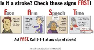
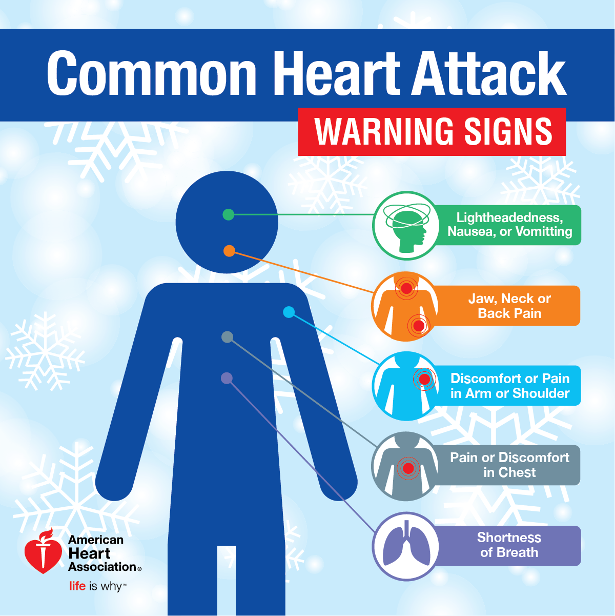
Chest pain is the most common symptom of a heart attack. The chest pain is usually a burning or pressure sensation beneath the mid or upper breast bone. The pain may radiate into the upper mid back, neck, jaw or arms. The pain may be severe but often is only moderate in severity.

There may be associated shortness of breath or sweating. If patients have had angina previously, the heart attack pain will feel the same as their usual angina only stronger and more prolonged. If you have a pain like this that lasts longer than 15 minutes, it is best to be evaluated immediately.

Calling your medic unit is the fastest and safest way to ask for help. If you have symptoms like this that wax and wane, this is often a warning sign that a heart attack is about to occur and prompt medical attention is needed.

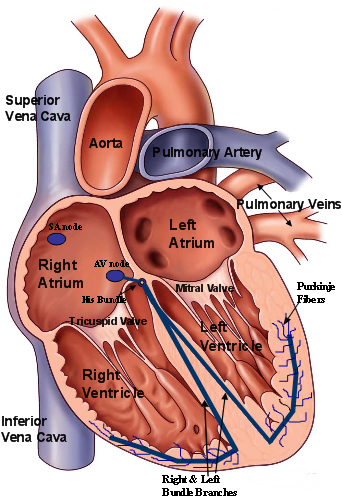
Once you are in an emergency room or a doctor’s office an electrocardiogram (ECG or EKG) will be obtained. This is often helpful in diagnosing a heart attack. Sometimes, however, the test is normal even when the heart injury is present but usually a great diagnostic tool.

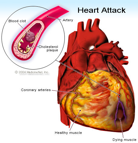
When heart cells die, certain enzymes present in heart cells are released into the bloodstream that serve as a marker of heart injury (troponin I and CPK or CK-MB). These enzymes can be measured by blood tests. The amount of enzyme released into the blood stream also helps assess how much heart damage has occurred.

How a heart attack is commonly treated:

The best way to limit the size of a heart attack is to restore the flow of blood to the heat muscle as fast as possible. There are two basic methods to do this.

1-Drugs called thrombolytics are quite effective.

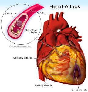
Because most heart attacks are caused by clots forming within the coronary artery, dissolving the clot quickly will restore blood flow and remove the symptoms.

The sooner these drugs are given, the quicker the blood flow will be restored.

2-Surgery

An alternative method involves the use of a angiogram– its a procedure that allows the visual display of the blockage on a computerized screen in the OR for the surgeon.  This is done through a special catheter with a camera at the end of it.

Commonly followed is a balloon angioplasty-this procedure is a balloon inflating and deflating breaking the blockage up on the computerized screen to make the blocked artery patent again to give good blood supply back to the area where the heart attack was.

Both angiogram and angioplasty is performed in the same surgical procedure.

This involves taking the heart attack victim promptly to the cardiac cath lab in the hospital.

An angiogram is performed to show the blocked blood vessel leading to the heart attack. Then a balloon catheter is placed across the blockage and flow is restored.

2-Stent – another common surgical procedure (this is a device that assists in holding the blood vessel open permanently or sometimes needs to be replaced). This is put in place where the artery needs to stay open, where the MI occurred, this creates a large channel for better blood supply to the area where the myocardial infarction occurred or close to it.

3-Bed rest and blood thinners – Smaller heart attacks, often those not producing significant abnormalities on the ECG are often treated with bedrest and blood thinners such as heparin or other meds to reduce the workload of the heart .  These heart attacks are called non-transmural myocardial infarctions. Before discharge, x-ray studies of the heart arteries are often carried out to see if angioplasty or surgery will be necessary.

Following thrombolytic (clot reducing) therapy, angiogram are often performed to outline the coronary anatomy to help determine if additional therapy such as angioplasty or bypass surgery is indicated. This may be done during the initial hospitalization or later as an outpatient procedure.

Following the treatment would be frequent cardiac visits to the cardiologist at first for follow up visits to than q3mths to q6mths to than just yearly if things all go well.  The M.D. will know the answer to that.

Complications that are often related to the damage done to your heart during a heart attack, which can lead to:

1-Abnormal heart rhythms (arrhythmias). Electrical “short circuits” can develop, resulting in abnormal heart rhythms, some of which can be serious, and may lead to death.

2-Heart failure. A heart attack might damage so much heart tissue that the remaining heart muscle can’t pump enough blood out of your heart. Heart failure can be temporary, or it can be a chronic condition resulting from extensive and permanent damage to your heart.

3-Sudden cardiac arrest. Without warning, your heart stops due to an electrical disturbance that causes an abnormal heart rhythm (arrhythmia). Heart attacks increase the risk of sudden cardiac arrest, which can cause death without immediate treatment.

QUOTE FOR MONDAY:

“A heart attack, also called a myocardial infarction, happens when a part of the heart muscle doesn’t get enough blood.

The more time that passes without treatment to restore blood flow, the greater the damage to the heart muscle.”

Center for Disease Control and Prevention (CDC)

What happens when someone has a heart attack or myocardial infarction:


A heart attack is a frightening experience. If you have experienced a heart attack, or are close with someone who has, you should know this: You are not alone. In fact, tens of thousands of people survive heart attacks and go on to lead productive, enjoyable lives.

What happens when a heart attach occurs:

First like striveforgoodhealth.com has stated this past week and before in previous articles remember ANY organ without 02-oxygen will die.  When this happens high risk a infarction will occur.  If this occurs in the brain its a brain infarction; if this occurs in the heart it is called a heart attack medically known as a myocardial infarction.

When you have a heart attack part of the heart tissue dies.  Your heart muscle needs oxygen to survive. A heart attack occurs when the blood flow that brings oxygen to the heart muscle is severely reduced or cut off completely.

A heart attack occurs when the flow of blood to an area of the heart is blocked. The blockage is most often a buildup of fat, cholesterol and other substances, which form a plaque in the arteries that feed that area of the heart (coronary arteries).

Sometimes, a plaque can rupture and form a clot that blocks blood flow. The interrupted blood flow can damage or destroy part of the heart muscle.

Also a condition called Atrial Fibrillation (AF), a type of a cardiac rhythm,  a very common type.  This AF can cause pooling of the blood in the chanmbers of the heart due to the irregular heart beat overtime not diagnosed or treated yet.  Overtime this pooling of blood can create a clot putting the heart at risk for complications.  The bulk of evidence in research demonstrates that AF patients hospitalized for AMI have serious adverse prognostic implications regarding in-hospital, but also long-term mortality.

A heart attack, also called a myocardial infarction, can be fatal, but treatment has improved dramatically over the years. It’s crucial to call 911 or emergency medical help if you think you might be saving a heart attack.

The sooner you get to an emergency room, the sooner you can get treatment to reduce the amount of damage to the heart muscle. At the hospital, health care professionals can run tests to find out if a heart attack is happening and decide the best treatment.

In some cases, a heart attack requires cardiopulmonary resuscitation (CPR) or an electrical shock (defibrillation) to the heart to get the heart pumping again. Bystanders trained to use CPR or a defibrillator may be able to help until emergency medical personnel arrive.

Remember, the chances of surviving a heart attack are better the sooner emergency treatment begins.

Common Symptoms of a Heart Attack:

  • Chest pain or discomfort. Most heart attacks involve discomfort in the center or left side of the chest that lasts for more than a few minutes or that goes away and comes back. The discomfort can feel like uncomfortable pressure, squeezing, fullness, or pain.
  • Feeling weak, light-headed, or faint. You may also break out into a cold sweat.
  • Pain or discomfort in the jaw, neck, or back.
  • Pain or discomfort in one or both arms or shoulders.
  • Shortness of breath. This often comes along with chest discomfort, but shortness of breath also can happen before chest discomfort.

*Other symptoms of a heart attack could include unusual or unexplained tiredness and nausea or vomiting. Women are more likely to have these other symptoms.*

CAUSES OF HEART ATTACKS:

A heart attack occurs when one or more of your coronary arteries becomes blocked. Over time, a buildup of fatty deposits, including cholesterol, form substances called plaques, which can narrow the arteries (atherosclerosis). This condition, called coronary artery disease, causes most heart attacks.

During a heart attack, a plaque can rupture and spill cholesterol and other substances into the bloodstream. A blood clot forms at the site of the rupture. If the clot is large, it can block blood flow through the coronary artery, starving the heart of oxygen and nutrients (ischemia).

You might have a complete or partial blockage of the coronary artery.

  • A complete blockage means you’ve had an ST elevation myocardial infarction (STEMI).
  • A partial blockage means you’ve had a non-ST elevation myocardial infarction (NSTEMI).

QUOTE FOR THE WEEKEND::

“Cardiomyopathy refers to diseases of the heart muscle. These diseases have many causes, signs and symptoms as well as treatments. In most cases, cardiomyopathy causes the heart muscle to become enlarged, thick or rigid. In rare instances, diseased heart muscle tissue is replaced with scar tissue.  Scar Tissue cannot be replaced anywhere in the body”

American Heart Association

Cardiomyopathy


NORNAL SIZE HEART


CARDIOMYOPATHY HEART Look at the second picture of the heart at the bottom right

CARDIOMYOPATHY:

Cardiomyopathy (kahr-dee-o-my-OP-uh-thee) is a disease of the heart muscle that makes it harder for your heart to pump blood to the rest of your body. Cardiomyopathy can lead to heart failure.

The main types of cardiomyopathy include dilated, hypertrophic and restrictive cardiomyopathy. Treatment — which might include medications, surgically implanted devices or, in severe cases, a heart transplant —this would all depend on which type of cardiomyopathy you have and how serious it is.

Symptoms

There might be no signs or symptoms in the early stages of cardiomyopathy. But as the condition advances, signs and symptoms usually appear, including:

  • Breathlessness with exertion or even at rest
  • Swelling of the legs, ankles and feet
  • Bloating of the abdomen due to fluid buildup
  • Cough while lying down
  • Fatigue
  • Heartbeats that feel rapid, pounding or fluttering
  • Chest discomfort or pressure
  • Dizziness, lightheadedness and fainting

Signs and symptoms tend to get worse unless treated. In some people, the condition worsens quickly; in others, it might not worsen for a long time.

When to see a doctor

See your doctor if you have one or more signs or symptoms associated with cardiomyopathy. Call 911 or your local emergency number if you have severe difficulty breathing, fainting or chest pain that lasts for more than a few minutes.

Because some types of cardiomyopathy can be hereditary, if you have it your doctor might advise that your family members be checked.

Causes

Often the cause of the cardiomyopathy is unknown. In some people, however, it’s the result of another condition (acquired) or passed on from a parent (inherited).

Contributing factors for acquired cardiomyopathy include:

  • Long-term high blood pressure
  • Heart tissue damage from a heart attack
  • Chronic rapid heart rate
  • Heart valve problems
  • Metabolic disorders, such as obesity, thyroid disease or diabetes
  • Nutritional deficiencies of essential vitamins or minerals, such as thiamin (vitamin B-1)
  • Pregnancy complications
  • Drinking too much alcohol over many years
  • Use of cocaine, amphetamines or anabolic steroids
  • Use of some chemotherapy drugs and radiation to treat cancer
  • Certain infections, especially those that inflame the heart
  • Iron buildup in your heart muscle (hemochromatosis)
  • A condition that causes inflammation and can cause lumps of cells to grow in the heart and other organs (sarcoidosis)
  • A disorder that causes the buildup of abnormal proteins (amyloidosis)
  • Connective tissue disorders

Types of cardiomyopathy include:

  • Dilated cardiomyopathy. In this type of cardiomyopathy, the pumping ability of your heart’s main pumping chamber — the left ventricle — becomes enlarged (dilated) and can’t effectively pump blood out of the heart.Although this type can affect people of all ages, it occurs most often in middle-aged people and is more likely to affect men. The most common cause is coronary artery disease or heart attack.
  • Hypertrophic cardiomyopathy. This type involves abnormal thickening of your heart muscle, particularly affecting the muscle of your heart’s main pumping chamber (left ventricle). The thickened heart muscle can make it harder for the heart to work properly.Hypertrophic cardiomyopathy can develop at any age, but the condition tends to be more severe if it becomes apparent during childhood. Most affected people have a family history of the disease, and some genetic mutations have been linked to hypertrophic cardiomyopathy.
  • Restrictive cardiomyopathy. In this type, the heart muscle becomes rigid and less elastic, so it can’t expand and fill with blood between heartbeats. This least common type of cardiomyopathy can occur at any age, but it most often affects older people.Restrictive cardiomyopathy can occur for no known reason (idiopathic), or it can by caused by a disease elsewhere in the body that affects the heart, such as when iron builds up in the heart muscle (hemochromatosis).
  • Arrhythmogenic right ventricular dysplasia. In this rare type of cardiomyopathy, the muscle in the lower right heart chamber (right ventricle) is replaced by scar tissue, which can lead to heart rhythm problems. It’s often caused by genetic mutations.
  • Unclassified cardiomyopathy. Other types of cardiomyopathy fall into this category.

Risk factors

There are a number of factors that can increase your risk of cardiomyopathy, including:

  • Family history of cardiomyopathy, heart failure and sudden cardiac arrest
  • Long-term high blood pressure
  • Conditions that affect the heart, including a past heart attack, coronary artery disease or an infection in the heart (ischemic cardiomyopathy)
  • Obesity, which makes the heart work harder
  • Long-term alcohol abuse
  • Illicit drug use, such as cocaine, amphetamines and anabolic steroids
  • Certain chemotherapy drugs and radiation therapy for cancer
  • Certain diseases, such as diabetes, an under- or overactive thyroid gland, or a disorder that causes the body to store excess iron (hemochromatosis)
  • Other conditions that affect the heart, such as a disorder that causes the buildup of abnormal proteins (amyloidosis), a disease that causes inflammation and can cause lumps of cells to grow in the heart and other organs (sarcoidosis), or connective tissue disorders

Complications

Cardiomyopathy can lead to other heart conditions, including:

  • Heart failure. Your heart can’t pump enough blood to meet your body’s needs. Untreated, heart failure can be life-threatening.
  • Blood clots. Because your heart can’t pump effectively, blood clots might form in your heart. If clots enter your bloodstream, they can block the blood flow to other organs, including your heart and brain.
  • Valve problems. Because cardiomyopathy causes the heart to enlarge, the heart valves might not close properly. This can lead to a backward flow of blood.
  • Cardiac arrest and sudden death. Cardiomyopathy can lead to abnormal heart rhythms. These abnormal heart rhythms can result in fainting or, in some cases, sudden death if your heart stops beating effectively.

Prevention

In many cases, you can’t prevent cardiomyopathy. Let your doctor know if you have a family history of the condition.

You can help reduce your chance of cardiomyopathy and other types of heart disease by living a heart-healthy lifestyle and making lifestyle choices such as:

  • Avoiding the use of alcohol or cocaine
  • Controlling high blood pressure, high cholesterol and diabetes
  • Eating a healthy diet
  • Getting regular exercise
  • Getting enough sleep
  • Reducing your stress