Archive | July 2019

QUOTE FOR WEDNESDAY:

Osteoporosis, which literally means porous bone, is a disease in which the density and quality of bone are reduced. As bones become more porous and fragile, the risk of fracture is greatly increased. The loss of bone occurs silently and progressively. Often there are no symptoms until the first fracture occurs.”

National Osteoporosis Foundation (www.nof.org)

Osteoporosis – What it is , its effects on the human body & the benefits of exercise.

 webmd_rm_photo_of_porous_bonesWNL vs diseased liveosteoporosis skeletal areas affectedtimthumb

How the neck of the foramen breaks due to Osteo.

Its a progressive bone disease that is characterised by a decrease in bone mass and density and that leads to an increased risk of fracture.  In osteoporosis, the bone mineral density (BMD) is reduced, bone microarchitecture deteriorates, and the amount and variety of proteins in bone are altered.

Osteoporosis causes bones to become weak and brittle —- so brittle that a fall or even mild stresses like bending over or coughing can cause a fracture. Osteoporosis-related fractures most commonly occur in the hip, wrist or spine. Bone is living tissue, which is constantly being absorbed and replaced. Osteoporosis occurs when the creation of new bone doesn’t keep up with the removal of old bone. Osteoporosis affects men and women of all races; but white and Asian women–especially after menopause–are at highest risk. Medications, healthy diet and weight bearing exercise can help prevent bone loss or strengthen already weak bones.

A weight bearing exercise is any exercise that has your legs and feet holding all of your weight. An example of this would be walking, yoga or even dancing.

The form of osteoporosis most common in women after menopause is referred to as primary type 1 or postmenopausal osteoporosis. Primary type 2 osteoporosis or senile osteoporosis occurs after age 75 and is seen in both females and males at a ratio of 2:1. Secondary osteoporosis may arise at any age and affect men and women equally. This form results from chronic predisposing medical problems or disease, or prolonged use of medications such as glucocorticoids, when the disease is called steroid- or glucocorticoid-induced osteoporosis.

The risk of osteoporosis fractures can be reduced with lifestyle changes and in those with previous osteoporosis related fractures medications. Lifestyle change includes diet, exercise, and preventing falls. The utility of calcium and vitamin D is questionable in most. Bisphosphonates are useful in those with previous fractures from osteoporosis but are of minimal benefit in those who have osteoporosis but no previous fractures. Osteoporosis is a component of the frailty syndrome.

Take the problem of Astronauts with osteoporosis:

Space travel has made it widely known that a stay outside the atmosphere – and thus outside the earth’s gravitational influence – disturbs the metabolism irreparably: the human body does not need any hard bones in zero-gravity, which leads to decalcification. A four year study of the ‘International Space Station’ showed that the bones of astronauts did not regenerate after even one year past their return to earth.

Anti-gravitational training is the key to osteoporosis if you can handle it (like jumping on a trampeline) Actual studies show that physical anti-gravitational activity helps the effected patients to regain their mobility and lessen the risk of bone fractures .

Osteoporosis in the U.S.A.

The National Osteoporosis Foundation (NOF) today released updated prevalence data estimating that a total of 54 million U.S. adults age 50 and older are affected by osteoporosis and low bone mass. Recently published online by the Journal of Bone and Mineral Research, the study, “The Recent Prevalence of Osteoporosis and Low Bone Mass in the United States Based on Bone Mineral Density at the Femoral Neck or Lumbar Spine,” includes the number of adults age 50 and over from the institutionalized and non-institutionalized population affected by osteoporosis and low bone mass and is an update to the prevalence data NOF released in 2013. Revealing that 10.2 million adults have osteoporosis and another 43.4 million have low bone mass, more than one-half of the total U.S. adult population is currently affected.

Assuming osteoporosis and low bone mass prevalence remain unchanged, the study projects that by 2020, the number of adults over age 50 with osteoporosis or low bone mass will grow from approximately 54 million to 64.4 million and by 2030, the number will increase to 71.2 million (a 29% increase from 2010); and it is anticipated that the number of fractures will grow proportionally.

“This disease causes an estimated two million broken bones each year and often results in immobility, pain, placement in a nursing home, isolation and other health problems, said Amy Porter, executive director and CEO of NOF. “Medicare (our tax dollars) pays for the cost for repair of 80 percent of broken bones that occur because of osteoporosis; these costs make osteoporosis the 10th ranked major illness among the top 5% highest cost Medicare beneficiaries (12% of all beneficiaries and 18% of high costs beneficiaries). We have to continue our efforts to eradicate this disease.”

The data is one of the first to look at the burden of osteoporosis using NOF’s criteria for diagnosing osteoporosis based on bone mineral density (BMD) at the hip or spine. Prior to 2005, the National Health and Nutrition Examination Survey (NHANES) only measured BMD at the hip. Spine BMD was added in 2005, providing the opportunity to estimate the burden of osteoporosis using BMD at either the hip or spine. The study estimates that among adults age 50 years and older, 10.2 million have osteoporosis at the femoral neck or lumbar spine and an additional 43.4 million have low bone mass at either skeletal site, placing them at increased risk for osteoporosis and broken bones.

Benefits of exercise

Women who have been physically active throughout their lives generally have stronger bones than do women who have led more sedentary lives. But it’s never too late to start exercising. For postmenopausal women, regular physical activity can:

  • Increase your muscle strength
  • Improve your balance
  • Make you better able to carry out daily tasks and activities
  • Maintain or improve your posture
  • Relieve or decrease pain
  • Improve your sense of well-being Before you start having osteoporosis.
  • Consult your doctor before starting any exercise program for osteoporosis. You may need some tests first, including:
  • Exercising if you have osteoporosis means finding the safest, most enjoyable activities for you given your overall health and amount of bone loss. There’s no one-size-fits-all prescription.
  • Bone density measurement
  • Fitness assessment

In the meantime, think about what kind of activities you enjoy most. If you choose an exercise you enjoy, you’re more likely to stick with it over time.

QUOTE FOR TUESDAY:

 
“Anemia is the most common blood disorder, and according to the National Heart, Lung, and Blood Institute, it affects more than 3 million Americans.”
 
American Society of Hematology (www.hematology.org)

Part 2 Anemia

Those at risk for anemia:

Anemia is a common condition. It occurs in all age, racial, and ethnic groups. Both men and women can have anemia. However, women of childbearing age are at higher risk for the condition because of blood loss from menstruation.

Anemia can develop during pregnancy due to low levels of iron and folic acid (folate) and changes in the blood. During the first 6 months of pregnancy, the fluid portion of a woman’s blood (the plasma) increases faster than the number of red blood cells. This dilutes the blood and can lead to anemia.

During the first year of life, some babies are at risk for anemia because of iron deficiency. At-risk infants include those who are born too early and infants who are fed breast milk only or formula that isn’t fortified with iron. These infants can develop iron deficiency by 6 months of age.

Infants between 1 and 2 years of age also are at risk for anemia. They may not get enough iron in their diets, especially if they drink a lot of cow’s milk. Cow’s milk is low in the iron needed for growth.

Drinking too much cow’s milk may keep an infant or toddler from eating enough iron-rich foods or absorbing enough iron from foods.

Older adults also are at increased risk for anemia. Researchers continue to study how the condition affects older adults. Many of these people have other medical conditions as well.

Major Risk Factors for anemia:

  • A diet that is low in iron, vitamins, or minerals
  • Blood loss from surgery or an injury
  • Long-term or serious illnesses, such as kidney disease, cancer, diabetes, rheumatoid arthritis, HIV/AIDS, inflammatory bowel disease (including Crohn’s disease), liver disease, heart failure, and thyroid disease
  • Long-term infections
  • A family history of inherited anemia, such as sickle cell anemia or thalassemia.

Complications of Anemia

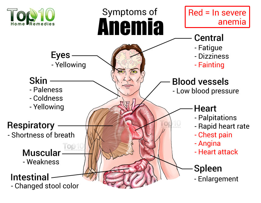
Some people who have anemia may have arrhythmias. Arrhythmias are problems with the rate or rhythm of the heartbeat. Over time, arrhythmias can damage your heart and possibly lead to heart failure.

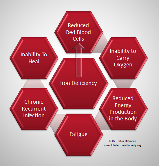
Anemia also can damage other organs in your body because your blood can’t get enough oxygen to them.

Anemia can weaken people who have cancer or HIV/AIDS. This can make their treatments not work as well.

Anemia also can cause many other health problems. People who have kidney disease and anemia are more likely to have heart problems. With some types of anemia, too little fluid intake or too much loss of fluid in the blood and body can occur. Severe loss of fluid can be life threatening.

QUOTE FOR MONDAY:

Anemia is a condition in which you don’t have enough healthy red blood cells to carry adequate oxygen to the body’s tissues. Having anemia may make you feel tired and weak. There are many forms of anemia, each with its own cause. Anemia can be temporary or long term, and it can range from mild to severe.

MAYO Clinic

Part I Anemia

anemia2

Anemia develops when you don’t have enough robust, healthy red blood cells to carry oxygen throughout your body. The blood cells may lack enough hemoglobin, the protein that gives blood its red color. Anemia affects one in 10 teen girls and women. It also develops in men and children and is linked to some illnesses.  Anemia is a condition that develops when your blood lacks enough healthy red blood cells or hemoglobin. Hemoglobin is a main part of red blood cells and binds oxygen. If you have too few or abnormal red blood cells, or your hemoglobin is abnormal or low, the cells in your body will not get enough oxygen. Symptoms of anemia — like fatigue — occur because organs aren’t getting what they need to function properly.

Anemia is the most common blood condition in the U.S. It affects about 3.5 million Americans. Women, young children, and people with chronic diseases are at increased risk of anemia.

There types of different anemia’s but today’s we’ll look more into is Iron deficiency anemia.

Iron deficiency anemia is a common type of anemia — a condition in which blood lacks adequate healthy red blood cells. Red blood cells carry oxygen to the body’s tissues.

As the name implies, iron deficiency anemia is due to insufficient iron. Without enough iron, your body can’t produce enough of a substance in red blood cells that enables them to carry oxygen (hemoglobin). As a result, iron deficiency anemia may leave you tired and short of breath.

You can usually correct iron deficiency anemia with iron supplementation. Sometimes additional tests or treatments for iron deficiency anemia are necessary, especially if your doctor suspects that you’re bleeding internally.

Initially, iron deficiency anemia can be so mild that it goes unnoticed. But as the body becomes more deficient in iron and anemia worsens, the signs and symptoms intensify.

Iron deficiency anemia symptoms may include:

  • Extreme fatigue, Pale skin, Weakness, Shortness of breath, Chest pain, Frequent infections
  • Headache, Dizziness or lightheadedness, Cold hands and feet, Inflammation or soreness of your tongue, Brittle nails, and Fast heartbeat.
  • Unusual cravings for non-nutritive substances, such as ice, dirt or starch.
  • Poor appetite, especially in infants and children with iron deficiency anemia.
  • An uncomfortable tingling or crawling feeling in your legs (restless legs syndrome).

Causes of iron deficiency anemia include:

  • Blood loss. Blood contains iron within red blood cells. So if you lose blood, you lose some iron. Women with heavy periods are at risk of iron deficiency anemia because they lose blood during menstruation. Slow, chronic blood loss within the body — such as from a peptic ulcer, a hiatal hernia, a colon polyp or colorectal cancer — can cause iron deficiency anemia. Gastrointestinal bleeding can result from regular use of some over-the-counter pain relievers, especially aspirin.
  • A lack of iron in your diet. Your body regularly gets iron from the foods you eat. If you consume too little iron, over time your body can become iron deficient. Examples of iron-rich foods include meat, eggs, leafy green vegetables and iron-fortified foods. For proper growth and development, infants and children need iron from their diet, too.
  • An inability to absorb iron. Iron from food is absorbed into your bloodstream in your small intestine. An intestinal disorder, such as celiac disease, which affects your intestine’s ability to absorb nutrients from digested food, can lead to iron deficiency anemia. If part of your small intestine has been bypassed or removed surgically, that may affect your ability to absorb iron and other nutrients.
  • Without iron supplementation, iron deficiency anemia occurs in many pregnant women because their iron stores need to serve their own increased blood volume as well as be a source of hemoglobin for the growing fetus.


QUOTE FOR THE WEEKEND:

“In cancer care, different types of doctors often work together to create a patient’s overall treatment plan that combines different types of treatments. This is called a multidisciplinary team.”

Cancer.Net (www.cancer.net)

QUOTE FOR FRIDAY:

“According to the American Cancer Society, about 11,280 people will be diagnosed with sarcoma this year and slightly more men than women develop soft tissue sarcoma. Sarcomas are cancers that are much more likely to affect children and young adults than many other more common cancers. Sarcoma is a very rare disease. Due to its rarity, it is crucial for patients to seek a cancer specialist in the treatment of their disease. Sarcoma arises in the connective tissue of the body.”

Rogel Cancer Center Michigan Medicine (www.rogelcancercenter.org)

QUOTE FOR THURSDAY:

“Gastrointestinal stromal tumor, GIST, is a relatively uncommon type of cancer that occurs in the gastrointestinal tract. GIST’s belong to a class of diseases called sarcomas. Experts estimate that 4,500 to 6,000 GIST’s are diagnosed each year in the United States. GIST’s can develop anywhere in the GI tract but occur most often in the stomach (approximately 60%).”

GIST Cancer Awareness Foundation (gistawareness.org)

Gastrointestinal stromal tumor (GIST)

Gastrointestinal stromal tumors (GISTs) are soft-tissue sarcomas that can be located in any part of the digestive system. Their most common sites are the stomach and small intestine.

GISTs start in specialized nerve cells located in the walls of your digestive system. These cells are part of the autonomic nervous system. A specific change in the DNA of one of these cells, which control such digestive processes as movement of food through the intestines, gives rise to a GIST.

Small GISTs may cause no symptoms, and they may grow so slowly that they have no serious effects. People with larger GISTs usually seek medical attention when they vomit blood or pass blood in their stool due to rapid bleeding from the tumor.

Other possible GIST symptoms include:

  • Anemia, caused by a slow-bleeding tumor
  • Abdominal pain
  • A growth you can feel in your abdomen
  • Nausea
  • Vomiting
  • Loss of appetite
  • Weight loss
  • Difficulty swallowing

GISTs can develop in people of all ages, but they are most common between age 50 and 70, and they almost never occur before age 40. In rare cases, an inherited genetic change (mutation) causes GISTs.

Diagnosis

After asking questions about your symptoms and medical history, your doctor will examine you carefully, checking for a growth in your abdomen. If signs and symptoms suggest you may have a GIST, tests to locate it and then determine its likelihood of spreading (metastasizing) to other organs will follow. These tests may include:

    • Contrast-enhanced computerized tomography (CT) scan. For this test, you swallow a liquid that makes your stomach and small intestine more visible on X-rays. You may also receive an injection of a similar substance. Then the scanner takes numerous X-rays as it moves over your abdomen. A computer combines the X-rays into detailed, cross-sectional images of your abdominal organs, showing the size and position of the tumor.
    • Upper endoscopy. The doctor examines the inner lining of the esophagus, stomach and the first part of the small intestine with a flexible, lighted tube (endoscope) passed down through your mouth. It may be possible to take small samples of abnormal tissue during an upper endoscopy. During this test, you’ll receive a mild relaxing medication (sedative) through an intravenous line.
    • Endoscopic ultrasound (EUS). This test also uses an endoscope, but with an ultrasound probe on the tip of the scope. As sound waves from the probe create echoes that bounce back to the probe, a computer translates the echoes into an image of the structures in the abdomen, showing the precise location of the tumor. If the tumor has metastasized to your liver or the lining of your abdomen, these areas may also be visible. An EUS also helps determine the depth of the tumor within the wall of the stomach or other locations in the gastrointestinal tract.
    • Fine-needle aspiration biopsy. A small sample of tissue from the tumor is necessary for a definite GIST diagnosis. The preferred method for taking a biopsy sample is endoscopic ultrasound with fine-needle aspiration. This procedure is the same as an EUS, but with a thin, hollow needle on the tip of the endoscope. The needle is used to remove small amounts of tissue for laboratory analysis.

Sometimes these tests aren’t possible, or their results are inconclusive, so a suspected GIST can’t be located without surgery. Tissue analysis then takes place after the tumor is removed.

  • Laboratory tests on biopsies. These tests provide information on the proteins tumor cells make. One of these tests, immunohistochemistry, detects specific proteins controlled by genes in GIST cells. Identification of these proteins helps guide treatment decisions. Sometimes, actual genetic testing of biopsy samples is necessary to locate GIST genes in tumor DNA. GIST cells are also examined under a microscope to see how many cells out of 50 different microscopic fields are actively dividing. This number is known as the mitotic rate. The higher the mitotic rate in a tumor, the more aggressive it is, and the greater is its likelihood of spreading to other organs.

Treatment

Small, asymptomatic GISTs found in the course of tests for another condition may be approached with watchful waiting in carefully selected cases.

Surgery

All large or symptomatic GISTs should be surgically removed unless they are too large or they involve too many organs and tissues for surgery (resection). Resection is also delayed or avoided in people whose general health makes any surgery too risky to undertake, as well as those likely to have metastatic GISTs.

It’s often possible to resect GISTs using minimally invasive surgery, which involves inserting a viewing tube (laparoscope) and surgical instruments through small incisions in the abdomen.

Targeted drug therapy

GISTs do not respond to traditional chemotherapy. Thanks to recently gained understanding of tumor genetic changes in GISTs, however, drugs that interrupt the process of tumor spread have greatly improved the outlook for people with the disease.

Imatinib (Gleevec) is the first line medical treatment used to prevent GIST recurrence after surgery. The drug is also used in situations where surgery isn’t possible, as well as in controlling recurrent GIST.

The current trend is to continue imatinib treatment as long as it’s tolerated and it remains effective. Unfortunately, GISTs tend to become resistant to imatinib over time. A different targeted drug, sunitinib malate (Sutent) often works on imatinib-resistant GISTs. A number of other targeted drugs now in development are expected to join imatinib and sunitinib in coming years.