Archive | September 2023

QUOTE FOR THE WEEKEND:

“Benign paroxysmal positional vertigo (BPPV) is the most common vertiginous disorder in the community. The cardinal symptom is sudden vertigo induced by a change in head position: turning over in bed, lying down in bed (or at the dentist or hairdresser), looking up, stooping, or any sudden change in head position. There is a wide spectrum of severity. Mild symptoms are inconsistent positional vertigo. Moderate symptoms are frequent positional attacks with disequilibrium between. When severe, vertigo is provoked by most head movements, giving an impression of continuous vertigo. The symptoms can last for days, weeks, months, or years, or be recurrent over many years.”

National Library of Medicine (https://www.ncbi.nlm.nih.gov/pmc/articles/PMC3144715/)

QUOTE FOR FRIDAY:

“BPPV is estimated to affect roughly 50% of all people at some time in their lives and becomes progressively more common with age. During periods when attacks are not occurring, the diagnosis is made from the characteristic history and by the exclusion of other disorders that can cause similar dizzy symptoms. When attacks are occurring, the Hallpike positional test is diagnostic. In the commonest form of BPPV the Hallpike test is positive (i.e. induces vertigo and nystagmus) when the affected ear is down most. Up to 10% of cases may involve both ears.”

Menieres Society (https://www.menieres.org.uk/information-and-support/symptoms-and-conditions/bppv)

QUOTE FOR THURSDAY:

“Benign paroxysmal positional vertigo (BPPV) is the most common cause of peripheral vertigo, accounting for over half of all cases. According to various estimates, a minimum of 20% of patients presenting to the provider with vertigo have BPPV. However, this figure could be an underestimation as BPPV is frequently misdiagnosed. It is crucial to distinguish BPPV from other causes of vertigo as the differential diagnosis includes a spectrum of disease processes ranging from benign to life-threatening. Because of the misleading and vague term ‘dizziness’ that patients commonly use, the provider must pin down what every patient means by it. It can be often achieved by asking the patient to describe what they are feeling without the use of the word ‘dizziness.

Barany first described BPPV in 1921. At that time, characteristic vertigo and nystagmus associated with postural changes were linked to the otolithic organs. In 1952, Dix and Hallpike, during their provocative testing, further described classic nystagmus and moved on to explain that the location of the pathology was the ear proper.”

National Library of Medicine (https://www.ncbi.nlm.nih.gov/books/NBK470308/)

QUOTE FOR WEDNESDAY:

“In 2021, an estimated 12.3 million adults seriously thought about suicide, 3.5 million made a plan, and 1.7 million attempted suicide. Many factors can increase the risk for suicide or protect against it. Suicide is connected to other forms of injury and violence. For example, people who have experienced violence, including child abuse, bullying, or sexual violence have a higher suicide risk. Being connected to family and community support and having easy access to healthcare can decrease suicidal thoughts and behaviors. So know there is a way to PREVENTION!”.

Centers for Disease Control – CDC (https://www.cdc.gov/suicide/facts/index.html)

Suicide in America


Suicide is a major public health problem and a leading cause of death in the United States. The effects of suicide go beyond the person who acts to take his or her life: it can have a lasting effect on family, friends, and communities. This fact sheet, developed by the National Institute of Mental Health (NIMH), can help you, a friend, or a family member learn about the signs and symptoms, risk factors and warning signs, and ongoing research about suicide and suicide prevention.

What Is Suicide?

Suicide is when people direct violence at themselves with the intent to end their lives, and they die because of their actions. It’s best to avoid the use of terms like “committing suicide” or a “successful suicide” when referring to a death by suicide as these terms often carry negative connotations.

A suicide attempt is when people harm themselves with the intent to end their lives, but they do not die because of their actions.

Who Is at Risk for Suicide?

Suicide does not discriminate. People of all genders, ages, and ethnicities can be at risk.

The main risk factors for suicide are:

  • A prior suicide attempt
  • Depression and other mental health disorders
  • Substance abuse disorder
  • Family history of a mental health or substance abuse disorder
  • Family history of suicide
  • Family violence, including physical or sexual abuse
  • Having guns or other firearms in the home
  • Being in prison or jail
  • Being exposed to others’ suicidal behavior, such as a family member, peer, or media figure
  • Medical illness
  • Being between the ages of 15 and 24 years or over age 60

Even among people who have risk factors for suicide, most do not attempt suicide. It remains difficult to predict who will act on suicidal thoughts.

Are certain groups of people at higher risk than others?

According to the Centers for Disease Control and Prevention (CDC), men are more likely to die by suicide than women, but women are more likely to attempt suicide. Men are more likely to use more lethal methods, such as firearms or suffocation. Women are more likely than men to attempt suicide by poisoning.

Also per the CDC, certain demographic subgroups are at higher risk. For example, American Indian and Alaska Native youth and middle-aged persons have the highest rate of suicide, followed by non-Hispanic White middle-aged and older adult males. African Americans have the lowest suicide rate, while Hispanics have the second lowest rate. The exception to this is younger children. African American children under the age of 12 have a higher rate of suicide than White children. While younger preteens and teens have a lower rate of suicide than older adolescents, there has been a significant rise in the suicide rate among youth ages 10 to 14. Suicide ranks as the second leading cause of death for this age group, accounting for 425 deaths per year and surpassing the death rate for traffic accidents, which is the most common cause of death for young people.

Why do some people become suicidal while others with similar risk factors do not?

Most people who have the risk factors for suicide will not kill themselves. However, the risk for suicidal behavior is complex. Research suggests that people who attempt suicide may react to events, think, and make decisions differently than those who do not attempt suicide. These differences happen more often if a person also has a disorder such as depression, substance abuse, anxiety, borderline personality disorder, and psychosis. Risk factors are important to keep in mind; however, someone who has warning signs of suicide may be in more danger and require immediate attention.

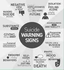
What Are the Warning Signs of Suicide?

The behaviors listed below may be signs that someone is thinking about suicide.

  • Talking about wanting to die or wanting to kill themselves
  • Talking about feeling empty, hopeless, or having no reason to live
  • Planning or looking for a way to kill themselves, such as searching online, stockpiling pills, or newly acquiring potentially lethal items (e.g., firearms, ropes)
  • Talking about great guilt or shame
  • Talking about feeling trapped or feeling that there are no solutions
  • Feeling unbearable pain, both physical or emotional
  • Talking about being a burden to others
  • Using alcohol or drugs more often
  • Acting anxious or agitated
  • Withdrawing from family and friends
  • Changing eating and/or sleeping habits
  • Showing rage or talking about seeking revenge
  • Taking risks that could lead to death, such as reckless driving
  • Talking or thinking about death often
  • Displaying extreme mood swings, suddenly changing from very sad to very calm or happy
  • Giving away important possessions
  • Saying goodbye to friends and family
  • Putting affairs in order, making a will

Do People Threaten Suicide to Get Attention?

Suicidal thoughts or actions are a sign of extreme distress and an alert that someone needs help. Any warning sign or symptom of suicide should not be ignored. All talk of suicide should be taken seriously and requires attention. Threatening to die by suicide is not a normal response to stress and should not be taken lightly.

If You Ask Someone About Suicide, Does It Put the Idea Into Their Head?

Asking someone about suicide is not harmful. There is a common myth that asking someone about suicide can put the idea into their head. This is not true. Several studies examining this concern have demonstrated that asking people about suicidal thoughts and behavior does not induce or increase such thoughts and experiences. In fact, asking someone directly, “Are you thinking of killing yourself,” can be the best way to identify someone at risk for suicide.

What Should I Do if I Am in Crisis or Someone I Know Is Considering Suicide?

If you or someone you know has warning signs or symptoms of suicide, particularly if there is a change in the behavior or a new behavior, get help as soon as possible.

Often, family and friends are the first to recognize the warning signs of suicide and can take the first step toward helping an at-risk individual find treatment with someone who specializes in diagnosing and treating mental health conditions. If someone is telling you that they are going to kill themselves, do not leave them alone. Do not promise anyone that you will keep their suicidal thoughts a secret. Make sure to tell a trusted friend or family member, or if you are a student, an adult with whom you feel comfortable. You can also contact the resources noted below.

Leading Cause of Death in the United States (2016)
Data Courtesy of CDC
Select Age Groups
Rank 10-14 15-24 25-34 35-44 45-54 55-64 All Ages
1 Unintentional
Injury
847
Unintentional
Injury
13,895
Unintentional
Injury
23,984
Unintentional
Injury
20,975
Malignant
Neoplasms
41,291
Malignant
Neoplasms
116,364
Heart
Disease
635,260
2 Suicide
436
Suicide
5,723
Suicide
7,366
Malignant
Neoplasms
10,903
Heart
Disease
34,027
Heart
Disease
78,610
Malignant
Neoplasms
598,038
3 Malignant
Neoplasms
431
Homicide
5,172
Homicide
5,376
Heart
Disease
10,477
Unintentional
Injury
23,377
Unintentional
Injury
21,860
Unintentional
Injury
161,374
4 Homicide
147
Malignant
Neoplasms
1,431
Malignant
Neoplasms
3,791
Suicide
7,030
Suicide
8,437
      CLRD
17,810

QUOTE FOR TUESDAY:

“A 12 lead EKG is a painless and noninvasive test that measures your heart’s electrical efficiency as it beats. As one of the fastest informational or diagnostic heart tests available, EKG testing can usually be completed in just five minutes.  To conduct an EKG test, our team attaches up to 12 small, flat, sticky patches called electrodes at various points on your chest, arms, and legs. The electrodes are connected to a monitor that registers your heart’s electrical activity over the course of the exam.Their is another device called telemetry monitoring that can be done in the hospital or even at home through what we call a holter monitor.  Both telemetry monitoring or holter monitoring are done with a 5 leads or electrodes on the upper chest (R and L side), mid chest (just under nipple line close to where the heart lies) and 2 more leads or electrodes just below the rib cage (on the R and L side). This holter device can be done for a couple of days and returned to the MD’s office.  An EKG or holter monitor test results that tell us whether electrical waves pass through your heart at a normal rate, faster than normal, slower than normal, or in an irregular pattern. Results that are fast, slow, or irregular, may be a sign that your heart is weak or overworked, or that it has some kind of structural (size or shape) abnormality.”

ECCA (https://eccacardiologists.com/2020/03/03/what-an-ekg-test-can-tell-your-doctor-about-your-heart/)

What cardiac rhythms tells your doctor about your heart!

Heart Beat symbol design element

Why cardiac monitoring can be vital important in quickly telling the doctors and nurses very important messages in what is going on with the patient’s heart and overall condition problem (Example A Myocardial Infarction or even to Cardiac Arrest).

Cardiac monitoring is a great way for doctors to understand a patients’ overall heart health, and can provide enough information to quickly and accurately helping the doctor or nurse as a diagnostic tool based on several details within a heart rhythm. While each arrhythmia monitoring device is a little different, these details are essential in diagnosing any underlying and potentially life-threatening events.

Your heart can have the best rhythm it can be in called Normal Sinus Rhythm which is a rhythm that is produced by the sinus node (SA node) that is the human pacemaker of the heart in out right upper atrium.  It starts a impulse (think of it as a message) that starts from the SA node and goes down the right atrium across to the left atrium (the upper chambers of the heart) with contiuing to send both impulses down to the bottom chambers of the heart which we call Ventricles creating the sound we all know the heart make called “lub dub”. This sound is creating when our heart valves open and close between the heart to allow complete fill up and release for the cardiac filling of our blood from top chambers to bottom and out of the heart to our circulation to send oxygenation out to all our tissues from feet to brain and back to lungs where our red blood cells carry the oxygen to tissues but take carbon dioxide back to the lungs for an exchange of new oxygen we breath in to exchange the carbon dioxide for new oxygen in the red blood cells and is send to the heart sent out back to our circulation to keep our body tissues oxygenated.  Without oxygenation that would be red blood cell starvation resulting into death for the human body.

There are times the SA node does not work for some reasons which causes the heart to start sending impulses from areas lower than where the SA node sits in the heart, in the upper right area of the heart.  Now some rhythms under the SA node can live a normal life with being checked up by a Cardiologist preferably or a Primary Care Doctor but know the Cardiologist will probably pick up before any other MD, if numerous years of experience.

Here is the basics to know about telemetry monitoring and your heart rate (also known as pulse):

1. Arrhythmias: Ambulatory heart monitors can be assigned for short-term use (24 to 72 hours) or for long-term use (up to 30 days or more) depending on what your doctor needs to know. Many cardiac monitoring devices record the ups and downs of your heartbeat to determine the presence of any irregularities in your rhythm that could be associated with an arrhythmia that’s new but possibly easily treatable or even curable to dangerous possibly or any underlying conditions.  There’s a device that we call holter monitor.  This device is what you wear for days and bring back to your doctor with leaving on 24hrs or  couple of days till you take off when the MD tells you too.  Than there is continuous telemetry monitoring in the hospital that records on the unit computer the patient usually is on.  This the MD reviews when you come back to his office with the holter monitor or the MD reviews daily or more when in the hospital.  This helps direct the MD in your care since it is a diagnostic tool for him or her.

2. Heart Rate: Your heart rate is the number of times your heart beats per unit of time, and can vary depending on your activities, sleep, and even what you eat. If it gets too low or too high when performing a specific activity, it’s essential that your doctor knows about it. A normal resting heart rate for adults ranges from 60 to 99 beats a minute.  The lower the better but usually not more than 50 if you have been in the heart rate or pulse of 50’s all your life due to being an athelete (some even in there 40’s) but if you have symptoms like dizzy, weakness, change in mental status, chest pain/discomfort, to indigestion that just won’t go away SEE THE MD; especially if the HR or pulse rate is new in a low rate that you are in.

3. P-wave analysis: On the telemetry monitor what MD’s, nurses and even technicians see are rhythm waves that is represented by names for each aspect of the wave we study.  The first wave if in normal sinus rhythm or some type of sinus rhythm we see what we call a p-wave represents the spread of electrical activity over the atrium, and normally lasts less than 0.11 seconds that derived from the SA node.  This is how sinus rhythms got their names.  An abnormally long p-wave occurs when it takes extra time for the electrical wave to reach the entire atrium.  This is the area right before that bigger wave we call QRS wave.   The prolongation for the PR interval signifies usually some type of AV block.  This occurs down at the valves between the upper chambers (atriums) and lower chambers (ventricles).  Ventricle rhythms means their is no impulse going through the atrium or we would see a atrium rhythm so now the ventricles take over to make a rhythm showing Ventricular Rhythms.  These are dangerous rhythms.

4. Morphology: This refers to the form of cardiac rhythms and how they differ depending on underlying conditions. The morphology of a heart rhythm can be observed as a series of deflections away from the baseline of an ECG, and can vary if you have any type of condition that could affect your heart  (Let’s say heart failure to even drugs like Cocaine which is famous for speeding the heart up commonly know for putting patients in atrial RVR; meaning atrial fibrillation but at a high heart rate in the atriums putting your pulse at a HR of 150 to 250 and can lead to a heart attack.

Cardiac and arrhythmia monitoring solutions means that you can start treatment much sooner. Your heart monitor provides your physician with data necessary for diagnoses for a wide range of populations including geriatric, diabetic and pediatric patients, all age groups.

QUOTE FOR THE WEEKEND:

“Childhood Cancer Awareness Month (CCAM) is recognized every September by childhood cancer organizations around the world.  Each year in the U.S., an estimated 15,780 are diagnosed with cancer, regarding children aged 0-19 are diagnosed with cancer.   20% of the children diagnosed with cancer in the U.S. will not survive.  Cancer remains the #1 cause of death for childen in America. Every 3 minutes a family hears the devastating words that their child has been diagnosed with cancer. ”

American Childhood Cancer Organization (https://www.acco.org/childhood-cancer-awareness-month/)


Childhood Cancer Awareness Month – Leukemia


Leukemia is the most common cancer in children and teens, accounting for almost 1 out of 3 cancers. Most childhood leukemias are acute lymphocytic leukemia (ALL). Most of the remaining cases are acute myeloid leukemia (AML). Chronic leukemias are rare in children.

A risk factor is anything that affects a person’s chance of getting a disease such as cancer. Different cancers have different risk factors.

Lifestyle-related risk factors such as tobacco use, diet, body weight, and physical activity play a major role in many adult cancers. But these factors usually take many years to influence cancer risk, and they are not thought to play much of a role in childhood cancers, including leukemias.

There are a few known RISK FACTORS for childhood leukemia.

Inherited syndromes

-Some inherited disorders increase a child’s risk of developing leukemia:

  • Down syndrome (trisomy 21): Children with Down syndrome have an extra (third) copy of chromosome 21. They are many times more likely to develop either acute lymphocytic leukemia (ALL) or acute myeloid leukemia (AML) than are other children, with an overall risk of about 2% to 3%. Down syndrome has also been linked with transient leukemia (also known as transient myeloproliferative disorder) – a leukemia-like condition within the first month of life, which often resolves on its own without treatment.
  • Li-Fraumeni syndrome: This is a rare condition caused by a change in the TP53 tumor suppressor gene. People with this change have a higher risk of developing several kinds of cancer, including leukemia, bone or soft tissue sarcomas, breast cancer, adrenal gland cancer, and brain tumors.

Other genetic disorders (such as neurofibromatosis and Fanconi anemia) also carry an increased risk of leukemia, as well as some other types of cancers.

Siblings (brothers or sisters) with leukemia have a slightly increased chance (2 to 4 times normal) of developing leukemia, but the overall risk is still low. The risk is much higher among identical twins. If one twin develops childhood leukemia, the other twin has about a 1 in 5 chance of getting leukemia as well. This risk is much higher if the leukemia develops in the first year of life.

Having a parent who develops leukemia as an adult does not seem to raise a child’s risk of leukemia.

Exposure to high levels of radiation is a risk factor for childhood leukemia. Japanese atomic bomb survivors had a greatly increased risk of developing AML, usually within 6 to 8 years after exposure. If a fetus is exposed to radiation within the first months of development, there may also be an increased risk of childhood leukemia, but the extent of the risk is not clear.

The possible risks from fetal or childhood exposure to lower levels of radiation, such as from x-ray tests or CT scans, are not known for sure. Some studies have found a slight increase in risk, while others have found no increased risk. Any risk increase is likely to be small, but to be safe, most doctors recommend that pregnant women and children not get these tests unless they are absolutely needed.

What is Leukemia?  First their are types of leukemia, which are cancers of the bone marrow and blood and this is the most common childhood cancers unfortunately.  They account for about 30% of all cancers in children.  The most common types that are found in children they are 1.) acute lymphocytic leukemia (ALL) 2.) acute myelogenous leukemia (AML).

1.)-Acute lymphocytic leukemia (ALL) is a type of cancer of the blood and bone marrow — the spongy tissue inside bones where blood cells are made.

The word “acute” in acute lymphocytic leukemia comes from the fact that the disease progresses rapidly and creates immature blood cells, rather than mature ones. The “lymphocytic” in acute lymphocytic leukemia refers to the white blood cells called lymphocytes, which ALL affects. Acute lymphocytic leukemia is also known as acute lymphoblastic leukemia.

Acute lymphocytic leukemia is the most common type of cancer in children, and treatments result in a good chance for a cure. Acute lymphocytic leukemia can also occur in adults, though the chance of a cure is greatly reduced.

2.)-Acute myelogenous leukemia (AML) is a cancer of the blood and bone marrow — the spongy tissue inside bones where blood cells are made.

The word “acute” in acute myelogenous leukemia denotes the disease’s rapid progression. It’s called myelogenous (my-uh-LOHJ-uh-nus) leukemia because it affects a group of white blood cells called the myeloid cells, which normally develop into the various types of mature blood cells, such as red blood cells, white blood cells and platelets.

Acute myelogenous leukemia is also known as acute myeloid leukemia, acute myeloblastic leukemia, acute granulocytic leukemia and acute nonlymphocytic leukemia.

2.) Acute myelogenous leukemia (AML) is a cancer of the blood and bone marrow — the spongy tissue inside bones where blood cells are made.

The word “acute” in acute myelogenous leukemia denotes the disease’s rapid progression. It’s called myelogenous (my-uh-LOHJ-uh-nus) leukemia because it affects a group of white blood cells called the myeloid cells, which normally develop into the various types of mature blood cells, such as red blood cells, white blood cells and platelets.

Acute myelogenous leukemia is also known as acute myeloid leukemia, acute myeloblastic leukemia, acute granulocytic leukemia and acute nonlymphocytic leukemia.


QUOTE FOR FRIDAY:

“The diagnosis of lymphoplasmacytic lymphoma/Waldenstrom macroglobulinemia is usually challenging due to the lack of specific morphologic, immunophenotypic, or chromosomal changes. This lack makes the differentiation of this disease entity from other small B cell lymphomas based on exclusion. Symptoms can be classified into two categories: neoplasmic organ involvement and IgM paraprotein-related symptoms. Patients may present with B-related symptoms such as fever, night sweats, weight loss. Because of the frequent involvement of bone marrow, most lymphoplasmacytic lymphoma patients present with weakness and/or fatigue related to anemia. Some patients may present with the involvement of spleen, liver, and other extranodal sites, including skin, stomach, and bowel. As a rule, the diagnosis of lymphoplasmacytic lymphoma should be considered in elderly individuals with unexplained weakness, bleeding, neurological deficits, neuropathies, and visual difficulties.”

National Library of Medicine (https://www.ncbi.nlm.nih.gov/books/NBK513356/)