Archive | July 2018

QUOTE FOR TUESDAY:

Anemia occurs when you do not have enough red blood cells or when your red blood cells do not function properly. It is diagnosed when a blood test shows a hemoglobin value of less than 13.5 gm/dl in a man or less than 12.0 gm/dl in a woman.

American Society of Hematology

QUOTE FOR MONDAY:

“Diffusion is by far the most important process involved in the transport of electrolytes and solutes in peritoneal dialysis (PD). Examples of such solutes include: Urea, Creatinine, K+, H+, HCO3, Phosphate, Albumin, proteins and toxins3,7. Diffusive transport of Na+ and Ca2+ is minimal.”

Advanced Renal Education (advancedrenaleducation.com)

QUOTE FOR THE WEEKEND:

Cardiomyopathy refers to diseases of the heart muscle. These diseases have many causes, signs and symptoms, and treatments. The heart muscle becomes enlarged, thick or rigid in cardiomyopathy, and in rare cases the muscle tissue is replaced with scar tissue.”

American Heart Associaton

Part I Cardiomyopathy

Cardiomyopathy (kahr-dee-o-my-OP-uh-thee) is a disease of the heart muscle that makes it harder for your heart to pump blood to the rest of your body. Cardiomyopathy can lead to heart failure.

The main types of cardiomyopathy include dilated, hypertrophic and restrictive cardiomyopathy. Treatment — which might include medications, surgically implanted devices or, in severe cases, a heart transplant —this would all depend on which type of cardiomyopathy you have and how serious it is.

Symptoms

There might be no signs or symptoms in the early stages of cardiomyopathy. But as the condition advances, signs and symptoms usually appear, including:

  • Breathlessness with exertion or even at rest
  • Swelling of the legs, ankles and feet
  • Bloating of the abdomen due to fluid buildup
  • Cough while lying down
  • Fatigue
  • Heartbeats that feel rapid, pounding or fluttering
  • Chest discomfort or pressure
  • Dizziness, lightheadedness and fainting

Signs and symptoms tend to get worse unless treated. In some people, the condition worsens quickly; in others, it might not worsen for a long time.

When to see a doctor

See your doctor if you have one or more signs or symptoms associated with cardiomyopathy. Call 911 or your local emergency number if you have severe difficulty breathing, fainting or chest pain that lasts for more than a few minutes.

Because some types of cardiomyopathy can be hereditary, if you have it your doctor might advise that your family members be checked.

Causes

Often the cause of the cardiomyopathy is unknown. In some people, however, it’s the result of another condition (acquired) or passed on from a parent (inherited).

Contributing factors for acquired cardiomyopathy include:

  • Long-term high blood pressure
  • Heart tissue damage from a heart attack
  • Chronic rapid heart rate
  • Heart valve problems
  • Metabolic disorders, such as obesity, thyroid disease or diabetes
  • Nutritional deficiencies of essential vitamins or minerals, such as thiamin (vitamin B-1)
  • Pregnancy complications
  • Drinking too much alcohol over many years
  • Use of cocaine, amphetamines or anabolic steroids
  • Use of some chemotherapy drugs and radiation to treat cancer
  • Certain infections, especially those that inflame the heart
  • Iron buildup in your heart muscle (hemochromatosis)
  • A condition that causes inflammation and can cause lumps of cells to grow in the heart and other organs (sarcoidosis)
  • A disorder that causes the buildup of abnormal proteins (amyloidosis)
  • Connective tissue disorders

Types of cardiomyopathy include:

  • Dilated cardiomyopathy. In this type of cardiomyopathy, the pumping ability of your heart’s main pumping chamber — the left ventricle — becomes enlarged (dilated) and can’t effectively pump blood out of the heart.Although this type can affect people of all ages, it occurs most often in middle-aged people and is more likely to affect men. The most common cause is coronary artery disease or heart attack.
  • Hypertrophic cardiomyopathy. This type involves abnormal thickening of your heart muscle, particularly affecting the muscle of your heart’s main pumping chamber (left ventricle). The thickened heart muscle can make it harder for the heart to work properly.Hypertrophic cardiomyopathy can develop at any age, but the condition tends to be more severe if it becomes apparent during childhood. Most affected people have a family history of the disease, and some genetic mutations have been linked to hypertrophic cardiomyopathy.
  • Restrictive cardiomyopathy. In this type, the heart muscle becomes rigid and less elastic, so it can’t expand and fill with blood between heartbeats. This least common type of cardiomyopathy can occur at any age, but it most often affects older people.Restrictive cardiomyopathy can occur for no known reason (idiopathic), or it can by caused by a disease elsewhere in the body that affects the heart, such as when iron builds up in the heart muscle (hemochromatosis).
  • Arrhythmogenic right ventricular dysplasia. In this rare type of cardiomyopathy, the muscle in the lower right heart chamber (right ventricle) is replaced by scar tissue, which can lead to heart rhythm problems. It’s often caused by genetic mutations.
  • Unclassified cardiomyopathy. Other types of cardiomyopathy fall into this category.

Risk factors

There are a number of factors that can increase your risk of cardiomyopathy, including:

  • Family history of cardiomyopathy, heart failure and sudden cardiac arrest
  • Long-term high blood pressure
  • Conditions that affect the heart, including a past heart attack, coronary artery disease or an infection in the heart (ischemic cardiomyopathy)
  • Obesity, which makes the heart work harder
  • Long-term alcohol abuse
  • Illicit drug use, such as cocaine, amphetamines and anabolic steroids
  • Certain chemotherapy drugs and radiation therapy for cancer
  • Certain diseases, such as diabetes, an under- or overactive thyroid gland, or a disorder that causes the body to store excess iron (hemochromatosis)
  • Other conditions that affect the heart, such as a disorder that causes the buildup of abnormal proteins (amyloidosis), a disease that causes inflammation and can cause lumps of cells to grow in the heart and other organs (sarcoidosis), or connective tissue disorders

Complications

Cardiomyopathy can lead to other heart conditions, including:

  • Heart failure. Your heart can’t pump enough blood to meet your body’s needs. Untreated, heart failure can be life-threatening.
  • Blood clots. Because your heart can’t pump effectively, blood clots might form in your heart. If clots enter your bloodstream, they can block the blood flow to other organs, including your heart and brain.
  • Valve problems. Because cardiomyopathy causes the heart to enlarge, the heart valves might not close properly. This can lead to a backward flow of blood.
  • Cardiac arrest and sudden death. Cardiomyopathy can lead to abnormal heart rhythms. These abnormal heart rhythms can result in fainting or, in some cases, sudden death if your heart stops beating effectively.

Prevention

In many cases, you can’t prevent cardiomyopathy. Let your doctor know if you have a family history of the condition.

You can help reduce your chance of cardiomyopathy and other types of heart disease by living a heart-healthy lifestyle and making lifestyle choices such as:

  • Avoiding the use of alcohol or cocaine
  • Controlling high blood pressure, high cholesterol and diabetes
  • Eating a healthy diet
  • Getting regular exercise
  • Getting enough sleep
  • Reducing your stress

QUOTE FOR FRIDAY:

“Retinal detachment separates the retinal cells from the layer of blood vessels that provides oxygen and nourishment. The longer retinal detachment goes untreated, the greater your risk of permanent vision loss in the affected eye.”

MAYO Clinic

Retinal Detachment

The retina is the light-sensitive tissue lining the back of our eye. Light rays are focused onto the retina through our cornea, pupil and lens. The retina converts the light rays into impulses that travel through the optic nerve to our brain, where they are interpreted as the images we see. A healthy, intact retina is key to clear vision.

The middle of our eye is filled with a clear gel called vitreous (vi-tree-us) that is attached to the retina. Sometimes tiny clumps of gel or cells inside the vitreous will cast shadows on the retina, and you may sometimes see small dots, specks, strings or clouds moving in your field of vision. These are called floaters. You can often see them when looking at a plain, light background, like a blank wall or blue sky.

As we get older, the vitreous may shrink and pull on the retina. When this happens, you may notice what look like flashing lights, lightning streaks or the sensation of seeing “stars.” These are called flashes.

Retinal tear and retinal detachment

Usually, the vitreous moves away from the retina without causing problems. But sometimes the vitreous pulls hard enough to tear the retina in one or more places. Fluid may pass through a retinal tear, lifting the retina off the back of the eye — much as wallpaper can peel off a wall. When the retina is pulled away from the back of the eye like this, it is called a retinal detachment.

The retina does not work when it is detached and vision becomes blurry. A retinal detachment is a very serious problem that almost always causes blindness unless it is treated with detached retina surgery.

A retinal tear or a detached retina is repaired with a surgical procedure. Based on your specific condition, your ophthalmologist will discuss the type of procedure recommended and will tell you about the various risks and benefits of your treatment options.

Torn retina surgery

Most retinal tears need to be treated by sealing the retina to the back wall of the eye with laser surgery or cryotherapy (a freezing treatment). Both of these procedures create a scar that helps seal the retina to the back of the eye. This prevents fluid from traveling through the tear and under the retina, which usually prevents the retina from detaching. These treatments cause little or no discomfort and may be performed in your ophthalmologist’s office.

Laser surgery (photocoagulation)
With laser surgery, your ophthalmologist uses a laser to make small burns around the retinal tear. The scarring that results seals the retina to the underlying tissue, helping to prevent a retinal detachment.

Freezing treatment (cryopexy)
Your eye surgeon uses a special freezing probe to apply intense cold and freeze the retina around the retinal tear. The result is a scar that helps secure the retina to the eye wall.

Detached retina surgery

Almost all patients with retinal detachments must have surgery to place the retina back in its proper position. Otherwise, the retina will lose the ability to function, possibly permanently, and blindness can result. The method for fixing retinal detachment depends on the characteristics of the detachment. In each of the following methods, your ophthalmologist will locate the retinal tears and use laser surgery or cryotherapy to seal the tear.

Scleral buckle
This treatment involves placing a flexible band (scleral buckle) around the eye to counteract the force pulling the retina out of place. The ophthalmologist often drains the fluid under the detached retina, allowing the retina to settle back into its normal position against the back wall of the eye. This procedure is performed in an operating room.

QUOTE FOR THURSDAY:

Healthcare-associated infections (HAIs) represent a huge problem and the figures from the CDC and ECDC give a sense of its scale: Respectively, they estimate that each year, 1.7 million people in the US and 4.2 million in Europe acquire a HAI.

Centers for Disease Control and Prevention

QUOTE FOR WEDNESDAY:

“Today, stem cells are mainly used in the treatment of disease and in tissue regeneration. They largely come from one of three sources – cord blood, bone marrow and peripheral blood. Cord blood stem cells are found in the blood of the umbilical cord.  BE THE MATCH (National Marrow Donor Program) in their efforts to reach expectant parents across the nation with useful facts and tips on why and how to save a newborn’s cord blood.”

AHC-AmericanHealthCouncil.org

QUOTE FOR TUESDAY:

“The cause of intussusception is not known. It may occur more frequently in people who have relatives who also had intussusception.”

Stanford Children’s Health/Lucile’s Packard Children’s Hospital

Intussusception in the small intestines!

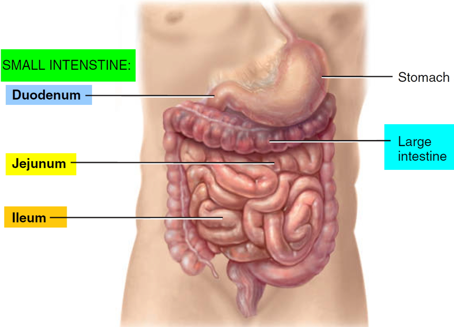
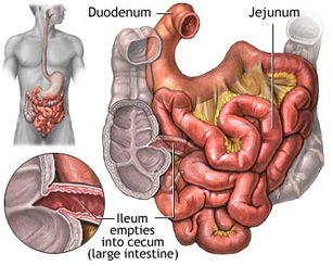
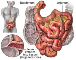
Intussusception is the most common cause of intestinal obstruction in children between ages 3 months and 3 years old. Intussusception occurs when a portion of the intestine folds like a telescope, with one segment slipping inside another segment. It can occur in the colon, the small bowel, or between the small bowel and colon. The result can be a blocked small bowel or colon. This causes an obstruction, preventing the passage of food that is being digested through the intestine.

Intussusception (in-tuh-suh-SEP-shun) is a serious condition in which part of the intestine slides into an adjacent part of the intestine.The walls of the 2 “telescoped” sections of intestine press on each other, causing irritation and swelling. Eventually, the blood supply to that area is cut off, which can cause damage to the intestine.   This “telescoping” often blocks food or fluid from passing through. Intussusception also cuts off the blood supply to the part of the intestine that’s affected, which can lead to a tear in the bowel (perforation), infection and death of bowel tissue.

Intussusception is the most common cause of intestinal obstruction in children younger than 3. The cause of most cases of intussusception in children is unknown. Though rare in adults, most cases of adult intussusception are the result of an underlying medical condition, such as a tumor.

In children, the intestines can usually be pushed back into position with an X-ray procedure. In adults, surgery is often required to correct the problem.

Symptoms:

Children

The first sign of intussusception in an otherwise healthy infant may be sudden, loud crying caused by abdominal pain. Infants who have abdominal pain may pull their knees to their chests when they cry.

The pain of intussusception comes and goes, usually every 15 to 20 minutes at first. These painful episodes last longer and happen more often as time passes.

Other frequent signs and symptoms of intussusception include:

  • Stool mixed with blood and mucus (sometimes referred to as “currant jelly” stool because of its appearance)
  • Vomiting
  • A lump in the abdomen
  • Lethargy
  • Diarrhea
  • Fever

Not everyone has all of the symptoms. Some infants have no obvious pain, and some children don’t pass blood or have a lump in the abdomen. Some older children have pain but no other symptoms.

Adults

Because intussusception is rare in adults and symptoms of the disorder often overlap with the symptoms of other disorders, it’s more challenging to identify. The most common symptom is abdominal pain that comes and goes. Nausea and vomiting may also occur. People sometimes have symptoms for weeks before seeking medical attention.

When to see a doctor

Intussusception requires emergency medical care. If you or your child develops the signs or symptoms listed above, seek medical help right away.

In infants, remember that signs of abdominal pain may include recurrent bouts of pulling the knees to the chest and crying.

Causes:

Your intestine is shaped like a long tube. In intussusception, one part of your intestine — usually the small intestine — slides inside an adjacent part. This is sometimes called telescoping because it’s similar to the way a collapsible telescope folds together.

In some cases, the telescoping is caused by an abnormal growth in the intestine, such as a polyp or a tumor (called a lead point). The normal wave-like contractions of the intestine grab this lead point and pull it and the lining of the intestine into the bowel ahead of it. In most cases, however, no cause can be identified for intussusception.

Children

In the vast majority of cases of intussusception in children, the cause is unknown. Because intussusception seems to occur more often in the fall and winter and because many children with the problem also have flu-like symptoms, some suspect a virus may play a role in the condition. Sometimes, a lead point can be identified as the cause of the condition — most frequently the lead point is a Meckel’s diverticulum (a pouch in the lining of the small intestine).

Adults

In adults, intussusception is usually the result of a medical condition or procedure, including:

  • A polyp or tumor
  • Scar-like tissue in the intestine (adhesions)
  • Weight-loss surgery (gastric bypass) or other surgery on the intestinal tract
  • Inflammation due to diseases such as Crohn’s disease

Risk factors:

Risk factors for intussusception include:

  • Age. Children — especially young children — are much more likely to develop intussusception than adults are. It’s the most common cause of bowel obstruction in children between the ages of 6 months and 3 years.
  • Sex. Intussusception more often affects boys.
  • Abnormal intestinal formation at birth. Intestinal malrotation is a condition in which the intestine doesn’t develop or rotate correctly, and it increases the risk for intussusception.
  • A prior history of intussusception. Once you’ve had intussusception, you’re at increased risk of developing it again.
  • A family history. Siblings of someone who’s had an intussusception are at a much higher risk of the disorder.

Complications:

Intussusception can cut off the blood supply to the affected portion of the intestine. If left untreated, lack of blood causes tissue of the intestinal wall to die. Tissue death can lead to a tear (perforation) in the intestinal wall, which can cause an infection of the lining of the abdominal cavity (peritonitis).

Peritonitis is a life-threatening condition that requires immediate medical attention. Signs and symptoms of peritonitis include:

  • Abdominal pain
  • Abdominal swelling
  • Fever

Peritonitis may cause your child to go into shock. Signs and symptoms of shock include:

  • Cool, clammy skin that may be pale or gray
  • A weak and rapid pulse
  • Abnormal breathing that may be either slow and shallow or very rapid
  • Anxiety or agitation
  • Profound listlessness

A child who is in shock may be conscious or unconscious. If you suspect your child is in shock, seek emergency medical care right away.