Archive | September 2025

QUOTE FOR TUESDAY:

“Chronic conditions are costly and major causes of death and disability. Addressing conditions earlier in adulthood can slow disease progression and improve well-being across the lifespan. We estimated, by life stage, 10-year trends among US adults in the prevalence of 1 or more chronic conditions, multiple chronic conditions (MCC; ≥2 conditions), and 12 selected chronic conditions.

Approximately 6 in 10 young, 8 in 10 midlife, and 9 in 10 older US adults report 1 or more chronic conditions. Trends in conditions worsened among young adults during 2013–2023. Recognizing the burden of chronic disease throughout life stages, especially earlier in life, practitioners and partners may consider prevention and management approaches critical for addressing costs, care, and health outcomes. Practitioners may also consider tailoring these approaches to unique roles, transitions, and challenges in different life stages.

Chronic conditions such as heart disease, cancer, stroke, and diabetes are costly and major causes of death and disability in the US (1,2). Over the past 20 years, the prevalence of chronic conditions has increased steadily, and this trend is expected to continue (3–5).

In 2018, more than half of US adults had at least 1 chronic condition, and more than one-quarter had 2 or more chronic conditions (6). In 2016, the total direct health care costs in the US for the treatment of chronic health conditions was $1.1 trillion (7). In addition to financial costs, increased death, and disability, the burden of chronic conditions includes decreased quality of life (8), increased health care utilization (8,9), lost productivity in the workforce (10), and loss in functioning (eg, social and leisure activities) (11).

In 2023, approximately 194 million American adults, and 6 in 10 young adults, 8 in 10 midlife adults, and 9 in 10 older adults reported 1 or more conditions. Prevalence of chronic conditions increased by 7.0 percentage points among young adults from 2013 to 2023.”

Centers for Disease Control and Prevention – CDC (Trends in Multiple Chronic Conditions Among US Adults, By Life Stage, Behavioral Risk Factor Surveillance System, 2013–2023)

The crisis is clear–chronic diseases are crushing healthcare in America.

Chronic diseases are crushing healthcare.

Our healthcare system is good at treating short-term problems, such as broken bones and infections. Medical advances are helping people live longer. But obesity is reaching epidemic proportions. The population is aging. We need to do a much better job managing chronic diseases.

Chronic conditions such as diabetes, heart disease, lung disease, and Alzheimer’s disease take a heavy toll on health. Chronic conditions also cost vast amounts of money. The trends are going in the wrong direction:

  • Obesity increases the risk of developing conditions, such as diabetes and heart disease. The rate of obesity in adults has doubled in the last 20 years. It has almost tripled in kids ages 2-11. It has more than tripled in children ages 12-19.

  • Without big changes, 1 in 3 babies born today will develop diabetes in their lifetime.

  • Average healthcare costs for someone who has one or more chronic conditions is 5 times greater than for someone without any chronic conditions.

  • Chronic diseases account for $3 of every $4 spent on healthcare. That’s nearly $7,900 for every American with a chronic disease.

  • These chronic diseases drive healthcare costs at an alarming annual rate:

  • Heart Disease and Stroke: $432 billion/year.

  • Diabetes: $174 billion/year.

  • Lung Disease: $154 billion/year.

  • Alzheimer’s Disease: $148 billion/year.The human cost of chronic diseases cannot be ignored:

  • The Human Cost

    • Chronic diseases cause 7 out of every 10 deaths.

    • Chronic diseases such as diabetes, cancer, and heart disease are the leading causes of disability and death in the US.

    • About 25% of people with chronic diseases have some type of activity limitation. This includes difficulty or needing help with personal tasks such as dressing or bathing. It may also mean being restricted from work or attending school.

    • Today, Americans suffering from chronic diseases face rising healthcare costs. They also receive lower quality care and have fewer options.

    • Health insurance co-pays and out-of-pocket expenses continue to rise. In many cases, choices and care are limited.

    • The disabling and long-term symptoms that often come with chronic diseases add to extended pain and suffering. This decreases the overall quality of life.The financial and human costs of chronic diseases can no longer be ignored.

    • There is a way we can prevent this and it would be keeping your weight ideal for your height within the therapeutic body mass index range (calculate it for free online).  If you need to lose weight you’ve come to the right blog.  Do it through diet, exercise balanced with rest and practicing routine healthy habits that prone you to having a healthy body overall which prevents disease.  So many diseases are due to these factors not practiced daily =  good diet with exercise, healthy habits and a therapeutic weight for your height.  If we had most of American citizens living this way certain diseases would be decreased terribly helping our country out with this economy of ours with the health care system.  If you need assistance in reaching these healthy practices   Do you want a better fit body or even an overall healthier family including grandchildren to even our country than take the action NOW.  For your goal in playing a part in living healthier and spreading the good news would benefit you and all around us.  Also, for the next decade & generation to be healthier will help Americans holistically in their lives all around (including our health care showing a spread of disease in lower percentage due to healthier dieting and activity choices by our people, who are so important in helping to decide where the health of the present and future of the US citizens lies.  Should it take our government to make a move (finally after so many years)?   We must face the epidemic of chronic diseases. If we don’t, the human costs will continue to soar. We might even face a lack of available or affordable care when it is needed most.


    • REFERENCES:
    • Centers for Disease Control and Prevention. Chronic Disease Overview: Costs of Chronic Disease. Centers for Disease Control and Prevention Web site. Available at http://www.cdc.gov/nccdphp/overview.htm. Accessed July 24, 2007.
    • Centers for Disease Control and Prevention. Overweight and Obesity. Centers for Disease Control and Prevention Web site. Available at http://www.cdc.gov/nccdphp/dnpa/obesity/trend/index.htm. Accessed July 24, 2007.
    • American Diabetes Association. The Dangerous Toll of Diabetes. American Diabetes Association Web site. Available at http://diabetes.org/diabetes-statistics/dangerous-toll.jsp. Accessed May 18, 2007.
    • Partnership for Solutions. Chronic Conditions: Making the Case for Ongoing Care, September 2004 Update. Partnership for Solutions Web site. Available at http://www.partnershipforsolutions.org/DMS/files/ chronicbook2004.pdf. Accessed July 24, 2007.
    • Mensah G, Brown D. An overview of cardiovascular disease burden in the United States. Health Aff 2007; 26:38-48.
    • American Diabetes Association. Direct and Indirect Costs of Diabetes in the United States. American Diabetes Association Web site. Available at http://www.diabetes.org/diabetes-statistics/ cost-of-diabetes-in-HYPERLINK “http://www.diabetes.org/diabetes-statistics/cost-of-diabetes-in-us.jsp”us.jsp. Accessed September 20, 2007.
    • Alzheimer’s Association. Alzheimer’s Disease Facts and Figures 2007. Alzheimer’s Association Web site. Available at http://www.alz.org/national/documents/Report_2007FactsAndFigures.pdf.

QUOTE FOR MONDAY:

“Cardiovascular diseases (CVDs) are the leading cause of mortality globally, responsible for a significant number of deaths and disabilities. In 2021 alone, CVDs accounted for 20.5 million deaths, comprising approximately one-third of all global deaths []. While cardiovascular conditions were traditionally considered diseases of affluence, this is no longer the case. Over three-quarters of CVD-related deaths occur in low- and middle-income countries (LMICs) [].

Moreover, these deaths are the primary contributor to premature non-communicable disease (NCD) mortality. Ischemic heart disease, specifically, stands as the leading cause of premature death in 146 countries for men and 98 countries for women []. ”

National Library of Medicine (The Heart of the World – PMC)

“Alone in the United States:

  • Heart disease is the leading cause of death for men, women, and people of most racial and ethnic groups.1
  • One person dies every 34 seconds from cardiovascular disease.1
  • In 2023, 919,032 people died from cardiovascular disease. That’s the equivalent of 1 in every 3 deaths.1
  • Heart disease cost about $417.9 billion from 2020to 2021.2This includes the cost of health care services, medicines, and lost productivity due to death.

Center of Disease Control and Prevention – CDC (Heart Disease Facts | Heart Disease | CDC)


World Heart Day – Learn about Cardiovascular Diseases – CVD, heart attack, the symptoms, and the how to go about the right treatment!

If you have been reading regularly on this blog than you know this day  is dedicated everywhere for the health topic “WORLD HEART DAY”.

Governments and non-government organizations celebrate and promote World Heart Day with activities such as fun runs, public talks, concerts, and sporting events. The World Heart Federation organizes awareness events in more than 100 countries. They include:

  • Health checks.
  • Sports events, including walks, runs and fitness sessions.
  • Public talks and science forums
  • Stage shows and concerts.
  • Exhibitions.

The World Heart Foundation organizes World Heart Day, an international campaign held on September 29 to inform people about cardiovascular diseases.

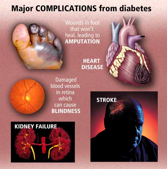
Cardiovascular Diseases:

Coronary heart disease is a common term for the buildup of plaque in the heart’s arteries that could lead to heart attack. But what about coronary artery disease? Is there a difference?

The short answer is often no — health professionals frequently use the terms interchangeably.

However, coronary heart disease , or CHD, is actually a result of coronary artery disease, or CAD, said Edward A. Fisher, M.D., Ph.D., M.P.H., an American Heart Association volunteer who is the Leon H. Charney Professor of Cardiovascular Medicine and also of the Marc and Ruti Bell Vascular Biology and Disease Program at the NYU School of Medicine.

With coronary artery disease, plaque first grows within the walls of the coronary arteries until the blood flow to the heart’s muscle is limited. View an illustration of coronary arteries. This is also called ischemia. It may be chronic, narrowing of the coronary artery over time and limiting of the blood supply to part of the muscle. Or it can be acute, resulting from a sudden rupture of a plaque and formation of a thrombus or blood clot.

The traditional risk factors for coronary artery disease are high LDL cholesterol, low HDL cholesterol, high blood pressure, family history, diabetes, smoking, being post-menopausal for women and being older than 45 for men, according to Fisher. Obesity may also be a risk factor.

“Coronary artery disease begins in childhood, so that by the teenage years, there is evidence that plaques that will stay with us for life are formed in most people,” said Fisher, who is former editor of the American Heart Association journal, ATVB. “Preventive measures instituted early are thought to have greater lifetime benefits. Healthy lifestyles will delay the progression of CAD, and there is hope that CAD can be regressed before it causes CHD.”

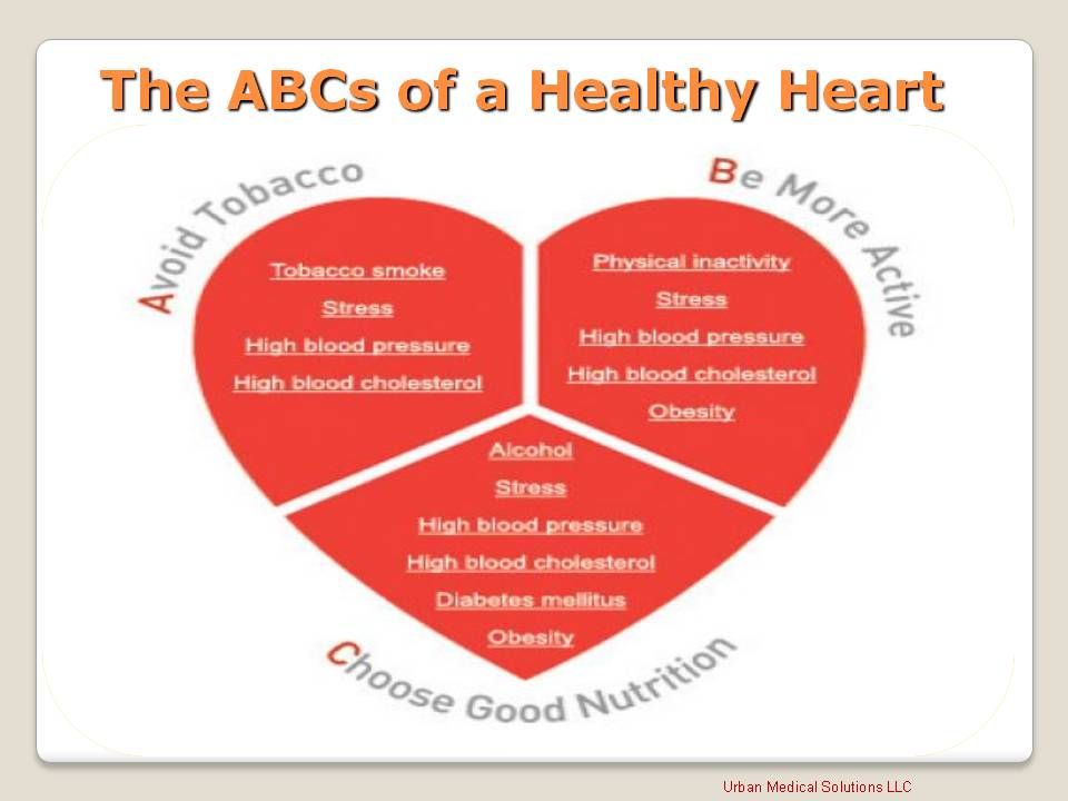
Living a healthy lifestyle that incorporates good nutrition, weight management and getting plenty of physical activity can play a big role in avoiding CAD.

“Coronary artery disease is preventable,” agreed Johnny Lee, M.D., president of New York Heart Associates, and an American Heart Association volunteer. “Typical warning signs are chest pain, shortness of breath, palpitations and even fatigue.”

What is a “widow maker”?  Well for starters, this is the deadliest heart attack.  The symptoms you need to know to possibly prevent the results of this widow maker.  It occurs when there is a complete blockage of the left artery feeding the heart with blood.  This causes a cut off of oxygen supply  to one of the large parts of the heart muscle, which can cause it to stop beating, causing you to die.

A heart attack is when one of the coronary arteries becomes blocked.

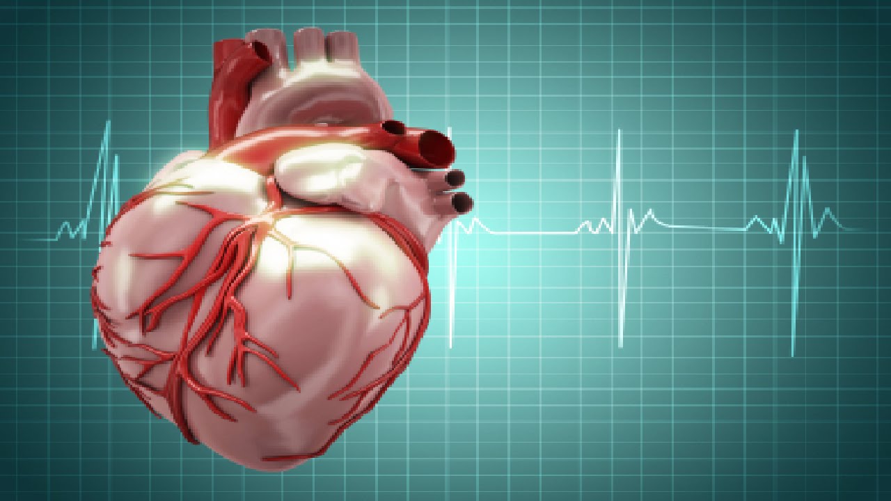
The heart muscle is then robbed of vital oxygenated blood, which if left untreated, can cause the heart muscle to begin to die.

A heart attack is a life-threatening emergency.

A widow maker heart attack is caused when the LAD artery becomes blocked.

It occurs when there’s a complete blockage of the left anterior descending (LAD) artery, one of two main arteries that brings OXYGENATED blood to the heart=FOOD TO THE HEART MUSCLE (O2).

When it is blocked due to a build up of plaque it is most often deadly, hence the name “widow maker”.  How to we get plaque build up?

Cholesterol plaques can be the cause of heart disease. Plaques begin in artery walls and grow over years. The growth of cholesterol!  The plaques slowly blocks blood flow in the arteries. Worse, a cholesterol plaque can suddenly rupture. The sudden blood clot that forms over the rupture then causes a heart attack or stroke.

Blocked arteries caused by plaque buildup and blood clots are the leading cause of death in the U.S. Reducing cholesterol and other risk factors can help prevent cholesterol plaques from forming. Occasionally, it can even reverse some plaque buildup.

“When the main artery down the front of the heart (LAD) is totally blocked or has a critical blockage, right at the beginning of the vessel, it is known as the Widow Maker. (The medical term for this is a proximal LAD lesion). No one knows exactly who came up with the term, but the reason they did is likely that if that artery is blocked right at the beginning of its course, then the whole artery after it goes down. This essentially means that the whole front wall of the heart goes down. As far as heart attacks go, this is a big one, with big consequences if not dealt with appropriately and FAST!”

myheart.net/Dr. Ahmed – an Interventional Cardiologist and Director of Structural Heart Disease at Princeton-Baptist Hospital.

Symptoms:

A widow maker heart attack has the same symptoms as any other heart attack.

They can be difficult to spot for sure, because they can vary from person to person.

The most common signs include:

  • chest pain, tightness, heaviness, pain or a burning feeling in your chest
  • pain in the arms, neck, jaw, back or stomach
  • for some people the pain and tightness will be severe, while for others it will just feel uncomfortable
  • sweating
  • feeling light-headed
  • becoming short of breath
  • feeling nauseous or vomiting

How is a heart attack treated?

The first port of call for treatment, is for doctors to treat the blocked artery.

There are two main procedures used to open up the blocked blood vessel.

The first, a primary percutaneous coronary intervention (PPCI) is an emergency coronary angioplasty.

It opens the blockage and helps restore blood supply to the heart.

The second treatment, is thrombosis, also known as a “clot buster”.

It involves injecting a drug into the vein to dissolve the blood clot and restore blood supply to the heart that way.  In some cases this procedure can be performed in the ambulance.

While these treatments are common, in some cases they will not be right for the patient and so won’t be performed.  The MD Cardiologist will know the right Rx.

Take good care of your HEART, the engine to the human body!  First do all preventative measures to prevent getting any cardiovascular diseases but if you have cardiovascular diseases then follow your M.D. instructions on any meds if he prescribed them for you, eat the proper foods for a cardiac diet, and balance rest with exercise.   See your cardiologist as he or she recommends.


QUOTE FOR THE WEEKEND:

“Gynecologic Cancers: The Numbers You Need to Know

  • Every 5 minutes, someone will be diagnosed with a gynecologic cancer
  • 5 types of gynecologic cancer, most with no early screening tests
  • Uterine cancer is the most common, followed by cervical and ovarian
  • Early detection saves lives but symptoms are often missed
  • We need more research, funding, and awareness”

Foundation for Women’s Cancer (Gynecologic Cancer Awareness Month – Foundation For Women’s Cancer)

September is Gynecological Cancer Awareness Month

According to the CDC, uterine cancer (cancer which develops in the uterus) is the most commonly diagnosed gynecological cancer in the U.S. and the fourth most common cancer in U.S. women overall.

Types of Gynecological Cancer

  • Cervical cancer.
  • Ovarian cancer.
  • Uterine cancer.
  • Vaginal cancer.
  • Vulvar cancer.

September is Gynecologic Cancer Awareness


It is estimated that within the past 2 years 98,000 women would be diagnosed with a gynecologic cancer and some 30,000 would die from the disease.

Gynecological Cancer Awareness Month provides an important opportunity to draw attention to this important women’s health issue and offer vital information on risk cancers, warning signs, and prevention strategies.

Uterine Cancer

Uterine cancer forms in the tissues of the uterus, the organ in which a fetus develops. The two types of uterine cancer are endometrial cancer and uterine sarcoma.

Endometrial cancer forms in the tissues of the endometrium – the lining of the uterus. Obesity, high blood pressure, and diabetes may increase the risk of endometrial cancer.

Uterine Sarcoma is a rare type of cancer that forms in the uterine muscles or in tissues that support the uterus. Exposure to X-rays during radiation therapy can increase the risk of uterine sarcoma.

Treatment with the breast cancer drug tamoxifen is a risk factor for both types of uterine cancer.

According to the CDC, uterine cancer (cancer which develops in the uterus) is the most commonly diagnosed gynecological cancer in the U.S. and the fourth most common cancer in U.S. women overall. While any woman can develop uterine cancer, it is most commonly diagnosed in women who have gone through menopause. Risk factors for uterine cancer include age (being age 50 or older), obesity, taking estrogen alone as hormone replacement, and having a family history of uterine, ovarian or colon cancer.

Routine testing is not recommended for uterine cancer, so it is important for women to be aware of symptoms—such as abnormal vaginal discharge or bleeding and/or pain and pressure in the pelvic area—and talk to a healthcare provider if they experience these.

Ovarian Cancer

While ovarian cancer accounts for about 3% of cancers among women, it causes more deaths than any other gynecological cancer, according to the American cancer Society. While the survival rates for ovarian cancer are excellent when the disease is diagnosed early, only about 20% of ovarian cancers are found at this early stage. One reason for this is that there is no recommended routine screening for ovarian cancer on women without symptoms. And the symptoms of ovarian cancer—including abnormal abdominal bloating, abdominal pain or pressure, and feeling full quickly when eating—can also be easily be ignored or mistaken for other problems.

For women who experience these symptoms, or who at at higher risk (including women who have had breast cancer or have a family history of ovarian, breast or colorectal cancer), a healthcare provider may recommending further screening. Testing may include rectovaginal pelvic exam, a transvaginal ultrasound,or a CA-125 blood test.

There are three types of ovarian cancer in adults, including ovarian epithelial cancer, which begins in the tissue covering the ovary, lining of the fallopian tube, or the peritoneum; ovarian germ cell tumors, which start in the egg or germ cells; and ovarian low malignant potential tumors, which begin in the tissue covering the ovary.

Cervical Cancer

According to the American Sexual Health Organization, the vast majority of cases of cervical cancer—cancer that develops on the cervix, the opening to the uterus—are linked to human papillomavirus (HPV) infection. The majority of women with an HPV infection will not develop cervical cancer, but regular screening is essential. In most cases cervical cancer can be prevented through early detection and treatment of abnormal cell changes that occur in the cervix years before cervical cancer develops. These changes are typically detected through a Pap test or an HPV test. HPV vaccines can also prevent cervical cancer.

In its early stages, cervical cancer typically doesn’t have any symptoms, which is why regular screening is so important. At later stages, symptoms may include abnormal vaginal discharge or bleeding or pain during sex. While these can also be signs of other health issues, if a woman experiences these symptoms, she should report them to her healthcare provider.

Cervical Cancer. Carcinoma of Cervix. Malignant neoplasm arising from cells in the cervix uteri. Vaginal bleeding. Vector diagram

Vaginal and Vulvar Cancer

There are two main types of vaginal cancer: squamous cell carcinoma and adenocarcinoma. Adenocarcinoma is more likely than squamous cell cancer to spread to the lungs and lymph nodes. A rare type of adenocarcinoma is linked to being exposed to diethylstilbestrol (DES) before birth.

Adenocarcinomas not linked with being exposed to DES are most common in women after menopause.

According to the American Sexual Health Organization, vaginal and vulvar cancers are rare—an estimated 1,000 women are diagnosed with vaginal cancer and 3,500 women with vulvar cancer each year. Like cervical cancer, vaginal and vulvar cancers are also associated with HPV infection, with up to 90% of vaginal cancers and pre-cancers and more than 50% of vulvar cancers linked to infection with the high-risk HPV types.

Vulvar cancer forms in a woman’s external genitalia. Vulvar cancer most often affects the outer vaginal lips.

Abnormal cells can grow on the surface of the vulvar skin for a long time. This condition is called vulvar intraepithelial neoplasia (VIN). Because it is possible for VIN to become vulvar cancer, it is important to get treatment.

Risk factors for vulvar cancer include having VIN, HPV infection, and having a history of genital warts.

HPV vaccines, which prevent some of the high-risk types of HPV, can also help prevent vaginal and vulvar cancers.


QUOTE FOR FRIDAY:

“Cardiac ablation is a treatment for irregular heartbeats, called arrhythmias. It uses heat or cold energy to create tiny scars in the heart. The scars block faulty heart signals and restore a typical heartbeat.

Cardiac ablation is most often done using thin, flexible tubes called catheters that are inserted through a blood vessel. Less commonly, ablation is done during heart surgery.

Cardiac ablation is a treatment to stop or prevent irregular heartbeats, called arrhythmias.

An arrhythmia happens when the electrical signals that tell the heart to beat don’t work as they should. The heart may beat too fast or too slow. Or the pattern of the heartbeat may be irregular.

Depending on the type of irregular heartbeat, cardiac ablation may be one of the first treatments. Other times, it’s done when medicines or other treatments don’t work.”

MAYO CLINIC (Cardiac ablation – Mayo Clinic)

Ablation, a great resolution for certain arrhythmias.

afib-with-rvr-7  afib-with-rvr-6afib

Ablation is used to treat abnormal heart rhythms, or arrhythmias. The type of arrhythmia and the presence of other heart disease will determine whether ablation can be performed surgically or non-surgically.

Ablation therapy using radio frequency waves on the heart is used to cure a variety of cardiac arrhythmiae such as supraventricular tachycardia, Wolff–Parkinson–White syndrome (WPW), ventricular tachycardia, and more recently as management of atrial fibrillation (especially when its newly diagnosed when medical management can’t change it back to normal sinus rhythm, which is the normal cardiac rhythm seen on a telemetry monitor or of an EKG taken on a patient).

An arrhythmia is a change in the heart’s normal rate or rhythm, normally between 60 and 100 beats per minute. Arrhythmias are classified by their location in the heart and by their speed or rhythm. An atrial arrhythmia is an abnormality that occurs in one of the two upper chambers of the heart, the left or right atrium. Arrhythmias are associated with aging and typically happen more frequently during middle age. At least 10 to 15 percent of people older than 70 years experience arrhythmias.  We have what we call our human pacemaker of the heart that naturally sends conduction for the heart to pump, which is called the sinus node.  This is in the upper left corner of the right chamber of the heart.  That is where the name sinus rhythm derives from (the sinus node) which is the best rhythm a human can be in as long as the pulse rate stays above 60 and stays under 100. Now if that sinus node for some reason breaks down and no longer works; so than the pace site starts somewhere in the right atrium below the sinus node (the heart is compensating for whatever is the reason the sinus node is not working).  So now the rhythms are called atrial rhythms because of where the new natural pacemaker site is in the heart.  This is where ablation comes into play if the type of atrial rhythm they have is detrimental to the patient; including if that patient is a candidate for this procedure.  Between our heart chambers on the top (called atriums) and below (called the ventricles) is a AV (meaning atrioventricular valve).  Rhythms above the ventricles are also grouped as supraventricular rhythms.  Which is what ablation is used for.

Types of rhythms a patient would be considered for ablation as a possible treatment:

Atrial fibrillation. The electrical signal that circles uncoordinated through the muscles of the atria (the upper chambers of the heart), causing them to quiver (sometimes more than 400 times per minute) without contracting. The ventricles (the lower chambers of the heart) do not receive regular impulses and contract out of rhythm, and the heartbeat becomes uncontrolled and irregular. It is the most common atrial arrhythmia, and 85 percent of people who experience it are older than 65 years.

Atrial fibrillation can cause a blood clot to form, which can enter the bloodstream and trigger a stroke. Underlying heart disease or hypertension increases the risk of stroke from atrial fibrillation as does age even without heart disease or hypertension.

Premature atrial contraction (PAC or premature atrial impulses). A common and benign arrhythmia, a PAC is a heartbeat that originates away from the sinus node, which sends electrical signals through the upper chamber. It typically occurs after the sinus node has initiated one heartbeat and before the next regular sinus discharge. A PAC can cause a feeling of a skipped heartbeat. Use of caffeine, tobacco, and/or alcohol, or stress can bring on PACs or increase their frequency.

Supraventricular tachycardia (SVT). Characterized by a rapid heart rate that ranges between 100 and 240 beats per minute, SVT usually begins and ends suddenly. SVT occurs when an electrical impulse ‘re-enters’ the atrial muscles. A disorder that a person may have at birth, SVT is commonly caused by a variation in the electrical system of the heart. SVT often begins in childhood or adolescence and can be triggered by exercise, alcohol, or caffeine. SVT is rarely dangerous, but can cause a drop in blood pressure, causing lightheadedness or near-fainting episodes, and, rarely, fainting episodes.

Atrial flutter. Differentiated from atrial fibrillation by its coordinated, regular pattern, atrial flutter is a coordinated rapid beating of the atria. Most who experience atrial flutter are 60 years and older and have some heart disorder, such as heart valve problems or a thickening of the heart muscle. Atrial flutter is classified into two types, according to the pathways responsible for it. Type I normally causes the heart rate to increase to and remain at 150 beats per minute. Rarely, the rate may reach 300 beats per minute; sometimes it decreases to 75 beats per minute. Type II increases the atrial rate faster, so the ventricular rate may be 160 to 170 beats per minute. As with atrial fibrillation, atrial flutter increases the risk of stroke.

Sick sinus syndrome (SSS). Common among older people, SSS is an improper firing of electrical impulses caused by disease or scarring in the sinus or Sinoatrial node (SA node). SSS normally causes the heart rate to slow, but sometimes it alternates between abnormally slow and fast. A progressive condition, with episodes increasing in frequency and duration, SSS can be caused by:

  • Degeneration of the heart’s electrical system; or
  • Diseases of the atrial muscle.                                                                                                                                                                                                                                                                                                                                                    Sinus tachycardia. The sinus node emits abnormally fast electrical signals, which increases the heart rate to between 100 beats per minute to 140 beats per minute at rest, and 200 beats per minute during exercise. A normal response to exercise or stress, it can also be caused by:
  • Adrenaline;
  • Consumption of caffeine, nicotine, or alcohol; and
  • Heart conditions.                                                                                                                                                                                                                                                                                                                                                                                   Wolff-Parkinson-White syndrome (WPW). WPW syndrome occurs when electrical signals fail to pause in the atrioventricular node because an extra pathway allows the impulse to “bypass” the normal pathway; and the syndrome is sometimes called bypass tract. WPW syndrome causes heart rates approaching 240 beats per minute.
  • Occasionally, impulses can go down one extra pathway and up another, creating a “loop” or “short circuit,” (called SVT because of WPW). Patients with WPW syndrome may develop atrial fibrillation and are at increased risk for developing a dangerous ventricular arrhythmia when this occurs. *
  • Sinus bradycardia. Associated with impaired impulse generation in the SA node, it causes the heart rate to decrease to fewer than 60 beats per minute. Commonly caused by SSS, drugs like beta-blockers and calcium-channel blockers can also cause sinus bradycardia. Occasionally sinus bradycardia can be caused by impaired conduction of impulses to the atrial muscles.

QUOTE FOR THURSDAY:

“Heart block is a problem with your heartbeat signal moving from the upper to lower part of your heart. The signal can only get through sometimes, or not at all. This makes your heart beat slowly or skip beats. People with second-degree or third-degree heart block may experience fainting, tiredness and shortness of breath.

Heart block is an issue with a heartbeat signal traveling from the top chambers of your heart to the bottom chambers of your heart. Normally, electrical signals (impulses) travel from your heart’s upper chambers (atria) to your lower chambers (ventricles). The signal moves through your AV node, a cluster of cells that link the electrical activity from your top to bottom chambers. If you have heart block, the signal only makes it to your ventricles some of the time, if at all.

The result is a heart that may not work well. Your heart may beat slowly or skip beats. In severe cases, heart block can affect your heart’s ability to pump blood, causing low blood flow to your entire body.

Other names for heart block are atrioventricular (AV) block or a conduction disorder.  The heart block is between the upper chambers (atriums) and the lower chamber (ventricles) with the electrical activity responsible to make your heart beat properly.”

Cleveland Clinc (Heart Block: Types, Symptoms & Causes)


The Heart Blocks with the heart: Type I HB, Type II HB= Mobitz I and Mobitz II, and lastly Type III HB. Learn the severity of the 4 A-V HBs and the Rxs.

HeartBlocks2

Than when AV nodes which can be blocked=atrio-ventricular block (AV heart block)=HB is a type of heart block in which the conduction between the atria and ventricles of the heart is impaired. Under normal conditions, the sinoatrial node (SA node) in the atria sets the intial pace for the heart, and these impulses travel down to the atriums than passing the AV valves into the ventricles and up the purkinje fibers . In an AV block, this message does not reach the ventricles or is impaired along the way. The ventricles of the heart have their own pacing mechanisms, which can maintain a lowered heart rate in the absence of SA stimulation.

The causes of pathological AV block are varied and include ischemia, infarction, fibrosis or drugs, and the blocks may be complete or may only impair the signaling between the SA and AV nodes.

Types of AV blocks: 1st degree HB which is NSR with a prolonged PR interval and can live a totally normal life with it. PR interval is the time from beginning of the p wave to the beginning of the qrs wave.  In all degrees of HB the PRI interval is prolonged.   You can live a normal life with 1st degree HB.

Mobitz type I is benign, and most people do not experience symptoms. A doctor may only discover the condition during a routine exam or when checking for other conditions using an ECG. Those who do not experience symptoms may require monitoring on an outpatient basis.  If people do experience symptoms, they may require treatment.

2nd degree HB-Mobitz I called Wenkebach is a NSR with a progressing increasing PR interval longer and longer till it actually drops a QRS and you see just the p wave without a QRS once and the rhythm restarts the pattern all over again.

Than 2nd degree HB-Mobitz II and this is a NSR with a prolonged PRI that measures the same amount each time but even though its not progressively getting longer its worse than Mobitz I always.  This is because with the same measurement prolonged PRI interval you only see 2 or 3 more  p waves without the QRS and these need immediate external subcutaneous pacing or just as effective if its not available (external subcutaneous pacing) than a dopamine IV drip 5-20 mcg/kg/min or epinephrine IV drip 2 to 10 mcg/min or epinephrine IV drip 2-10 mcg/min (not kg based).

Than there is complete HB 3rd degree heart block where no conduction from atriums to ventricles is going through so the atriums are contracting the way they want and the ventricles contracting the way they want.  The problem is there is a total disassociation between the upper and lower chambers. Treatment for most patients with acquired complete heart block will require a permanent pacemaker or an implantable cardioverter defibrillator (ICD).  Temporarily till OR the pt may have subcutaneous pacing but may not work at in some cases.