Part I January National Radon Month Awareness

 National Radon Month   National Radon Month4

Radon is a naturally occurring gas that seeps out of rocks and soil. Radon comes from uranium that has been in the ground since the time the earth was formed, and the rate of radon seepage is very variable, partly because the amounts of uranium in the soil vary considerably. Radon flows from the soil into outdoor air and also into the air in homes from the movement of gases in the soil beneath homes. Outside air typically contains very low levels of radon, but it builds up to higher concentrations indoors when it is unable to disperse. Some underground mines, especially uranium mines, contain much higher levels of radon.

Radon is present outdoors and indoors. It is normally found at very low levels in outdoor air and in drinking water from rivers and lakes. It can be found at higher levels in the air in houses and other buildings, as well as in water from underground sources, such as well water.

Radon breaks down into solid radioactive elements called radon progeny (such as polonium-218, polonium-214, and lead-214). Radon progeny can attach to dust and other particles and can be breathed into the lungs. As radon and radon progeny in the air break down, they give off radiation that can damage the DNA inside the body’s cells.

 How are people exposed to radon?

1-At home and in other buildings.

For both adults and children, most exposure to radon comes from being indoors in homes, offices, schools, and other buildings. The levels of radon in homes and other buildings depend on the characteristics of the rock and soil in the area. As a result, radon levels vary greatly in different parts of the United States, sometimes even within neighborhoods. Elevated radon levels have been found in every state.

Radon gas given off by soil or rock can enter buildings through cracks in floors or walls; construction joints; or gaps in foundations around pipes, wires, or pumps. Radon levels are usually highest in the basement or crawl space. This level is closest to the soil or rock that is the source of the radon. Therefore, people who spend much of their time in basement rooms at home or at work have a greater risk for being exposed.

Small amounts of radon can also be released from the water supply into the air. As the radon moves from the water to air, it can be inhaled. Water that comes from deep, underground wells in rock may have higher levels of radon, whereas surface water (from lakes or rivers) usually has very low radon levels. For the most part, water does not contribute much to overall exposure to radon.

Radon exposure can also occur from some building materials if they are made from radon-containing substances. Almost any building material made from natural substances, including concrete and wallboard, may give off some level of radon. In most cases these levels are very low, but in a few instances these materials may contribute significantly to radon exposure.

Some granite countertops may expose people to different levels of radon. Most health and radiation experts agree that while a small portion of granite countertops might give off increased levels of radon, most countertops give off extremely low levels.

According to the US Environmental Protection Agency (EPA), it’s very unlikely that a granite countertop in a home would increase the radiation level above the normal, natural background level that comes from nearby soil and rocks. Still, people concerned about radon from countertops and from other household sources can test these levels using home detection kits or can hire a professional to do the testing (see the section “How can I avoid exposure to radon?”).

According to the EPA, the average indoor radon level is about 1.3 picocuries per liter (pCi/L). People should take action to lower radon levels in the home if the level is 4.0 pCi/L or higher. The EPA estimates that nearly 1 out of every 15 homes in the United States has elevated radon levels.

Outdoors, radon generally disperses and does not reach high levels. Average levels of radon outdoors, according to the EPA, are about 0.4 pCi/L.

2-At certain jobs

 In the workplace, people working underground, such as some types of miners, are among the most likely to be exposed to high levels of radon. High death rates from lung problems among miners in some parts of the world were first noted hundreds of years ago, long before people knew what radon was. Studies of radon-exposed miners during the 1950s and 1960s confirmed the link between radon exposure and lung cancer.

Higher levels of radon exposure are also more likely for people who work in uranium processing factories or who come in contact with phosphate fertilizers, which may have high levels of radium (an element that can break down into radon).

Does radon cause cancer?

 Being exposed to radon for a long period of time can lead to lung cancer. Radon gas in the air breaks down into tiny radioactive elements (radon progeny) that can lodge in the lining of the lungs, where they can give off radiation. This radiation can damage lung cells and eventually lead to lung cancer.

Cigarette smoking is by far the most common cause of lung cancer in the United States, but radon is the second leading cause. Scientists estimate that about 20,000 lung cancer deaths per year are related to radon.

Exposure to the combination of radon gas and cigarette smoke creates a greater risk for lung cancer than either factor alone. Most radon-related lung cancers develop in smokers. However, radon is also thought to cause a significant number of lung cancer deaths among non-smokers in the United States each year.

Although radon is chemically inert and electrically uncharged, it is radioactive, which means that radon atoms in the air can spontaneously decay, or change to other atoms. When the resulting atoms, called radon progeny, are formed, they are electrically charged and can attach themselves to tiny dust particles in indoor air. These dust particles can easily be inhaled into the lung and can adhere to the lining of the lung. The deposited atoms decay, or change, by emitting a type of radiation called alpha radiation, which has the potential to damage cells in the lung. Alpha radiations can disrupt DNA of these lung cells. This DNA damage has the potential to be one step in a chain of events that can lead to cancer. Alpha radiations travel only extremely short distances in the body. Thus, alpha radiations from decay of radon progeny in the lungs cannot reach cells in any other organs, so it is likely that lung cancer is the only potentially important cancer hazard posed by radon.

For centuries, it has been known that some underground miners suffered from higher rates of lung cancer than the general population. In recent decades, a growing body of evidence has causally linked their lung cancers to exposure to high levels of radon and also to cigarette smoking. The connection between radon and lung cancer in miners has raised concern that radon in homes might be causing lung cancer in the general population, although the radon levels in most homes are much lower than in most mines. The National Research Council study, which has been carried out by the sixth Committee on Biological Effects of Ionizing Radiation (BEIR) VI, has used the most recent information available to estimate the risks posed by exposure to radon in homes.

You can’t see, smell or taste radon, but it could be present at a dangerous level in your home. Radon is the leading cause of lung cancer deaths among nonsmokers in America and claims the lives of about 21,000 Americans each year. In fact, the EPA and the U.S. Surgeon General urge all Americans to protect their health by testing their homes, schools and other buildings for radon.

Exposure to radon is a preventable health risk and testing radon levels in your home can help prevent unnecessary exposure. If a high radon level is detected in your home, you can take steps to fix the problem to protect yourself and your family.

Radon, being naturally occurring, cannot be entirely eliminated from our homes. Of the deaths that attributes to radon (both independently and through joint action with smoking), perhaps one-third could be avoided by reducing radon in homes where it is above the “action guideline level” of 148 Bqm-3 (4 pCiL-1) to below the action levels recommended by the Environmental Protection Agency.4

The risk of lung cancer caused by smoking is much higher than the risk of lung cancer caused by indoor radon. Most of the radon-related deaths among smokers would not have occurred if the victims had not smoked. Furthermore, there is evidence for a synergistic interaction between smoking and radon.

QUOTE FOR FRIDAY:

“The CDC says that all women of reproductive age consume 400 mcg of folic acid, and pregnant women consume 600 mcg, each day to prevent two types of neural tube defects. During Folic Acid Awareness Week, observed January 3-9, 2021 (as well as the entire month for National Birth Defects Prevention Month), WIC staff are encouraged to take the opportunity to enhance their efforts to educate moms about the role folic acid plays in preventing congenital disabilities, & how their WIC food packages include folic acid-rich foods.  WIC staff can brush up on folic acid basics and find educational resources via Eye on Nutrition: Folate and Folic Acid.”

USDA WIC Works Resource System/U.S. Department of Agriculture

Folic Awareness Month – how it impacts a women childbearing years to geriatric, cautions/concerns on this vitamin and how it protects your future!

 folicacid2  folicacid1

What is folic acid? Folic acid is a type of B vitamin that is normally found in foods such as dried beans, peas, lentils, oranges, whole-wheat products, liver, asparagus, beets, broccoli, brussels sprouts, and spinach.

What does folic acid do for us?

1-Supports Normal Fetal Development. Folate plays an integral role in fetal development and the benefits for pregnant women and their offspring cannot be understated. Folate deficiency during early pregnancy can lead to neural tube defects. This is a serious problem that can lead to pregnancy termination or a baby born with spina bifida. The good news? Studies have found increased folate levels from one month prior to conception to 3 months afterward can reduce the chance of these defects by 50%.

2-Folic acid plays a role with our nervous system.  This B vitamin is required for proper brain function and aids in maintaining healthy emotional and mental health.

3-Cell growth is also significantly impacted by folic acid. This vitamin assists in the production and maintenance of DNA, RNA, and blood cells. This is especially notable during adolescence and pregnancy, when cell health and growth is particularly important.

4-It reduces the effects of our aging process.  Folic acid is not only useful for early development and pregnancy, but it may also prevent age related conditions later in life. One study found that elderly individuals who had high levels of homocysteine and reduced folate levels benefited from folic acid supplementation. Patients showed a reduction in the rate of hearing loss as they aged. Another study showed that patients who took 2,500 mcg of folic acid in addition to 500 mg of vitamin B6 and 1,000 mcg of vitamin B12 daily showed a reduced risk of developing AMD (a condition leading to loss of vision). Having proper levels of folic acid can assist health at any stage of life.

5-Reducing the risk of heart disease, so vital in America.  Heart disease is a major concern in the United States and folic acid may help lower the rate of occurrence. Through regulating possibly harmful substances, such as the amino acid homocysteine, folates may reduce the risk of heart disease. Studies have shown that patients with high levels of homocysteine have an increased risk of heart disease by 1.7 times and an increased risk of stroke by 2.5 times. Although more research is required to make a definitive link between this amino acid and heart disease, regulating it via folate supplementation takes little effort.

Folic Acid Deficiency

Even if one is not interested in preventative benefits of this vitamin, it is important to maintain proper levels. This can be challenging for some as there are several factors that reduce folic acid levels in the body. Alcoholism, inflammatory bowel disease (IBD), celiac disease, and some medications such as tetracycline (an antibiotic), methotrexate (used for treating cancer, rheumatoid arthritis, and psoriasis), and chemotherapy all significantly hamper folic acid levels by inhibiting absorption.

Deficiency of this B vitamin can lead to:

  • Inhibited growth
  • Loss of appetite leading to weight loss
  • Diarrhea
  • Forgetfulness
  • Fatigue and weakness
  • Irritability
  • Sores and inflammation in or around the mouth

Cautions and Concerns of Supplementation

Folic acid, and B vitamins in general, are not associated with any severe side effects. Because they are water soluble any excess left over is simply excreted through urine. However, extreme dosages may cause:

  • Stomach problems
  • Sleep problems
  • Skin reactions
  • Confusion
  • Loss of appetite
  • Nausea
  • Seizures

Consult a physician if you intend to take more than 800mcg of folic acid. Those undergoing treatment for seizures or cancers should speak to their physician before taking folate supplements.

Protect your Future with Folic Acid

One of the best ways to ensure that you have adequate levels of folic acid in your diet is taking a folate supplement. Although, one usually acquires enough vitamin B9 through a balanced diet, dietary restrictions, food allergies, and food availability may impact one’s ability to ingest the requisite amount of folic acid. Mega B, produced by HoltraCeuticals, provides the needed daily amount of folic acid as well as a supporting cast of B vitamins to ensure the greatest impact. As an easy-to-digest form of B vitamins, Mega B is particularly beneficial for the elderly, vegetarians, and those who have chronic conditions as they are at greater risk of deficiency.

Even if you’re not at risk of deficiency, increasing B vitamin, or more specifically folate intake, can help prevent a slew of debilitating conditions and keep your body running at its best. Because there is relatively little danger associated with vitamin B supplementation, there are very few reasons not to utilize one. By taking a folate supplement and further spreading awareness during Folic Acid Awareness Week and throughout the year, you can fight against B vitamin deficiency and help reduce the occurrence of life-altering conditions.

QUOTE FOR THURSDAY:

“Tetralogy of Fallot is a congenital heart defect that occurs when a baby’s heart does not form correctly during pregnancy.  It is characterized by 4 defects: a ventricular septal defect (VSD), pulmonary stenosis, an overriding aorta, and right ventricular hypertrophy.  These defects affect the way the blood flows through the heart and lungs, potentially causing symptoms like cyanosis due to low oxygen levels in the blood.  TOF is a critical congenital heart defect, and when surgery is often necessary for repair, lifelong follow ups with a cardiac doctor are essential for monitoring and managing the condition.

The causes of tetralogy of Fallot among most babies are unknown. Some babies have heart defects because of changes in their genes or chromosomes. A combination of genes and other risk factors may increase the risk for tetralogy of Fallot. These factors can include things in a mother’s environment, what she eats or drinks, or the medicines she uses.”

Center for Disease Control and Prevention – CDC (Tetralogy of Fallot | Congenital Heart Defects (CHDs) | CDC)


Part III Congenital Birth Defects Cardiac-Tetralogy of Fallot

What is it?

A heart defect that features four problems.

They are:

  • a hole between the lower chambers of the heart
  • an obstruction from the heart to the lungs
  • The aorta (blood vessel) lies over the hole in the lower chambers
  • The muscle surrounding the lower right chamber becomes overly thickened

More information for parents of children with Tetralogy of Fallot

What causes it?

In most children, the cause of tetralogy of Fallot isn’t known. It’s a common type of heart defect. It may be seen more commonly in children with Down syndrome or DiGeorge syndrome. Some children can have other heart defects along with tetralogy of Fallot.

How does it affect the heart?

Normally the left side of the heart only pumps blood to the body, and the heart’s right side only pumps blood to the lungs. In a child with tetralogy of Fallot, blood can travel across the hole (VSD) from the right pumping chamber (right ventricle) to the left pumping chamber (left ventricle) and out into the body artery (aorta). Obstruction in the pulmonary valve leading from the right ventricle to the lung artery prevents the normal amount of blood from being pumped to the lungs. Sometimes the pulmonary valve is completely obstructed (pulmonary atresia).

How does tetralogy of Fallot affect my child?

Infants and young children with unrepaired tetralogy of Fallot are often blue (cyanotic). The reason is that some oxygen-poor blood is pumped to the body through the hole in the wall between the right and left ventricle instead of being pumped to the lungs.

What can be done about tetralogy of Fallot?

Tetralogy of Fallot is treated surgically. A temporary operation may be done at first if the baby is small or if there are other problems. Complete repair comes later. Sometimes the first operation is a complete repair.

Tetralogy of Fallot patch diagramTetralogy of Fallot shunt diagram

Temporary Operation

In some infants, a shunt operation may be done first to provide adequate blood flow to the lungs. This is not open-heart surgery and doesn’t fix the inside of the heart. The shunt is usually a small tube of synthetic material sewn between a body artery (or the aorta) and the pulmonary artery. The shunt is closed when a complete repair is done later.

Complete Repair

Complete repair tends to be done early in life. The surgeon closes the ventricular septal defect with a patch and opens the right ventricular outflow tract by removing some thickened muscle below the pulmonary valve, repairing or removing the obstructed pulmonary valve and, if needed, enlarging the branch pulmonary arteries that go to each lung.

Sometimes a tube is placed between the right ventricle and the pulmonary artery. This is sometimes called a Rastelli repair. It’s similar to the type of repair used for some other heart defects.

Will my child’s activities be limited?

Your child may need to limit physical activity, particularly for competitive sports, if there is leftover obstruction or leak in the pulmonary valve, which is common after repair. Children with decreased heart function or rhythm disturbances may need to limit their activity more.

If the tetralogy has been repaired with surgery, and there’s no obstruction or leak in the pulmonary valve, your child may be able to participate in normal activities without much increased risk.

Your child’s pediatric cardiologist will help decide if your child needs limits on physical activity.

What will my child need in the future?

If your child has had tetralogy of Fallot repaired, he or she will need regular follow-up with a pediatric cardiologist. As an adult, your child will need lifelong regular follow-up with a cardiologist who’s had special training in congenital heart defects.

Some long-term problems can include leftover or worsening obstruction between the right pumping chamber and the lung arteries. Children with repaired tetralogy of Fallot have a higher risk of heart rhythm disturbances called arrhythmias. Sometimes these may cause dizziness or fainting.

Generally, the long-term outlook is good, but some children may need medicines, heart catheterization or even more surgery.

What about preventing endocarditis?

Children with tetralogy of Fallot are at increased risk for endocarditis. Some children, including those have had a valve replacement, still have a shunt or have leaks around surgical patches, and need to take antibiotics before certain dental procedures to help prevent endocarditis.

QUOTE FOR WEDNESDAY:

“An unclosed hole in the main body artery (aorta).

Before a baby is born, the fetus’s blood does not need to go to the lungs to get oxygenated. The ductus arteriosus is a hole that allows the blood to skip the circulation to the lungs. However, when the baby is born, the blood must receive oxygen in the lungs and this hole is supposed to close. If the ductus arteriosus is still open (or patent) the blood may skip this necessary step of circulation. The open hole is called the patent ductus arteriosus.”

American Heart Association (Patent Ductus Arteriosus (PDA) | American Heart Association)

“Truncus arteriosus occurs when the two large arteries carrying blood away from the heart don’t form properly and one large artery is present instead. This artery (the truncus) sits over a large opening or hole in the wall between the two pumping chambers (ventricular septal defect). With only one artery, there is no specific path to the lungs for oxygen before returning to the heart to deliver oxygen to the body.”

American Heart Association (Truncus Arteriosus | American Heart Association)

Part II Congenital Cardiac Defects Awareness Month-Patent ductus arteriosus & Truncus arteriosus

The duct should close in the first hours after birth. If it does not, the blood begins to shunt from the aorta into the pulmonary artery and hyperperfuse the lungs. The left side of the heart will have an increase in blood return and become volume overloaded. Too much blood is going to the lungs. RA – RV – Lungs _ LA – LV – Aorta now blood shunts backwards because pressure in L side higher than R so pressure in aorta in higher it backflows (it is already oxygenated) and prevents the blood that needs to be oxyenate doesn’t get there. Dispalces blood that needs to be oxygenated. Mixed blood in oxygenation. L sided heart failure. L to R shunt. THIS IS CALLED A LEFT-TO-RIGHT SHUNT.

-1 Patent ductus arteriosus (PDA).

Simply put, this is a hole in your baby’s aorta that doesn’t close.

Aorta is the largest artery in the body, the aorta arises from the left ventricle of the heart, goes up (ascends) a little ways, bends over (arches), then goes down (descends) through the chest and through the abdomen to where ends by dividing into two arteries called the common iliac arteries that go to the legs which is called the femoral artery than.

Anatomically, the aorta is traditionally divided into the ascending aorta, the aortic arch, and the descending aorta. The descending aorta is, in turn, subdivided into the thoracic aorta (that descends within the chest) and the abdominal aorta (that descends within the belly).

The aorta gives off branches that go to the head and neck, the arms, the major organs in the chest and abdomen, and the legs. It serves to supply them all with oxygenated blood. The aorta is the central conduit from the heart to the body. A hole in the aorta causes many problems.

During pregnancy, the hole allows your baby’s blood to bypass his lungs and get oxygen from your umbilical cord. After he’s born, he starts to get oxygen from his own lungs, and the hole has to close.

If it doesn’t, it’s called patent ductus arteriosus, or PDA. Small PDAs may get better on their own. A larger one could need surgery.

 2-Truncus arteriosus.

This is when your baby is born with one major artery instead of two that carry blood to the rest of his body. He will need surgery as an infant to repair the defect, and may need more procedures later in life.

I-transposition of the great arteries. This means that the right and left chambers of your baby’s heart are reversed. His blood still flows normally, but over time, his right ventricle doesn’t work as well because it must pump harder.

D-transposition of the great arteries. In this condition, the two main arteries of your baby’s heart are reversed. His blood doesn’t move through the lungs to get oxygen, and oxygen-rich blood doesn’t flow throughout his body. He will have to have surgery to repair this condition, usually within the first month of his life.

Single ventricle defects. Babies are sometimes born with a small lower chamber of the heart, or with one valve missing. Different types of single ventricle defects include:

  • Hypoplastic left heart syndrome: Your baby has an undeveloped aorta and lower left chamber, or ventricle.
  • Pulmonary atresia/intact ventricular septum: Your baby has no pulmonary valve, which controls blood flow from the heart to the lungs.
  • Tricuspid atresia: Your baby has no tricuspid valve, which should be between the upper and lower chambers of the right side of his heart.

Which Type?

In some cases, your doctor can spot congenital heart problems when your baby is still in the womb. But he can’t always diagnose the defect until after birth and until your baby shows signs of a problem.

Many mild congenital heart defects are diagnosed in childhood or even later because they don’t cause any obvious symptoms. Some people don’t find out they have them until they’re adults.

Whatever the type of congenital heart defect, rest assured that with advances in diagnostic tools and treatments, there’s a much greater chance of a long, normal life than ever before.

Heart transplants are recommended for children who have serious heart problems. These children are not able to live without having their heart replaced. Illnesses that affect the heart in this way include complex congenital heart disease, present at birth at times. They also include heart muscle disease (cardiomyopathy).


QUOTE FOR TUESDAY:

“A congenital heart defect (CHD) results when the heart, or blood vessels near the heart, don’t develop normally before birth. Such defects result when a mishap occurs during heart development soon after conception — often before the mother is aware that she is pregnant.

There are several categories of possible childhood heart problems: defects from faulty embryo development, misplaced structures, structures that don’t develop properly and heart rhythm disturbances. These defects are usually, but not always, diagnosed early in life. Congenital heart defects range in severity from simple problems, such as “holes” between chambers of the heart, to very severe malformations, such as the complete absence of one or more chambers or valves.

Such problems may or may not have a disruptive effect on a person’s circulatory system. But having a congenital heart defect can increase your risk of developing certain medical conditions.”

American Heart Association (About Congenital Heart Disease | American Heart Association)


Part I Congenital Cardiac Defects Awareness Month-Hole defects, valve defects, atresia defects, and Epstein’s anomaly of the heart.

Your heart has 4 chambers=

2 uppers=atriums & 2 lowers=ventricles.

Congenital heart defects:

1.  Types of Hole Defects in the heart:

This a problem with your heart that you’re born with.

-They’re the most common kind of birth defect.

There are at least 18 different types of congenital heart defects. Most affect the walls, valves, or blood vessels of your heart. Some are serious and may need several surgeries and treatments.

1-Heart Septal Defects=Hole(s) in the Heart

This means you’re born with a hole in the wall, or septum of the heart that separates the left and right sides of your heart. The hole lets blood from the two sides mix.  This allows more oxygenated (L side of the heart) and more carbon dioxide blood (Rt side of the heart) mix together causing many problems.

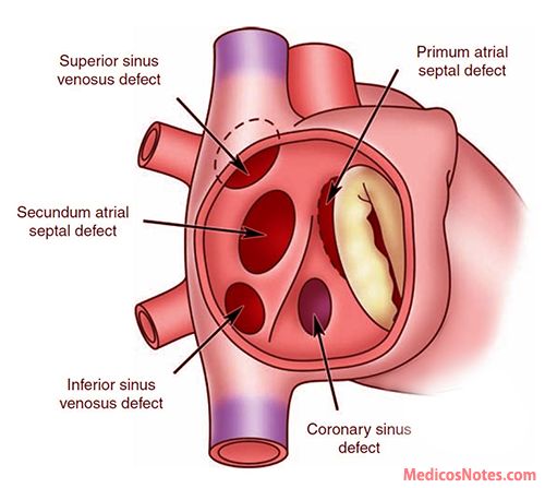
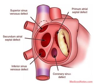
a Atrial Septal Defect (ASD)

An ASD is a hole in the wall between the upper chambers or the right and left atrium, of your heart.

Some ASDs close on their own. Your doctor may need to repair a medium or large ASD with open-heart surgery or another procedure.

The cardiology surgeon might seal the hole with a minimally invasive catheter procedure. The MD inserts a small tube, or catheter, in your blood vessel all the way to your heart. Then he can cover the hole with a variety of devices.

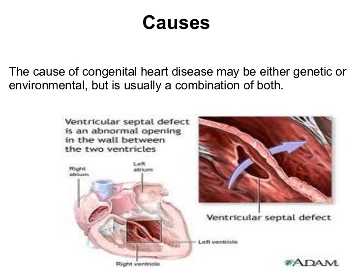
b Ventricular Septal Defect (VSD)

A ventricular septal defect (pronounced ven·tric·u·lar sep·tal de·fect) (VSD) is a birth defect of the heart in which there is a hole in the wall (septum) that separates the two lower chambers (ventricles) of the heart. This wall also is called the ventricular septum.

A VSD is a hole in the part of your septum that separates your heart’s lower chambers that we call ventricles. If you have a VSD, blood gets pumped back to your lungs instead of to your body.

A small VSD may also close on its own. But if yours is larger, you may need surgery to repair it.  Similar to ASD surgery, just a different spot on the chambers.

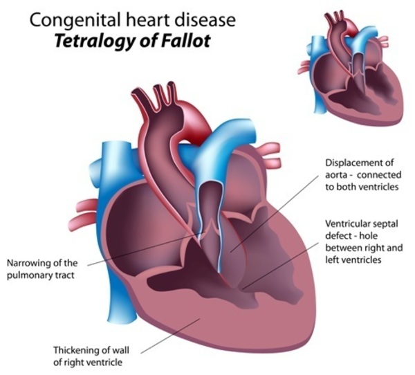
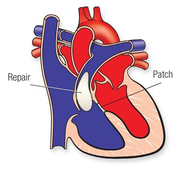
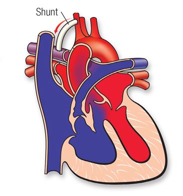
c Complete Atrioventricular Canal Defect (CAVC)

This is the most serious septal defect. It’s when you have a hole in your heart that affects all four chambers.

Complete atrioventricular canal (CAVC) defect is a severe congenital heart disease in which there is a large hole in the tissue (the septum) that separates the left and right sides of the heart. The hole is in the center of the heart, where the upper chambers and lower chambers meet.

As the heart formed abnormally, the valves that separate the upper and lower chambers also developed abnormally. In a normal heart, two valves separate the upper and lower chambers of the heart: the tricuspid valve separates the right chambers and the mitral valve the left chambers. In a child with a complete atrioventricular canal defect, there is one large valve, and it may not close correctly.

As a result of the abnormal passageway between the two sides of the heart, blood from both sides mix, and too much blood circulates back to the lungs before it travels through the body. This means the heart works harder than it should have to, often becoming enlarged and damaged if the problems aren’t repaired.

A CAVC prevents oxygen-rich blood from going to the right places in your body. Your doctor can repair it with patches. But some people need more than one surgery to treat it.

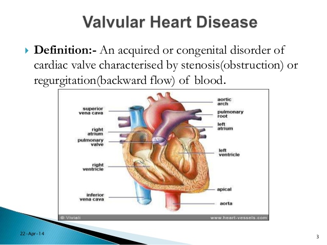
2-Types of Heart Valve Defects

Valves control the flow of blood through your heart’s ventricles and arteries. And some minor heart defects can involve the valves.  Your heart has four valves  aortic, pulmonic, tricuspid, and mitral valve.  All four of them can develop different kinds of heart valve disease, this would include stenosis, regurgitation, and atresia.

a Stenosis.

When your cardiac valves become involved with stenosis where they narrow or stiffen, and won’t open or allow properly from a little amount to completely closed.  Ranging the blood to pass easily to not at all depending on the extent of the stenosis to the valve.  This makes treatment range from none and just medication if needed at all to surgery later in life or immediately born, again depending on the extent of cardiac valvular stenosis.

Aortic Valve Stenosis:

Aortic valve stenosis is most common in the world and in elderly, becoming more and more common after age 65. Several diseases can also cause it to develop in people when they reach middle age. The bicuspid aortic valve is the most common cause of aortic stenosis in patients less than the age of 70 years in developed countries. Rheumatic valve disease is the most common cause in developing countries.  Although some people have aortic stenosis because of a congenital heart defect called a bicuspid aortic valve, this condition more commonly develops during aging as calcium or scarring damages the valve, that is a common cause for this condition in elderly pts since it takes time to develop calcium build up in causing the stenosis.  This in turn restricts the amount of blood flowing getting through the aorta to deliver that oxygenated blood to the body.  The calcium build up takes time to put an affect on the aorta with symptoms as well; so this is why this is commonly seen in elders.

Aortic stenosis is one of the most common and serious valve disease problems. Know the aorta is the main artery bringing blood from the heart to the body’s tissues/organs. Aortic stenosis is a narrowing of the aortic valve opening, and can sometimes be referred to as a failing heart valve. Aortic stenosis restricts the blood flow from the left ventricle to the aorta and may also affect the pressure in the left atrium due to the blood flow regurgitating backwards from the stenosis to the L Ventricle up to the L atrium.

The condition may range from mild to severe; the longer you have it the more severe the symptoms and intensity it would have on the heart, especially left untreated.

Over time, aortic valve stenosis causes your heart’s left ventricle to pump harder to push blood through the narrowed aortic valve. The extra effort may cause the left ventricle to thicken, enlarge and weaken. If not addressed, this form of heart valve disease may lead to heart failure known as CHF.  This heart failure would start on the Left Side of the Heart first=L CHF and if left untreated it would in time effect the Right Side of the Heart=R CHF as well.  The symptoms of L CHF versus R CHF would be different at first and in another topic later this year (over extends this topic for today).

Pulmonary Valve Stenosis:

Pulmonary valve stenosis is a heart valve disorder that involves the pulmonary valve. This is the valve separating the right lower chamber or the right ventricle (one of the chambers in the heart) and the pulmonary artery.  Pulmonary artery is one of the few arteries with high carbon dioxide and low oxygen level in the blood stream. This blood flow normally is being sent to the lungs for more oxygen where it will send the blood flow to the left side of the heart to go through the aorta and sent throughout the body to give high oxygen to all organs.  This is how we survive; with out oxygen we would be going through cellular starvation and die.   The pulmonary artery carries oxygen-poor blood to the lungs  as its ending function. Stenosis, or narrowing in the pulmonary valve occurs when the valve cannot open wide enough to its normal capacity or in some cases completely stenosis, not able to open or close at all.

Treatment surgery at some time in the person’s lifetime.  It would all depend on the severity of the stenosis condition.

Mitral Valve Stenosis:

Mitral valve stenosis — sometimes called mitral stenosis — is a narrowing of the valve between the two left heart chambers. The narrowed valve reduces or blocks blood flow into the heart’s main pumping chamber. The heart’s main pumping chamber is the lower left heart chamber, also called the left ventricle.

There are two types of mitral valve regurgitation:

  • Degenerative mitral regurgitation: This occurs when the mitral valve itself is dysfunctional. The flaps may droop or bulge and do not close tightly.
  • Functional mitral regurgitation: Functional mitral regurgitation happens when an issue outside of the valve (such as diseases of the left ventricle) causes the leakage. You may have normal valve flaps and still be diagnosed with functional mitral regurgitation.

Mitral valve stenosis can make you tired and short of breath. Other symptoms may include irregular heartbeats, dizziness, chest pain or coughing up blood. Some people don’t notice symptoms.

Mitral valve stenosis can be caused by a complication by a sore throat with strep throat called rheumatic fever. Rheumatic fever is now rare in the United States.

Treatment for mitral valve stenosis may include medication or mitral valve repair or replacement surgery.  Some people only need regular health checkups. Treatment depends on the severity of the condition and whether it’s getting worse. Untreated, mitral valve stenosis can lead to serious heart complications.

Tricuspid Valve Stenosis:

Tricuspid stenosis (TS) is narrowing of the tricuspid orifice that obstructs blood flow from the right atrium to the right ventricle. Almost all cases result from rheumatic fever. Symptoms include a fluttering discomfort in the neck, fatigue, cold skin, and right upper quadrant abdominal discomfort. Jugular pulsations are prominent, and a presystolic murmur is often heard at the left sternal edge in the fourth intercostal space and is increased during inspiration. Diagnosis is by echocardiography. TS is usually benign, requiring no specific treatment, but symptomatic patients may benefit from surgery.

b Regurgitation.

A damaged or diseased valve can severely compromise the ability of the heart in inefficiently which in turn does not let  oxygen or carbon dioxide to be used or removed from the body in the way its suppose to be and in time effects the heart and can lead to heart failure if left untreated. Instead of the valve having stenosis it is now not closing completely when it should allowing regurgitation to occur.

Your valves don’t close tightly, the valve affected will allow regurgitation due to the valve not working efficiently with opening and closing the way it should during your heart beat (lub dub-the sound you hear of the heart when auscultating through the stethoscope, the RN or doctor uses.) Instead this lets your blood flow or leak backward r/t the valve partially open or to completely opened when the valve should be closely closed at that time and results into regurgitation.  Remember the cardiac valves open and close as their function to allow blood flowing with a heart pumping to deliver oxygen to the blood from red blood cells that carry O2 and remove carbon dioxide blood through red blood cells carrying it out of the blood stream back to the lungs to get more O2 for the RBCs to carrying it to organs / tissues that need it to survive.  Depending on the extent of valvular regurgitation will decide on treatment which ranges from surgery later in life or immediately when born.

It does the opposite of valve stenosis in that stenosis is a narrowing of the vessel whereas regurgitation is due to a valve remaining open partially or completely when the valve is suppose to be closing to allow blood fill up in the chamber of the heart and common one is mitral valve regurgitation but there are others like tricuspid, pulmonary and ventricular valve regurgitation.

Mitrial Valve Regurgitation:

Mitral valve regurgitation is the most common type of heart valve disease. In this condition, the valve between the left heart chambers doesn’t close fully. Blood leaks backward across the valve. If the leakage is severe, not enough blood moves through the heart or to the rest of the body. Mitral valve regurgitation can make you feel very tired or short of breath.

Other names for mitral valve regurgitation are:

  • Mitral regurgitation (MR).
  • Mitral insufficiency.
  • Mitral incompetence.

Treatment of mitral valve regurgitation may include regular health checkups, medicines or surgery. You may not need treatment if the condition is mild.

Severe mitral valve regurgitation often requires a catheter procedure or heart surgery to repair or replace the mitral valve. Without proper treatment, severe mitral valve regurgitation can cause heart rhythm problems or heart failure.

Tricuspid Valve Regurgitation:

Tricuspid valve disease is a type of heart valve disease (valvular heart disease). The valve between the two right heart chambers (right atrium and right ventricle) doesn’t work properly. As a result, the heart must work harder to send blood to the lungs and the rest of the body.

Tricuspid valve disease often occurs with other heart valve problems.

Symptoms and treatments of tricuspid valve disease vary, depending on the specific valve condition. Treatment may include monitoring, medication, or valve repair or valve replacement.

The most common cause of tricuspid regurgitation is enlargement of the right ventricle. Pressure from heart conditions, such as heart failure, pulmonary hypertension and cardiomyopathy, cause the ventricle to expand. The result is a misshapen tricuspid valve that is leaky and cannot close properly.

Aortic Valve Regurgitation:

Aortic valve regurgitation — also called aortic regurgitation — is a type of heart valve disease. The valve between the lower left heart chamber and the body’s main artery doesn’t close tightly. As a result, some of the blood pumped out of the heart’s main pumping chamber, called the left ventricle, leaks backward.

The leakage may prevent the heart from doing a good enough job of pumping blood to the rest of the body. You may feel tired and short of breath.

Aortic valve regurgitation can develop suddenly or over many years. Once the condition becomes severe, surgery often is needed to repair or replace the valve.

Pulmonary Valve Regurgitation:

Pulmonary valve disease affects the valve between the heart’s lower right chamber and the artery that delivers blood to the lungs. That artery is called the pulmonary artery. The valve is called the pulmonary valve.

A diseased pulmonary valve doesn’t work properly. Pulmonary valve disease changes how blood flows from the heart to the lungs.

The pulmonary valve usually acts like a one-way door from the lower right heart chamber to the lungs. Blood flows from the chamber through the pulmonary valve. It then goes to the pulmonary artery and into the lungs. Blood picks up oxygen in the lungs to take to the body.

Many types of pulmonary valve disease are due to heart conditions present at birth. Treatment depends on the type and severity of pulmonary valve disease.

c Atresia.

Tricuspid Atresia:

This happens when your valve isn’t formed right or has no opening to let your blood pass through. It causes more complicated heart problems. Tricuspid atresia (pronounced try-CUSP-id uh-TREE-zhuh) is a birth defect of the heart where the valve that controls blood flow from the right upper chamber of the heart to the right lower chamber of the heart doesn’t form at all.  In babies with this defect, blood can’t flow correctly through the heart and to the rest of the body.

Aortic Atresia:

Aortic valvular atresia is a congenital condition in which the aortic valvular cusps are fused at birth. It frequently forms as a spectrum of malformations of the left ventricular outflow tract (LVOT). The atresia can be characterized as sub-valvular, valvular, or supra-valvular, depending on the site of the anomaly. Most commonly, the defect presents as aortic stenosis, though in rare cases, it can manifest as complete atresia.

Pulmonary Atresia

Pulmonary atresia is a birth defect of the pulmonary valve, which is the valve that controls blood flow from the right ventricle (lower right chamber of the heart) to the main pulmonary artery (the blood vessel that carries blood from the heart to the lungs). Pulmonary atresia is when this valve didn’t form at all, and no blood can go from the right ventricle of the heart out to the lungs. Because a baby with pulmonary atresia may need surgery or other procedures soon after birth, this birth defect is considered a critical congenital heart defect (critical CHD). Congenital means present at birth.

In a baby without a congenital heart defect, the right side of the heart pumps oxygen-poor blood from the heart to the lungs through the pulmonary artery. The blood that comes back from the lungs is oxygen-rich and can then be pumped to the rest of the body. In babies with pulmonary atresia, the pulmonary valve that usually controls the blood flowing through the pulmonary artery is not formed, so blood is unable to get directly from the right ventricle to the lungs.

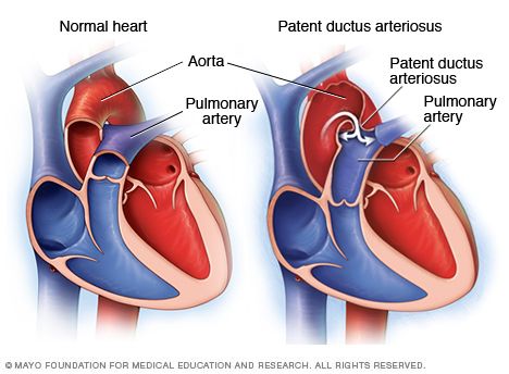
In pulmonary atresia, since blood cannot directly flow from the right ventricle of the heart out to the pulmonary artery, blood must use other routes to bypass the unformed pulmonary valve. The foramen ovale, a natural opening between the right and left upper chambers of the heart during pregnancy that usually closes after the baby is born, often remains open to allow blood flow to the lungs. Additionally, doctors may give medicine to the baby to keep the baby’s patent ductus arteriosus open after the baby’s birth. The patent ductus arteriosus is the blood vessel that allows blood to move around the baby’s lungs before the baby is born and it also usually closes after birth.

d Ebstein’s anomaly.

This is a defect in another heart valve, the tricuspid valve-between the top and bottom of the right side of the heart, which may keep it from closing tightly. Ebstein anomaly is a rare heart problem present at birth. This means it’s a congenital heart defect. The tricuspid valve is incorrectly formed and located lower than usual in the heart. The condition may occur with a hole between the two upper chambers of the heart, called an atrial septal defect.Babies who have Ebstein’s also often have an atrial septal defect (ASD).

In people with Ebstein anomaly, the heart can grow larger. The condition can lead to heart failure.

Treatment of Ebstein anomaly depends on the symptoms. Some people without symptoms only need regular health checkups. Others may need medicines and surgery.

Keep in mind a baby can be born with more than one cardiac defect depending on the pt’s heart formation during pregnancy or at birth or in some cases symptoms arise later in life and that could be the time the patient is diagnosed with the heart condition they have.  Symptoms help tell the doctor there is a problem so diagnosing gets involved until the etiology is found.  If it is your heart your MD will find the defect no problem.

Part II tomorrow on other defects!


QUOTE FOR MONDAY:

Key highlights on Hypothyroidism today

“Genetic VariantsGWAS have identified genetic variants that influence thyroid hormone production, paving the way for personalized med.

Environmental FactorsEndocrine disruptors are recognized as significant contributors to thyroid dysfunction.

Novel BiomarkersBiomarkers like reverse T3 & deiodinase  enzymes are improving diagnostic accuracy.

Therapeutic ApproachesLevothyroxine remains the primary treatment, but new combinations and controlled-release formulations are being explored.

                      Lifestyle and NutritionDiet, exercise, stress management, and complementary medicine are being integrated into treatment plans.

Immunotherapies and Stem Cell TherapyThese emerging therapies are being studied for their potential to improve outcomes in hypothyroidism. 

These highlights reflect the current state of research and treatment strategies for hypothyroidism, emphasizing the need for multidimensional approach to patient care.”

Journal of Chemical Risks-JCHR (Understanding Hypothyroidism: A Clear Look at Current Research and Treatment | Journal of Chemical Health Risks)

Key Highlights on Hyperthyroidism today:

“Approximately 1.2 percent of the United States population has hyperthyroidism.

Women are 2 to 10 times more likely to develop hyperthyroidism than men.

The prevalence of hyperthyroidism is approximately 0.5 percent in the general population for overt cases.

Graves’ disease accounts for 60 to 80 percent of all hyperthyroidism cases.

Toxic multinodular goiter accounts for 15 to 20 percent of hyperthyroidism cases.

Toxic adenoma accounts for approximately 3 to 5 percent of hyperthyroidism cases.

Approximately 25 to 50 percent of people with Graves’ disease will develop thyroid eye disease (ophthalmopathy).

Atrial fibrillation occurs in 10 to 15 percent of patients with hyperthyroidism.

Thyroid storm has a mortality rate of 10 to 30 percent if not treated immediately.

Remission rates for Graves’ disease after 12-18 months of antithyroid drug therapy are about 40 to 50 percent.

Radioactive iodine therapy cures hyperthyroidism in approximately 90 percent of patients with a single dose.

Hypothyroidism develops in more than 80 percent of patients treated with radioactive iodine for Graves’ disease within 10 years.

Overt hyperthyroidism complicates 0.1 to 0.4 percent of all pregnancies.

Subclinical hyperthyroidism is estimated to occur in up to 15 percent of elderly subjects aged 80 and over.

Neonatal Graves’ disease occurs in 1 to 5 percent of infants born to mothers with Graves’ disease.”

GITNUX (Hyperthyroidism Statistics Statistics: Market Data Report 2025)