Archive | August 2016

QUOTE FOR THE WEEKEND:

“More action is needed at every level of public health and health care to improve patient safety and eliminate infections that commonly threaten hospital patients.”

CDC (Center for disease control and prevention).

Part 3 Actual types of health care infections (HCIs) we deal with in health care facilities.

HAI88 HAI0

HAI7  HAI22

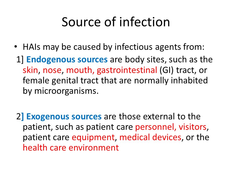
1-C-DIFF or C-difficile, know as Clostridium difficile infection (CDI), or Clostridium difficile–associated disease (CDAD), which is an infection of the intestines caused by the anaerobic, spore-forming, gram-positive bacillus C. difficile.   This microbe was first identified in 1935 when it was isolated from the stools of neonates. C. difficile produces heat-resistant spores that can remain viable on fomites in the environment for years, becoming a source of outbreaks in healthcare facilities. This bacillus also produces two types of toxins: Toxin A (an enterotoxin) and Toxin B (a cytotoxin). These toxins are responsible for the inflammatory responses of the colon, which results in loss of epithelial integrity and the production of watery diarrhea. C. difficile is the most common cause of antibiotic-associated diarrhea and pseudomembranous colitis and has proved extremely difficult to control due to new, more resistant strains.

  1. difficile is found in the intestinal tract of up to 70% of healthy infants, 1–3% of healthy adults, and in higher rates (up to 20%) in persons on antibiotic therapy. Prevalence, severity, colectomy rates, and mortality rates due to C. difficile have all risen in the past two decades, proving that this microbe has become an extremely virulent superbug that is both persistent and difficult to treat. Surveillance data from the CDC show that between 2000 and 2007 mortality from C. difficile increased by 400% due to a more virulent strain of this organism.

Studies of hospital-acquired CDI have shown that the infection is independently associated with increased risk of in-hospital death: For every 10 hospital patients who acquire a CDI, 1 patient died. Data from the CDC show that C. difficile is responsible for 337,000 infections and 14,000 deaths in the United States each year.

The greatest risk factor for CDI is the use of antibiotics, such as cephlasporins, clindamycin, or the penicillins, because these antibiotics kill the normal flora of the colon, causing overgrowth of C. difficile. Risk is increased for those taking multiple antimicrobials and those who take antimicrobials for longer time periods. Other risk factors for CDI include advanced age. Although almost half of the infections occur in persons younger than 65, most CDI-related deaths occur in the elderly. People with HIV infection, compromised immune systems, and compromised physical status are also at increased risk for CDI. Hospital admission increases one’s chance of acquiring CDI, as does gastrointestinal surgery.

Transmission of CDI occurs by the fecal-oral route.

The time between exposure to C. difficile and infection is 2 to 3 days. Symptoms of CDI vary greatly, ranging from asymptomatic to mild (fever, malaise, and gastrointestinal symptoms, including abdominal pain and cramps, and mild to moderate foul-smelling diarrhea that is rarely bloody) to extremely severe toxic megacolon, septic shock, and even death. Complications of C. difficile include pseudomembranous colitis or fulminant colitis.

Diagnosis is based on clinical history (antibiotic use in the previous 2 months, diarrhea after 72 or more hours of hospitalization), and presence of C. difficile in the stool. Stool culture is the most sensitive test and is often used for diagnosis in the hospital setting. Colonoscopy revealing histopathology with pseudomembranous colitis is also diagnostic but not necessary in most cases.

Treatment for CDI begins with discontinuation of the antibiotic causing the infection. In many cases, this step is the only necessary treatment since normal flora can reestablish in the colon. If mild to moderate diarrhea persists, patients can be treated with either metronidazole or vancomycin. In cases of severe diarrhea, vancomycin is the drug of choice for treatment due to its history of rapid symptom resolution and overall fewer treatment failures. Although antibiotic treatment will clear the infection, it will not kill the bacterial spores. In 27% of cases, relapse occurs within 3 weeks of antibiotic termination. In extreme cases, colectomy with end ileostomy may be necessary. Treatment for asymptomatic cases is not recommended.

An innovative CDI treatment may be on the horizon. Researchers have shown that C. difficile infection arises as the result of the disruption of natural flora in the intestines, a condition known as dysbiosis. New research in the treatment of CDI involves isolating specific gut bacteria in the fecal matter of healthy individuals and incorporating it into the gut of a person with CDI to restore normal flora and cure the infection.

CDI can be catastrophic to patients and indeed to entire healthcare facilities if an outbreak occurs. To prevent CDI, follow these guidelines from the CDC:

  • Immediately isolate patients with confirmed C. difficile infection and use contact precautions for the duration of diarrhea. Consider extending precautions beyond the duration of diarrhea and using presumptive contact precautions for patients with diarrhea pending confirmed C. difficile diagnosis.
  • Educate healthcare personnel, patients, their families, and any visitors about C. difficile and help them maintain contact precautions.
  • Follow proper handwashing techniques. Hand hygiene for C. difficile must include vigorous washing of hands with soap and water to mechanically remove spores. Alcohol-based hand rubs are not effective against C. difficile.
  • Because C. difficile spores can survive on objects for long periods of time be sure to thoroughly clean and disinfect equipment and objects in the environment. Consider use of sodium hypochlorite (bleach)–containing agents or EPA-registered disinfectants with sporicidal claim for environmental cleaning.
  • Enact a laboratory-based alert system for immediate notification of positive test results.


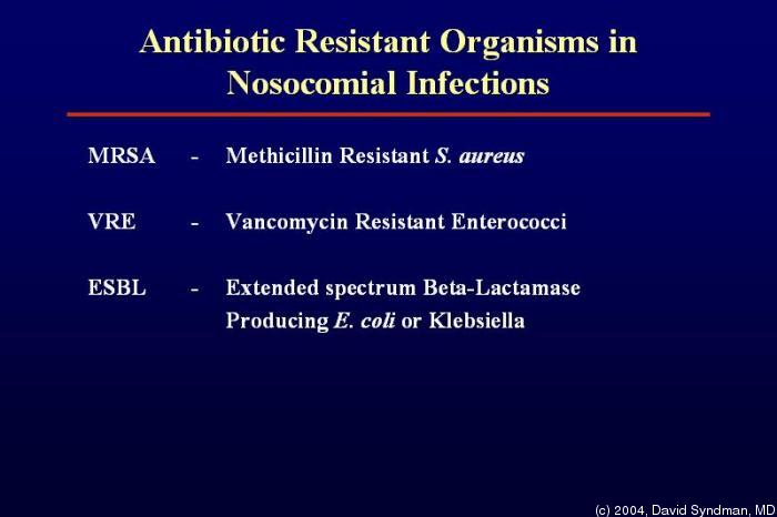
2- MRSA Methicillin-resistant Staphylococcus aureus (MRSA), also known as multidrug resistant S. aureus, includes any strain of S. aureus that has become resistant to the group of antibiotics known as beta-lactam antibiotics. Included in this group are the penicillins (methicillin, amoxicillin, oxacillin) and cephalosporins. Staphylococcus aureus includes gram-positive, nonmotile, non-spore-forming cocci that can be found alone, in pairs, or in grapelike clusters.

When penicillin was first introduced in the early 1940s, it was considered to be a wonder drug because it reduced the death rate from Staphylococcus infection from 70% to 25%. Unfortunately, by 1944, drug resistance was beginning to occur, so methicillin was synthesized, and, in 1959, it became the world’s first semisynthetic penicillin. Shortly thereafter in 1961, staphylococcal resistance to methicillin began as well, and the name “methicillin-resistant S. aureus” and the acronym MRSA were coined. Although methicillin was discontinued in 1993, the name and acronym have remained because of MRSA history.

MRSA is now the most common drug-resistant infection acquired in healthcare facilities. In addition to becoming more problematic as a top HAI in recent years, transmission of MRSA has also become more common in children, prison inmates, and sports participants. Community-associated MRSA (CA-MRSA) most often presents in the form of skin infections. Hospital-acquired MRSA (HA-MRSA) infections manifest in various forms, including bloodstream infections, surgical site infections, and pneumonia. Although approximately 25–30% of persons are colonized in the nasal passages with Staphylococcus, less than 2% are colonized with MRSA. MRSA are extremely resistant and can survive for weeks on environmental surfaces. Transfer of the pathogen can occur directly from patient contact with a contaminated surface or indirectly as healthcare workers touch contaminated surfaces with gloves or hands and then touch a patient.

Risk factors for healthcare-acquired MRSA infection include advanced age, young age, use of quinolone antibiotics, and extended stay in a healthcare facility. Those with diabetes, cancer, or a compromised immune system are also at increased risk of infection.

Symptoms of MRSA infection vary depending on the type and stage of infection and the susceptibility of the organism. Skin infections may appear as painful, red, swollen pustules or boils; as cellulitis; or as a spider bite or bump. They can be found in areas where visible skin trauma has occurred or in areas covered by hair. Patients may also have fever, headaches, hypotension, and joint pain. Complications of MRSA-related skin infections include endocarditis, necrotizing fasciitis, osteomyelitis, and sepsis.

Patient history of admission to a healthcare facility is useful in diagnosing HA-MRSA. Definitive diagnosis of MRSA is made by oxacillin/methicillin resistance that is shown by lab culture and susceptibility testing. Specimens submitted for testing vary depending on the site of suspected infection and may include tissue, wound drainage, sputum, respiratory secretions, and blood or urine cultures.

Treatment for MRSA infections varies based on site of infection, stage of infection, and age of the individual. Treatment includes drainage of abscesses, surgical debridement, decolonization strategies, and antimicrobial therapy with antibiotics such as vancomycin, clindamycin, daptomycin, linezolid, rifampin, trimethoprim-sulfamethoxazole (TMP-SMX), quinupristin-dalfopristin, telavancin, and tetracyclines (limited use). MRSA is rapidly becoming resistant to rifampin; therefore, this drug should not be used alone in the treatment of MRSA infections. Consultation with an infectious disease specialist is recommended for treatment of severe MRSA infections.

3-VRE- Vancomycin-Resistant Enterococci Infection (VRE) or Enterococci (formerly known as Group D streptococci). VRE are non-spore-forming, gram-positive cocci that exist in either pairs or short chains. They are commonly found in the human intestine or the female genital tract. The most common organism associated with vancomycin-ressistant enterococci (VRE) infection in hospitals is Enterococcus faecium. Enterococcus faecalis is also a cause of human disease. VRE infections can occur in the urinary tract, in wounds associated with catheters, in the bloodstream, and in surgical sites. Enterococci are a common cause of endocarditis, intra-abdominal infections, and pelvic infections.

VRE was first reported in Europe in 1986, followed in 1989 by the first report in the United States. Since then it has spread rapidly. Between 1990 and 1997, the prevalence of VRE in hospital patients increased from less than 1% to 15%.

VRE, which is found predominantly in hospitalized or recently hospitalized patients, are difficult to eliminate because they are able to withstand extreme temperatures, can survive for long periods on environmental surfaces, and are resistant to vancomycin. Transmission of VRE occurs most commonly in the form of person-to-person contact by the hands of healthcare workers after contact with the blood, urine, or feces an infected individual. VRE is also spread from contact with environmental surfaces, or through contact with the open wound of an infected person.

People most at risk for infection with VRE include the elderly and those with diabetes, those with compromised immune systems, and those who are already colonized with the bacteria. Prolonged hospitalization, catheterization (urinary and intravenous), and long-term use of vancomycin or other antibiotics also increase a person’s risk of infection.

Symptoms of VRE infection vary depending on the site of infection and may include erythema, warmth, edema, fever, abdominal pain, pelvic pain, and organ pain. Definitive diagnosis is made by culture and susceptibility testing with specimens obtained from suspected sites of infection


QUOTE FOR FRIDAY:

“Children and geriatrics are two groups with the highest risk for infection. In addition to age but there are a number of other key risk factors that can contribute to the likelihood of getting a hospital infection. HAIs are one of the biggest public health issues facing society today. Every year, an estimated 1.7 million Americans get a healthcare associated infection, and 99,000 people die as a result of these infections.”

Pennsylvania Dept. of Health


Part 2 Know what puts people at risk for health acquired infections (HAIs):

Sepsis (HAI)1   HAI5HAI6

Factors and types of procedures that put an individual at risk for HAIs:

*Healthcare facilities, including hospitals, acute care facilities, and long-term care facilities, contain many organisms and methods of transfer of bacteria; however, certain infections occur more frequently than others in healthcare environment.

*The most common pathogens causing catheter associated urinary tract infections (CAUTIs) are E. coli, Candida spp., Enterococcus spp., Pseudomonas aeruginosa, Klebsiella pneumoniae, and Enterobacter spp. Pathogens may gain access to the urinary system during insertion, manipulation, maintenance, or removal of a urinary catheter. Pathogens enter the urinary tract via the extra-luminal route, by moving along the outside of the catheter getting up in the urethra back up in the urinary bladder and something not sterile or even clean got up into a area that is causing risk of infection.

Studies show that by the 30th day of catheterization, which is also considered the demarcation between short- and long-term catheterization the daily risk, of bacteria in the urine approaches to 100%. In a acute hospital if a patient has a catheter applied for surgery it is out the next day or two and if not many facilities have MDs having to renew the order in the patient’s chart or its removed if the intial order stated the time it starts and the time the orders ends (usually in 48 hrs), again if it is a simple operation (ex. A pacemaker insertion with no post-op difficulties as opposed to a patient who has CABG-coronary artery bypass graft surgeRemember in most cases there’s several factors putting many people at higher risk for the UTIs, here are some examples:

*Certain women – One major reason for this is because the urethra in all women is shorter than in men, this cuts down on the distance for urine in the urinary bladder to reach the end of the urethra where the urine comes out. This allows bacteria to take a shorter route to enter a women in reaching her bladder going back up the urethra and putting her at higher risk of infection.      Also women more sexually active is at higher risk for UTIs also cause of the same reason again due to a shorter urethra.

`*AGE -the older adults and those with prolonged catheterization (infection begins when an organism successfully colonizes by entering the body, growing and multiplying). Medical conditions that increase the risk of a CAUTIs include diabetes, diarrhea, renal insufficiency, and a compromised immune system. Colonization of the catheter drainage bag can also increase a patient’s risk for a CAUTIs (the reason to get it out of the patient as soon as possible, if the patient is a candidate for removal). A patient for example that may not be a candidate and leaving it in for 2 weeks or more is a patient who is incontinent but let’s say is in the hospital with a open wound to the lower back or buttock or hip but if urine gets into the wound it will just infect it so to prevent urine incontinency from touching the wound the foley catheter is inserted as a diversion. When the wound heals it’s immediately discontinued and taken out of the patient, for example.

If a bacteria infection is presented (a UTI) than the patient should be started on a antibiotic that only kills bacterial infections. The patient should stay on the medication 7 to 14 days.

* Central Line-Associated Bloodstream Infections-Perhaps the most deadly HAI, central line–associated bloodstream infections (CLABSIs) are transmitted via a central venous catheter (CVC) directly to a patient’s bloodstream. A CVC is any intravascular catheter that terminates at or close to the heart or in one of the great vessels and is used for infusion of medications, nutrition, and blood; withdrawal of blood; hemodialysis access; and hemodynamic monitoring. CVCs may be placed in the large veins of the chest (through axillary or subclavian veins), the neck (through the internal jugular vein), or the groin (through the femoral vein). The infection cannot be related to any other infection the patient might have and must not have been present or incubating when the patient was admitted to the healthcare facility. Tens of thousands of CLABSIs occur in U.S. hospitals each year, with deadly results: Up to 25% of those diagnosed with a CLABSI will die from the infection. Unfortunately a significant number of these infections continue to occur in inpatient settings and in hemodialysis facilities, even though most CLABSIs, especially those occurring in ICUs, are preventable. The longer the patient has a central line, the more likely he or she is to acquire a bloodstream infection via the line. The type of material the catheter is made from also affects infection transmission. Infection risk is also increased in patients with concurrent infection and those treated in the ICU.

*Surgical Skin Infections (SSIs) – Anytime the skin barrier is broken, the risk for infection rises, so it is only logical that surgery will increase a patient’s risk for a HAI. Surgical site infections (SSIs) occur at the location where a surgery was previously performed. To be diagnosed as a SSI, the infection must occur within 30 days of surgery if no implant is left in place or within 1 year if the implant is in place and the infection appears to be related to the operative procedure.

*Ventilator-associated pneumonia (VAP) is an infection of the lungs that develops after a person has been on a ventilator for longer than 48 hours. The most common type of HAI contracted in the ICU, VAP occurs in as many as 28% of patients who have had mechanical ventilation. Infection occurs because the endotracheal or tracheostomy tube allows passage of microbes into the lungs. These organisms may originate from the patient’s aspirate, from the oropharynx and digestive tract, or from external sources, such as contaminated equipment and medications.

Although any microbe can be the causative agent, certain microbes are most often implicated due to increasing drug resistance. Pseudomonas aeruginosa is the most common multidrug-resistant organism responsible for VAP. Other microbes that cause VAP include Staphlococcus aureus, Klebsiella spp., Escherichia coli, Enterobacter spp., Actinobacter spp., MRSA, and Serratia marcescens. Pneumonia is considered early in onset if it occurs within the first 4 days after hospital admission. Multidrug resistant organisms are more likely to be the cause of late-onset pneumonia, defined as 5 or more days post-admission.

In addition to recent ventilation, other risk factors increase a patient’s chance of acquiring VAP, such as hospitalization or antibiotic use within the past 90 days, hospital stay greater than 5 days, hemodialysis within the past 30 days, and known circulation of multidrug-resistant organisms in the facility. Immuno-compromised residents and those who reside in a nursing home or long-term care facility are also at greater risk for VAP.   Empiric treatment is vital in patients with suspected VAP and can be based on patient risk factors for multidrug-resistant organisms, known local prevalence of resistant organisms, severity of infection, and total number of days the patient was hospitalized before the onset of pneumonia. Unless diagnostic testing shows otherwise, initial empiric therapy should not be changed in the first 48 to 72 hours because clinical response to antibiotic therapy is not likely during this time frame. Patients should be treated with antibiotic therapy for at least 72 hours after a clinical response is attained. To prevent VAP, the CDC recommends the following strategies:

Strategies to Prevent Aspiration

  • Maintain patients in a semirecumbent position.
  • Avoid gastric overdistention.
  • Avoid unplanned extubation and reintubation.
  • Use a cuffed endotracheal tube with inline or subglottic suctioning.
  • Maintain an endotracheal cuff pressure of at least 20 cm water.

Strategies to Reduce Colonization of the Aerodigestive Tract

  • When possible, use orotracheal intubation rather than nasotracheal intubation.
  • Avoid histamine receptor 2 (H2)–blocking agents and proton pump inhibitors for patients who are not at high risk for developing a stress ulcer or stress gastritis.
  • Perform regular oral care with an antiseptic solution.

Strategies to Minimize Contamination of Equipment

  • Use sterile water to rinse reusable respiratory equipment.
  • Remove condensate from ventilatory circuits, keeping the ventilatory circuit closed while you do so.
  • Change the ventilatory circuit only when visibly soiled or malfunctioning.
  • Store and disinfect respiratory therapy equipment properly.

In addition to these strategies, healthcare professionals should perform daily assessments of readiness to wean to minimize the duration of ventilation. Whenever possible, use noninvasive ventilation methods.

See tomorrow’s Part 3 on HAIs discussing actual names of infections that we deal with in health facilities and learn about them to be prepared if, unfortunately, you, your family or friends have to go to one.


QUOTE FOR THURSDAY:

“Infections prolong hospital stays, create long-term disability, increase resistance to antimicrobials, represent a massive additional financial burden for health systems, generate high costs for patients and their family, and cause unnecessary deaths”

World Health Organization

Part 1 Health Acquired Infections (HAIs)

HAIs2HAIs

HAIs3 HAIs4


The purpose of this article is to help broaden the public in knowing about HAIs including how their family or friend should be cared for when in a hospital with what they can do when visiting a loved one in a health facility for both the patient’s and visitor’s benefit.  

History of HAIs 

Let us start with some history. In England in the 1830s, the term hospitalism was coined by Sir James Simpson to describe HAIs. In those days, it was believed that infection was spread because of inadequate ventilation and stagnant air. To prevent infection, windows were opened, and whenever possible care was taken to prevent overcrowding of hospital rooms. Little was known about microbes and their pathogenicity, and consequently little was done about personal hygiene. In Victorian society, the idea of one’s personal hygiene being connected to infection was taken personally and was met with great resistance.

Despite the efforts of medical personnel, many patients died of overwhelming sepsis following preventable infections. In the late 1860s after much persistence, Joseph Lister, a British surgeon, introduced the concept of antisepsis, which significantly decreased death from postoperative infection. After penicillin was introduced in 1941, postsurgical infection rates and deaths from postsurgical pneumonia were both dramatically decreased.

Today, modern medicine has brought a more thorough understanding of pathogens and the epidemiology of the diseases they cause. Unfortunately, in spite of the vast amounts of medical advances that have occurred over the years, the healthcare industry is still faced with the enormous task of preventing and reducing the risk of HAIs.

Today HAIs are defined as nosocomial infections which are infections that are acquired in hospitals and other healthcare facilities (like a nursing home or sub-acute rehab facility). To be classified as a nosocomial infection the patient must have been admitted for reasons other than an infection. He or she must also have shown no signs of active or incubating infection upon admission.

On average, nosocomial patients stay in the hospital 2.5 times longer than patients without infection.   An estimated 40 percent of nosocomial infections are caused by poor hand hygiene (WHO). Hospital staff can significantly reduce the number of cases with regular hand washing. They should also wear protective garments and gloves when working with patients.

Invasive procedures increase the risk of nosocomial infections. Noninvasive procedures are recommended when possible. Most nosocomial infections are due to bacteria. Since antibiotics are frequently used within hospitals, the types of bacteria and their resistance to antibiotics is different than bacteria outside of the hospital. Nosocomial infections can be serious and difficult to treat, especially if it’s a multi-resistant bacteria.

How do infections even occur?

Harmful microbes are all around us, and although infection poses a threat to everyone, certain people are more at a potential of infection. For example, people in healthcare facilities are more at risk than those in the community simply because they are exposed to others who are infected with disease-causing organisms. These people are exposed to so many other peoples germs and bacteria as opposed to a private home simply puts you at potential for picking up them if not proper prevention is carried out by all that come in contact with you, starting simply with hand washing by the patient and those that see the patient (medical staff to visitors).

Even more at risk are special populations or conditions of patients, such as those with compromised immune systems, those who have undergone recent surgery, those with poor nutritional status, and those with open wounds. Patients undergoing certain medical procedures, such as intubations and central lines, are also at increased risk. Medical devices also carry a risk of infection. Urinary catheters, central lines, mechanical ventilation equipment, and surgical drains all put patients at risk for infection. Any foreign object in the body or any unnatural opening of the body (Ex. surgical wounds or trauma wounds) puts that individual at risk for local infection to that area and if left untreated goes to general infection (temp greater than 100.5 or 101 F). We will go into more detail on risk factors tomorrow in Part 2.

Further, certain medications and various chemotherapies weaken patients’ immune systems, leaving patients more vulnerable to infection. The length of time spent in a healthcare facility also affects the risk of infection: The longer the stay, the longer that person is at potential for exposure of bacteria. A person in a hospital versus in a home puts greater risk for that person’s chances of acquiring a HAI since there is more bacteria and germs exposure in a hospital compared to a home due to the population. Over 25 years ago and further back the doctors kept patients in the hospital longer than needed; they were thinking this was the best care for the patient but now it’s get the patient out as soon as possible when clearance from the MD’s for discharge is reached. The logic behind this is to get that patient back at home exposed to less bacteria or germs where they can reach their optimal level of functioning since they no longer need a hospital care but follow up with their MD at their office.

How does infection travel from one object to another or even person to person?

The most common method of transmission is by direct contact with an infectious microorganism. Sputum, blood, and feces are common vehicles for microbe transmission. Healthcare workers and patients spread microbes via droplets generated by talking, sneezing, or coughing. Small particles of evaporated droplets (droplet nuclei) and dust particles carry microorganisms and spread infection over long distances.

Infection can also be spread through inanimate objects known as fomites; which are any object or substance capable of carrying infectious organisms, such as germs or parasites, and hence transferring them from one individual to another. Fomites can be skin cells, hair, clothing, and bedding which are common hospital sources of contamination including improperly sterilized medical equipment that is used on more than one patient. Healthcare workers who move from patient to patient carry infectious organisms on their clothes, stethoscopes, and phones. Other modes of transmission include the spread of infectious agents through food and water or through vectors, such as mosquitoes, flies, and rats.

Though hospitals throughout America have this problem to face but so do hospitals elsewhere; it’s a worldwide problem. Know these facilities in most places have developed infection control people who continuously make policies/procedures in their facilities to prevent the spread of infection through all routes with the knowledge we know today as opposed to 25 years ago and further back regarding infection spreading in the hospital.

Turn into Part 2 and read about what puts people at risk for health acquired infections HAIs.


QUOTE FOR TUESDAY:

Researchers from the University of Dundee and University College London in the UK found that some common medications made to dissolve in water contain such high levels of sodium that taking the maximum dose each day would cause you to exceed the recommended daily limits, and put you at risk of heart attack and stroke.

University of Dundee and College of London in the UK


Part 2 Iron Deficiency Anemia

anemia2   Anemia Part 2

Anemia Part II

This form of iron deficiency anemia is treated with changes in your diet and iron supplements.

If the underlying cause of iron deficiency is loss of blood — other than from menstruation — the source of the bleeding must be located and stopped. This may involve surgery.

    • Rapid growth cycles (infancy, adolescence)
    • Heavy menstrual bleeding or chronic blood loss from the GI tract
    • Pregnancy
    • Diets that contain insufficient iron (rare in the United States)
    • Breastfed infants who have not started on solid food after six months of age
    • Babies who are given cow’s milk prior to age 12 months
  • Alcoholism-Most often healthy red blood cells last between 90 and 120 days. Parts of your body then remove old blood cells. A hormone called erythropoietin (epo) made in your kidneys signals your bone marrow to make more red blood cells.To first diagnose the person with any anemia the following needs to be done to help the doctor in diagnostic tooling , which is tests to rule out and rule in what the actual problem isn’t or is. With the MD knowing the results of these tests it will guide the doctor knowing the correct diagnosis to use the best treatment to either cure or get the problem under control (Ex. What is curable is iron deficiency anemia but sickle cell anemia is not).


  • Hemoglobin is the oxygen-carrying protein inside red blood cells. It gives red blood cells their red color. People with anemia do not have enough hemoglobin.                                                                                                                                                                                                  
  • Although many parts of the body help make red blood cells, most of the work is done in the bone marrow. Bone marrow is the soft tissue in the center of bones that helps form all blood cells.

The diagnosis tests that are usually done by a doctor are the following:

Physical exam. During a physical exam, your doctor may listen to your heart and your breathing. Your doctor may also place his or her hands on your abdomen to feel the size of your liver and spleen. He would look at the color of the skin and the eyes to look for paleness.

Blood Tests. Your doctor would do the basis blood tests being a CBC which is used to count the number of blood cells in a sample of your blood. For anemia, your doctor will be interested in the levels of the red blood cells contained in the blood particularly your hematocrit (the solids of the blood) and the hemoglobin (the liguid of your blood) in your bloodstream. If anemic both of these will be low and hematocrit below 7.0 down to 6.0 is critical.

Normal adult hematocrit values vary from one medical practice to another but are generally between 40 and 52 percent for men and 35 and 47 percent for women. Normal adult hemoglobin values are generally 14 to 18 grams per deciliter for men and 12 to 16 grams per deciliter for women.

Additional testing maybe ordered as well; like the following to help determine what the person has with the what treatment to tell the MD is needed to help the individual get better.

This could be: . A test to determine the size and shape of your red blood cells. Some of your red blood cells may also be examined for unusual size, shape and color. Doing so can help pinpoint a diagnosis. For example, in iron deficiency anemia, red blood cells are smaller and paler in color than normal. In vitamin deficiency anemias, red blood cells are enlarged and fewer in number.

If you receive a diagnosis of anemia, your doctor may order additional tests to determine the underlying cause. For example, iron deficiency anemia can result from chronic bleeding of ulcers, benign polyps in the colon, colon cancer, tumors or kidney problems.

Occasionally, it may be necessary to study a sample of your bone marrow to diagnose anemia.

 ***Treatment for iron-deficiency anemia will depend on its cause and severity. Treatments may include dietary changes and supplements, medicines, and surgery.

Severe iron-deficiency anemia may require a blood transfusion, iron injections, or intravenous (IV) iron therapy. Treatment may need to be done in a hospital.

The goals of treating iron-deficiency anemia are to treat its underlying cause and restore normal levels of red blood cells, hemoglobin, and iron.*****

Watching your sodium is not just in your diet but checking sodium in your meds also!

sodium      what sodium doessodium4

The researchers, led by Jacob George, MD, senior clinical lecturer and honorary consultant in clinical pharmacology at the University of Dundee, tracked more than 1.2 million patients in the United Kingdom for more than seven years, and compared the patients taking the soluble forms of drugs, such as acetaminophen, ibuprofen, aspirin and others, to patients taking the traditional pill form. They found that the patients taking the soluble forms were at a 16 percent higher risk of heart attack or stroke, and at a 28 percent higher risk of developing high blood pressure, which researchers attributed to the high levels of sodium in these medications.

“These tablets all fizz when you put them in water,” Dr. George said. “The manufacturers use the salt to create that fizz, but it does much more harm than good.”

The dissolvable form of acetaminophen, better known as Tylenol, contained the most sodium, at 427 milligrams per dose, according to the study. If you took the maximum amount daily, you’d be getting more than 3,400mg of sodium, far more than the USDA recommended daily allowance of 2,000mg before you factor in your diet.

Soluble ibuprofen and aspirin contained far less sodium, with 202mg and 149mg per pill respectively, according to the study, but George said the levels are still far from acceptable.

“There’s no legal regulation anywhere in the U.S. or the UK to limit the amount of sodium per tablet,” he said. “The sodium levels in these medications should be clearly labeled on the packages.”

Diets high in sodium are linked to high blood pressure, heart attack and stroke, but many people are unaware just how much salt they get in their diet, let alone from their medication, according to the study.

Soluble acetaminophen and ibuprofen are often given by prescription and are much more common in the UK than in America, but soluble aspirin is frequently sold in U.S. drug stores under the brand Alka Seltzer, said Patrick Fratellone, MD, a New York City-based cardiologist.

“The U.S. should start looking at its soluble medications and determine whether the amount of salt in them should be allowed,” Dr. Fratellone said. “There’s no restrictions on the amount manufacturers can add.”

Merle Myerson, MD, director of the cardiovascular disease prevention and pre-exercise heart screening program at St. Luke’s and Roosevelt Hospital in New York City agreed, saying that even though salt is sometimes necessary in these medications, it’s not necessary for people to take them.

“We often tell patients that salt is hidden in foods,” Dr. Myerson said. “Processed foods and fast foods have a lot of hidden salt. Here again, we’re seeing another source of hidden salt that may contribute to a person’s risk for hypertension.”

“Salt helps make the medication more digestible and dissolve better,” she added. “But if it contains too much salt, it could add up to amounts that could be dangerous.”

These medications are typically used by people who have trouble swallowing pills, George said, but anyone who does should speak to their doctor.

“There are groups of people who legitimately need these medications, but anyone who takes them should realize the cardiovascular risk of these in the long term,” he said. “They need to speak to their physician to come to an informed decision of whether they should actually be taking them.”

Fratellone said that he recommends that patients avoid soluble medications, and added that he refuses to prescribe them.

“Patients need to avoid them,” he said. “There needs to be information on the front of the packaging telling patients how much salt in is their medicine.”

“We’re trying to get patients to reduce the amount of salt in their diet,” Fratellone added, “and manufacturers are putting it in our medication without giving us fair warning.”

Decongestants may raise your blood pressure.

People with high blood pressure should be aware that the use of decongestants may raise blood pressure or interfere with the effectiveness of some prescribed blood pressure medications. Many over-the-counter cold and flu preparations contain decongestants such as:

  • Ephedrine
  • Levmetamfetamine
  • Naphazoline
  • Oxymetazoline
  • Phenylephrine
  • Phenylpropanolamine
  • Propylhexedrine
  • Pseudoephedrine
  • Synephrine
  • Tetrahydrozoline
  • Check the sodium content.

Some OTCs are high in sodium, which can also raise blood pressure. Look at the active and inactive ingredients lists for words like “sodium” or “soda.” Note the amount of

sodium in the medication. People with high blood pressure should consume less than 1,500 mg of sodium per day from all sources; one dose of some OTCs can contain more than a whole day’s allowance.

Those who prescribe medications may be unaware that they may contain substantial amounts of sodium. References such as package inserts, the Physicians’ Desk Reference, and Drug Information 1 do not always provide information about sodium, and the sodium content, when it does appear, may be given in various units of measurement or may need to be calculated.

Certain medications containing one or more drugs are high in sodium content regardless of the formulation used, whereas others vary according to the formulation (Sodium Content of Various Drug Products and Formulations.). High-sodium and low-sodium formulations of the same drug products should not be used interchangeably in patients who are following sodium-restricted diets.

The Food and Drug Administration (FDA) has proposed a rule for the labeling of the sodium content of over-the-counter drugs,4 and it will be working with pharmaceutical organizations to develop voluntary sodium labeling for prescription drugs. The FDA will recommend that makers of prescription drugs declare the sodium content if it exceeds 5 mg (0.22 mmol) per single recommended dose and that they issue a warning if the sodium content exceeds 140 mg (6.09 mmol) per maximal daily dose. Sodium contained in active ingredients, excipients, and any recommended diluent will be included. These measures will simplify the calculation of the total sodium content of any product prescribed for patients whose sodium intake is restricted. Until these initiatives have been implemented, this information should be obtained from the manufacturer.

References:

1.) American Heart Association

2.) The New England Journal of Medicine

3.) EverydayHealth.com