Archives

Gastrointestinal stromal tumo – GIST.


Cancer starts when cells in the body begin to grow out of control. Cells in nearly any part of the body can become cancer, and then can spread to other areas of the body.

Gastrointestinal stromal tumors (GISTs) are not common, and the exact number of people diagnosed with these tumors each year is not known. Until the late 1990s, not much was known about these tumors (and doctors didn’t have good ways of identifying them with lab tests), so many of them ended up being classified as other kinds of cancers.

Current estimates for the total number of GIST cases diagnosed each year in the United States range from about 4,000 to about 6,000.

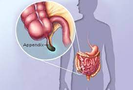
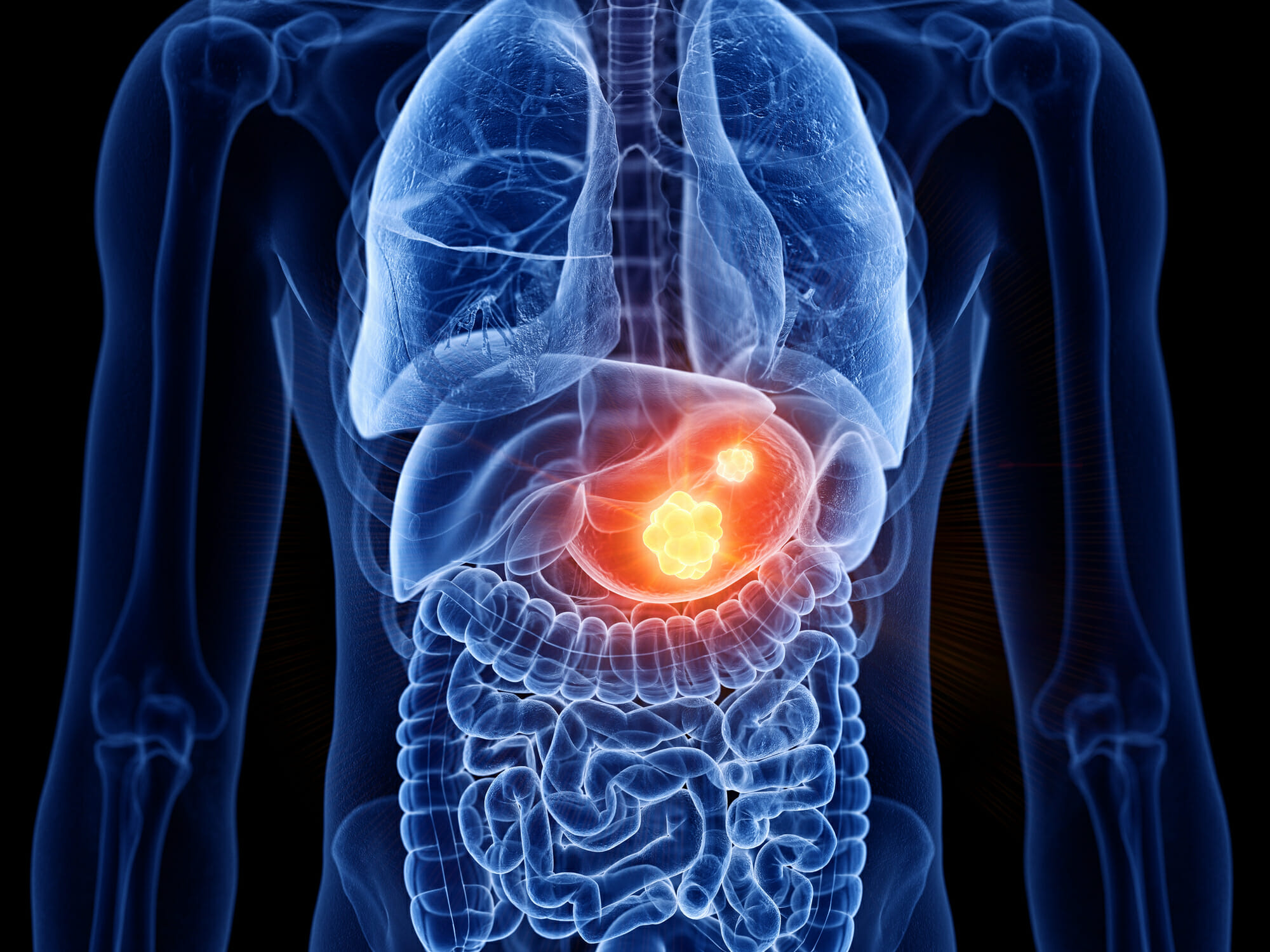
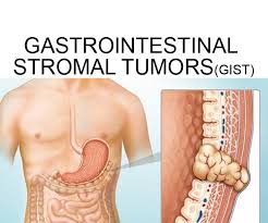
A gastrointestinal stromal tumor (GIST) is a type of cancer that begins in the digestive system. GISTs happen most often in the stomach and small intestine.

A GIST is a growth of cells that’s thought to form from a special type of nerve cells. These special nerve cells are in the walls of the digestive organs. They play a part in the process that moves food through the body.

The GI tract processes food for energy and rids the body of solid waste. After food is chewed and swallowed, it goes through the esophagus, a tube that carries food down the throat and chest to the stomach. The esophagus joins the stomach just beneath the diaphragm (the thin band of muscle below the lungs).

The stomach is a sac-like organ that helps the digestive process by mixing the food with gastric juices. The food and gastric juices are then emptied into the small intestine. The small intestine, which is about 20 feet long, continues breaking down the food and absorbs most of the nutrients into the bloodstream.

The small intestine joins the large intestine, the first part of which is the colon, a muscular tube about 5 feet long. The colon absorbs water and mineral nutrients from the remaining food matter. The waste left after this process (stool) goes into the rectum, where it is stored until it passes out of the body through the anus.

Gastrointestinal stromal tumors (GISTs) are uncommon cancers that start in special cells in the wall of the gastrointestinal (GI) tract, also known as the digestive tract. To understand GISTs, it helps to know something about the structure and function of the GI tract.

Gastrointestinal stromal tumors (GISTs) start in very early forms of special cells in the wall of the GI tract called the interstitial cells of Cajal (ICCs). ICCs are sometimes called the “pacemakers” of the GI tract because they signal the muscles in the GI tract to contract to move food and liquid along.

More than half of GISTs start in the stomach. Most of the others start in the small intestine, but GISTs can start anywhere along the GI tract. A small number of GISTs start outside the GI tract in nearby areas such as the omentum (an apron-like layer of fatty tissue that hangs over the organs in the abdomen) or the peritoneum (the thin lining over the organs and walls inside the abdomen).

Some GISTs seem to be much more likely than others to grow into other areas or spread to other parts of the body. Doctors look at certain factors to help tell whether a GIST is likely to grow and spread quickly, such as:

  • The size of the tumor
  • Where it’s located in the GI tract
  • How fast the tumor cells are dividing (its mitotic rate, described in Tests for Gastrointestinal Stromal Tumors)

Small GISTs may cause no symptoms, and they may grow so slowly that they don’t cause problems at first. As a GIST grows, it can cause signs and symptoms. They might include:

  • Abdominal pain
  • A growth you can feel in your abdomen
  • Fatigue
  • Nausea
  • Vomiting
  • Cramping pain in the abdomen after eating
  • Not feeling hungry when you would expect to
  • Feeling full if you eat only a small amount of food
  • Dark-colored stools caused by bleeding in the digestive system

GISTs can happen in people at any age, but they are most common in adults and very rare in children. The cause of most GISTs isn’t known. A small number are caused by genes passed from parents to children.

GISTs are not the same as other, more common types of GI tract cancers that develop from other types of cells.

Cancers can occur anywhere in the GI tract − from the esophagus to the anus. Most cancers that start in the GI tract, including most esophagus cancers, stomach cancers, and colon and rectum cancers, start in the gland cells that line almost all of the GI tract. The cancers that develop in these cells are called adenocarcinomas.

Cancers can also start in squamous cells, which are flat cells that line some parts of the GI tract, like the upper part of the esophagus and the end of the anus. Cancers starting in these cells are called squamous cell carcinomas.

The GI tract also has neuroendocrine cells. These cells have some features in common with nerve cells but also have other features in common with hormone-producing (endocrine) cells. Cancers that develop from these cells are called neuroendocrine tumors (NETs). These cancers are rare in the GI tract. Carcinoid tumors are an example of a neuroendocrine tumor found in the GI tract.

Other rare types of cancer in the GI tract include different types of soft tissue sarcomas, such as:

  • Leiomyosarcomas: cancers of smooth muscle cells
  • Angiosarcomas: cancers of blood vessel cells
  • Malignant peripheral nerve sheath tumors (MPNSTs): cancers of cells that support and protect nerves

GISTs are different from these other types of GI tract cancers. They start in different types of cells, need different types of treatment, and have a different prognosis (outlook). This is why doctors need to figure out whether a person with a tumor in the GI tract has a GIST, some other type of cancer, or a non-cancerous condition.

How it is diagnosed:

First you M.D. will do a physical exam.

They may also do the following tests:

  • Upper endoscopy.
  • Computed tomography (CT) scan.
  • Positron emission tomography (PET).
  • Biopsy to obtain tissue for a pathologist to examine under a microscope.

Treatment:

The most common treatment is surgery to remove the tumors.

Survival:

Survival rates vary depending on tumor size and activity at the time of diagnosis, your overall health and the tumor’s response to treatment. Overall, data from the U.S. National Cancer Institute (NCI) show that 85% of people with GIST were alive five years after diagnosis.

QUOTE FOR THURSDAY:

The entire digestive tract helps with our immune system, but some scientists and doctors think the appendix may be a place for our body to store certain healthy types of gut bacteria that otherwise could be altered or changed during an intestinal illness or with overuse of antibiotics.

Appendicitis is an inflammation of the appendix. The appendix is a finger-shaped pouch that sticks out from the colon on the lower right side of the belly, also called the abdomen.

Appendicitis causes pain in the lower right part of the belly. However, in most people, pain begins around the belly button and then moves. As inflammation worsens, appendicitis pain typically increases and eventually becomes serious.

Although anyone can develop appendicitis, most often it happens in people between the ages of 10 and 30. Treatment of appendicitis is usually antibiotics and, in most instances, surgery to remove the appendix.”

MAYO CLINIC (https://www.mayoclinic.org/diseases-conditions/appendicitis/symptoms-causes/syc-20369543)

The appendix of the human body & Appendicitis.

The appendix is a small pouch attached to the large intestine. The appendix is a small, finger-shaped pouch of intestinal tissue located between the small intestine (cecum) and large intestine (colon). The appendix is a small finger-shaped tube that branches off the first part of the large intestine.

Appendicitis (means append=appendix and itis=inflammation.  Appendicitis causes pain in your lower right abdomen. However, in most people, pain begins around the navel and then moves. As inflammation worsens, appendicitis pain typically increases and eventually becomes severe.

Although anyone can develop appendicitis, most often it occurs in people between the ages of 10 and 30. Standard treatment is surgical removal of the appendix.

Signs and symptoms of appendicitis may include:

  • Sudden pain that begins on the right side of the lower abdomen
  • Sudden pain that begins around your navel and often shifts to your lower right abdomen
  • Pain that worsens if you cough, walk or make other jarring movements
  • Nausea and vomiting
  • Loss of appetite
  • Low-grade fever that may worsen as the illness progresses
  • Constipation or diarrhea
  • Abdominal bloating
  • Flatulence

The site of your pain may vary, depending on your age and the position of your appendix. When you’re pregnant, the pain may seem to come from your upper abdomen because your appendix is higher during pregnancy.

Strongly suggestive of appendicitis is pushing down on the R lower quadrant and upon letting go the pain is severe compared to when pushing down.  It is called “rebound effect”.

Diagnostic Tests for confirming appendicitis:

  • Blood test. This allows your doctor to check for a high white blood cell count, which may indicate an infection.
  • Urine test. Your doctor may want you to have a urinalysis to make sure that a urinary tract infection or a kidney stone isn’t causing your pain.
  • Rectal exam
  • Imaging tests. Your doctor may also recommend an abdominal X-ray, an abdominal ultrasound, computerized tomography (CT) scan or magnetic resonance imaging (MRI) to help confirm appendicitis or find other causes for your pain.

Treatment:

Appendicitis treatment usually involves surgery to remove the inflamed appendix. Before surgery you may be given a dose of antibiotics to treat infection.  There are times though antibiotics may only be used and the MD see’s if the appendicitis is resolved; it depends on the MD and the severity of the appendicitis.

Surgery to remove the appendix (appendectomy)

Appendectomy can be performed as open surgery using one abdominal incision about 2 to 4 inches (5 to 10 centimeters) long (laparotomy). Or the surgery can be done through a few small abdominal incisions (laparoscopic surgery). During a laparoscopic appendectomy, the surgeon inserts special surgical tools and a video camera into your abdomen to remove your appendix.

In general, laparoscopic surgery allows you to recover faster and heal with less pain and scarring. It may be better for older adults and people with obesity.

But laparoscopic surgery isn’t appropriate for everyone. If your appendix has ruptured and infection has spread beyond the appendix or you have an abscess, you may need an open appendectomy, which allows your surgeon to clean the abdominal cavity.

Expect to spend one or two days in the hospital after your appendectomy.

Draining an abscess before appendix surgery

If your appendix has burst and an abscess has formed around it, the abscess may be drained by placing a tube through your skin into the abscess. Appendectomy can be performed several weeks later after controlling the infection.


QUOTE FOR WEDNESDAY:

“A widowmaker heart attack happens when you have a blockage in the biggest artery in your heart. That means blood can’t move through your left anterior descending (LAD) artery, which provides 50% of your heart muscle’s blood supply. Immediate treatment is crucial for a chance at survival.

A widowmaker heart attack is a type of heart attack in which you have a full blockage in your heart’s biggest artery. This artery, the left anterior descending (LAD) artery, sends oxygen-rich blood to your heart’s left ventricle. This lower chamber pumps oxygen-rich blood to your aorta, which sends it to your body.

Healthcare providers call a heart attack a “myocardial infarction,” which means you don’t have enough blood going to your heart muscle (myocardium). Your heart muscle can die without enough blood flow.

A heart attack is a medical emergency. Call 911 or your local emergency number if you think you’re having a heart attack.”

Cleveland Clinic (https://my.clevelandclinic.org/health/diseases/24507-widowmaker-heart-attack)


What is the Black Widow?

When it comes to the heart, time is critical. Blood carries oxygen to the body. When blood flow is blocked, cells can die. The more time that passes before treatment, the bigger the risk of death.

A complete blockage in the main artery that feeds the front and side walls of the heart and the septum, a muscular boundary between the left and right chambers.

“That’s a big deal” ,says Dr. Todd Caulfield, the Providence interventional cardiologist who operates on patient’s like this. “People call this the widow maker.”, he states.  Nationwide about 300,000 people suffer cardiac arrest outside a hospital every year, according to the federal Centers for Disease Control and Prevention. Only 8 percent survive.

The heart pumps blood to the body and brain. The heart also needs blood so it pumps blood to itself, too. The heart’s blood supply is essentially three main arteries: one that goes to the front; the other that goes to the back; and the final artery that connects to the side of the heart. When one of those arteries is blocked, the heart doesn’t get enough blood and oxygen, causing a heart attack. This is simply due to the block that doesn’t allow oxygen to the heart tissue that that artery feeds oxygenated blood.  This causes ischemia (lack of oxygen perfusion to tissue).  It causes pain first (chest pain=angina) and if the block continues than it will lead to infarction meaning death of the tissue not receiving enough (partial block) or no oxygen (a complete block).  The exact same thing happens in the brain, which causes a stroke; or the foot causing first pain leading to blackish skin of the foot tissue=necrotic tissue which ends up being amputated (like in some patients with  diabetes or peripheral vascular disease).  So lack of oxygen to our tissue parts anywhere can cause death to the tissue and depending on the tissue area cause death (like with the heart, due to the heart being the engine of the human body).

Interventional Cardiologist Randy Goodroe says patients are getting treatment quicker for heart attacks, more now than ever before. So the once grim reality of suffering from “the widowmaker” – a heart attack that results in immediate death – has become somewhat of a misnomer.

It’s called a “widowmaker”, Dr. Goodroe added, because it would typically kill you if the artery doesn’t get opened fast enough.

There are two different kinds of heart attacks. One is called an ST-elevation myocardial infarction, or STEMI, where the artery is totally blocked (i.e. the widowmaker), and we are on the clock to get the artery opened as soon as possible. There’s also a non-ST-elevation myocardial infarction (NSTEMI) where an artery is partially or temporarily blocked, and is still urgent to get the artery cleared, but not emergent.

Common Risk Factors:

Age is definitely a risk factor. We start seeing coronary disease in people who are between 50 and 60-years-old, and obviously, the older the person, the older the arteries, the higher the risk. Diabetes is another one of the greatest risk factors. Smoking is the strongest modifiable risk factor, meaning it’s the worst thing people can do to themselves to accelerate the disease process. High blood pressure and family history are also risk factors for heart attacks.

How to detect it if in the ER with symptoms & than repair it:

If a patient enters the ER with heart attack symptoms, quickly performed is an electrocardiogram (EKG) including cardiac enzymes to and echocardiogram or stress test to detect a totally obstructed artery. The artery is like a pipe or a tube., so we put a tiny wire through the blockageThose patients are very quickly taken to the cardiac catheterization lab where an angiogram is done to detect if there is and how bad the blockage is, if present.  Than with an angioplasty (balloons) at the end of the angiogram catheter break up the blockage and stents inserted in the area of blockage to get the blood flowing to the heart again.  The balloon is taken out and a stent is slid over that also has a balloon on it and once the stent goes up, it props open the blockage.  Blockage is repaired.


QUOTE FOR TUESDAY:

“Fibrodysplasia ossificans progressiva (FOP) is a genetic condition where people are born with bunions and their body’s muscle tissue and connective tissues, like tendons and ligaments, turn into bone on the outside of their skeleton. This condition restricts movement and can cause a loss of mobility over time in people diagnosed with the condition.”

Cleveland Clinic (https://my.clevelandclinic.org/health/diseases/24476-fibrodysplasia-ossificans-progressiva”)

Fibrodysplasia Ossificans Progressiva/Stone Man Syndrome

Fibrodysplasia ossificans progressiva (FOP) 

It is a disorder in which skeletal muscle and connective tissue, such as tendons and ligaments, are gradually replaced by bone (ossified). This condition leads to bone formation outside the skeleton (extra-skeletal or heterotopic bone) that restricts movement.

This process generally becomes noticeable in early childhood, starting with the neck and shoulders and moving down the body and into the limbs. People with FOP are born with abnormal big toes (hallux valgus) which can be helpful in making the diagnosis. Trauma, such as a fall or invasive medical procedure, or a viral illness may trigger episodes of muscle swelling and inflammation (myositis). These flareups lasts for several days to months and often result in permanent bone growth in the injured area.

FOP is almost always caused by a mutation at the same place in the ACVR1 gene (The ACVR1 gene provides instructions for making the activin receptor type-1 (ACVR1) protein, which is a member of a protein family called bone morphogenetic protein (BMP) type I receptors.)  and is inherited in an autosomal dominant manner. This condition occurs in about 1 in 1,600,000 newborns and about 800 people worldwide are known to have FOP.

SIGNS AND SYMPTOMS

-Fibrodysplasia ossificans progressiva (FOP) is characterized by the gradual replacement of muscle tissue and connective tissue (such as tendons and ligaments) by bone, restricting movement. This process generally becomes noticeable in early childhood, starting with the neck and shoulders and proceeding down the body and into the limbs.

-The formation of extra-skeletal bone causes progressive loss of mobility as the joints become affected. Speaking and eating may also become difficult as the mouth becomes affected. Over time, people with FOP may become malnourished because of the inability to eat. They may also develop breathing difficulties as a result of extra bone formation around the rib cage that restricts expansion of the lungs.

-Any trauma to the muscles of an individual with FOP (a fall or an invasive medical procedure) may trigger episodes of muscle swelling and inflammation followed by more rapid ossification in the injured area. Flare-ups may also be caused by viral illnesses such as the flu.

-Affected individuals may also have short thumbs and other skeletal abnormalities.

Inheritance

-Fibrodysplasia ossificans progressiva is inherited in an autosomal dominant pattern, which means one copy of the altered gene in each cell is sufficient to cause the disorder.

-Most cases of fibrodysplasia ossificans progressiva result from new mutations in the gene. These cases occur in people with no history of the disorder in their family. In only a small number of cases, an affected person has inherited the mutation from one affected parent.

How this disease is diagnosed:

-Making a diagnosis for a genetic or rare disease can often be challenging. Healthcare professionals typically look at a person’s medical history, symptoms, physical exam, and laboratory test results in order to make a diagnosis. The following resources provide information relating to diagnosis and testing for this condition. If you have questions about getting a diagnosis, you should contact a healthcare professional.

-The Genetic Testing Registry (GTR) provides information about the genetic tests for this condition. The intended audience for the GTR is health care providers and researchers. Patients and consumers with specific questions about a genetic test should contact a health care provider or a genetics professional.

TREATMENT:

There is currently no definitive treatment.  However, a brief course of high-dose corticosteroids, such as Prednisone, started within the first 24 hours of a flare-up, may help reduce the intense inflammation and tissue swelling seen in the early stages of fibrodysplasia ossificans progressiva.  Other medications, such as muscle relaxants, mast cell inhibitors, and aminobisphosphonates, if appropriate, should be closely monitored by a physician.  Surgery to remove heterotopic and extra-skeletal bone is risky and can potentially cause painful new bone growth.

References:


  • Fibrodysplasia ossificans progressiva. Genetics Home Reference (GHR). August 2007; http://ghr.nlm.nih.gov/condition/fibrodysplasia-ossificans-progressiva.
  • FOP Fact Sheet. International Fibrodysplasia Ossificans Progressiva Association. http://www.ifopa.org/what-is-fop/overview.html. Accessed 6/5/2014.
  • Pignolo R, Kaplan F. Pediatric Fibrodysplasia Ossificans Progressiva. E-medicine. July 30, 2009; http://emedicine.medscape.com/article/1007104-overview. Accessed 3/17/2011


QUOTE FOR MONDAY:

“Auto-brewery syndrome (ABS), also known as gut fermentation syndrome, describes the condition in which the concentration of ethanol increases to a noticeable level in the setting of little or no alcohol consumption []. It is a rare condition that is more prevalent in patients with underlying gut problems [,]. The suggested mechanism of this unique phenomenon revolves around the fermentation of carbohydrates in the human body by microorganisms [].

ABS stems from the widespread proliferation of gut microorganisms, which, in turn, leads to endogenous production of ethanol. This phenomenon is pre

ceded mostly by the intake of carbohydrate-rich meals or antibiotic use, which can disturb the gut ecosystem [,]. It is also frequently associated with underlying pathology. This syndrome is also found to be in patients suffering from other disorders such as Crohn’s disease, short bowel syndrome, and chronic intestinal pseudo-obstruction [].

The causative organisms implicated in ABS include fungi and bacteria, with the most common yeasts being Saccharomyces and Candida species.”

National Library of Medicine NIH  (https://www.ncbi.nlm.nih.gov/pmc/articles/PMC7667719/)

QUOTE FOR THE WEEKEND:

“Never.

Never do we loose our loved ones.

They accompany us; they don’t disappear from our lives.

We are merely in different rooms, temporarily.”

Paulo Coelo (South American author, who has sold over 165 million books in his lifetime.)

Life is precious-how we look at aging today and the next step in heaven!

Many look at aging as it sucks but believe or not some look at it as a privilege but how?  It’s how you look at it and how you as a child looking up to parents to mentors around you looked at aging.   Of course there are those that say “Aging sucks” I have even said that.  2. Aging is a privilege.   How is aging a privilege let me explain.  A paradox of living is that striving to age healthfully overall can lead to increased longevity, meaning you’ll have more experiences with death the older you get. As we age, so do the people around us. This is just as much a part of the “aging is a privilege” perspective as any of the rest of it.  Keep in mind those you die young miss out on those great memories or great experiences you may have had 20 to 50 years of being with that person who died.

Living longer has both joys and consequences. We will lose friends and loved ones to accidents, illnesses, and, as we reach our later years, old age—though we desperately want to know the “whys” of old age losses, seeking answers on how to avoid them.

In Stephen King’s fantasy The Green Mile, the unjustly executed main character passes some of his miraculous life-extending gifts onto a death row guard, forever changing the rest of the guard’s life. Its bittersweet conclusion leaves burning questions as well as heavy food for thought. The guard now possesses a form of near immortality—something he sees as punishment for not being able to stop the prisoner’s execution even though he knew him to be innocent. And he goes on to outlive all the people he loves.

While death I say frequently at the age I am and even before with being a RN about 40 years that its a natural part of life and an unavoidable consequence of aging, it doesn’t mean you won’t be deeply affected by it.

“There but for the grace of God go I” is often a saying on the tops of our minds as we join other mourners. But it is the very fact that we are there with others that defines our affirmation that life is indeed precious.

While you may not be able to predict how grief will affect you, having a support system in place and the skills necessary to care for your mental health will offer a foundation from which to continue this amazing thing called life.

Our unique personalities and experiences often influence how we think about death, but other factors enter into our approach to it. The culture in which we were raised can shape our beliefs and perceptions of death. How? Because the way the people around us perceive and react to grief affects our feelings as well.

Why is it that we feel differently about the loss of a person we know depending on how they left us? A sudden fatal car accident, a long-drawn-out illness, or the fact that they decided to end their own suffering—any of these conjure up feelings and opinions in us that are hard to shake—most of which were passed on to us by our parents. “She lived a long time.” “What a tragedy to see a life end that early.” “It’s too bad he didn’t take better care of himself.” “At least she is no longer in pain.” No matter what words we use to justify, judge, or comfort ourselves, the fact remains that that person is no longer around.  The key is to acceptance and to know how to deal with the emotions you’re having instead of analyzing the timing or the circumstances that led to it. In that way, you can begin to celebrate that person’s time here on Earth in the spirit of appreciation.

Some of us have cared for loved ones like myself and still am, watching the physical part of the process happen before our eyes. We may have gently washed a loved one’s face, moved them around to change their bedding, and noticed the many physical changes that took place. The thing is, fear (of the unknown) is simply a normal human reaction for both the caregiver and the one ailing.  Though I must say being a RN helped in so many ways in the acceptance of death with having religious background and still practicing to this day.

You can think you’re doing well with the losses happening around you, but don’t be surprised that you can phase in and out of the feelings you experience both now and in the future.  You may have remember something due to something you see, smell, hear, or touch.

Some say they may be “at peace” with a particular loss.  I say that especially when someone was sick and in a terminal drawn out diagnosis.  I have seen too often.   But even getting there doesn’t necessarily spell relief once it’s reached. The fact is, we won’t all experience death and dying in the same way. Our unique experiences may even change as we age and are confronted with death more often.

So, the best we can say about it is that the physical, emotional, and spiritual aspects of dying that are distressing and confusing are just plain outright normal. especially for humans since we have a heart and feelings. The thing to remember is that we are not alone. Don’t hesitate to reach out to someone you know and trust to “vent” awhile over the loss of someone you knew, whether it’s a long-distance loss or one you witnessed personally. Our friends and family often don’t know how to act around us even though they want to help.

Perhaps that’s the way we all need to look at this phenomenon of the frequency of funerals we must attend as we age. Life is a precious gift, and whether your spiritual beliefs help you cope with them or not, be sure to find something that works for you.