Archives

Part II ALS Awareness Month-Stages of ALS

Once ALS starts, it almost always progresses, eventually taking away the ability to walk, dress, write, speak, swallow, and breathe, and shortening the life span. The onset of ALS often involves muscle weakness or stiffness as early symptoms. Progression of weakness, wasting, and paralysis of the muscles of the limbs and trunk, as well as those that control vital functions such as speech, swallowing, and breathing, generally follows.

How fast and in what order this occurs is very different from person to person. While the average survival time is three years, about 20 percent of people with ALS live five years, 10 percent will survive 10 years and 5 percent will live 20 years or longer.

End stages of ALS often include trouble swallowing and speaking. Weak and paralyzed vocal cords make speaking difficult and eventually impossible. Trouble swallowing occurs gradually in some patients, but can occur suddenly in others.

Stages of ALS

ALS is a relentlessly progressive disorder. The rate of progression between individuals is variable and the history generally reflects gradual and progressive worsening over time until death occurs.

Early stages:

Muscles

  • Muscles may be weak and soft, or they may be stiff, tight, and spastic. Muscle cramping and twitching (fasciculation) occurs, as does loss of muscle bulk (atrophy).
  • Symptoms may be limited to a single body region or mild symptoms may affect more than one region.

Physical effects

  • The person may experience fatigue, poor balance, slurred words, a weak grip, tripping when walking, or other minor symptoms.
  • Sometimes this stage occurs before a diagnosis is made.

Middle stages:

Muscles

  • Symptoms become more widespread.
  • Some muscles are paralyzed, while others are weakened or unaffected. Fasciculations may continue.

Physical effects

  • Unused muscles may cause contractures, in which the joints become rigid, painful, and sometimes deformed.
  • If a fall occurs, the person may not be able to stand back up alone.
  • Driving is relinquished.
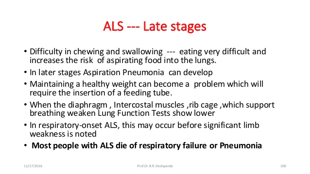
  • Weakness in swallowing muscles may cause choking and greater difficulty eating and managing saliva.
  • Weakness in breathing muscles can cause respiratory insufficiency, especially when lying down.
  • Some people experience bouts of uncontrolled and inappropriate laughing or crying (pseudobulbar affect). Despite how it seems, the person usually doesn’t feel particularly sad or happy.

Late stages:

Muscles

  • Most voluntary muscles are paralyzed.
  • The muscles that help move air in and out of the lungs are severely compromised.

Physical effects

  • Mobility is extremely limited, and help is needed in caring for most personal needs.
  • Poor respiration may cause fatigue, fuzzy thinking, headaches, and susceptibility to pneumonia. (Respiratory insufficiency is a leading cause of death in ALS.)
  • Speech, or eating and drinking by mouth, may not be possible
  • Assistance needed if not needed in the previous stage yet; in the home care you would need:

    • Power wheelchair, hospital bed, mechanical lift, and switches that enable any moving body part to operate computers, environmental control units, and communication devices.
    • Assisted ventilation, either noninvasive or invasive (tracheostomy).
    • Feeding tube.
    • Possibly urinary catheters aren’t required but can make toileting easier.
    • The type of home assistance you need:

    1.) Caregivers should:

    • Find and train caregiving help.
    • Oversee 24-hour care operations.
    • Find ways to help the person with ALS stay socially and mentally active.
    • Get into a routine that supports themselves as well as the person with ALS.
    • Know that although this stage is all-consuming, surprisingly many caregivers report great stability and satisfaction in their daily lives at this later stage of the disease.

       2.) Visiting RN (Nurse) who basically follows up on the care and decline or no change in pt    with letting the attending M.D. in charge be kept informed on pt’s status who makes any change with orders on the pt’s care.  It’s a whole team effect to make sure the pt gets the best care!

  • Progression is not always a straight line in an individual, either. It is not uncommon to have periods lasting weeks to months where there is very little or no loss of function. There are even very rare examples in which there is significant improvement and recovery of lost function. These ALS “arrests” and “reversals” are, unfortunately, usually transient. Less than 1 percent of people with ALS will have significant improvement in function lasting 12 months or longer

End stage

  • The vast majority of deaths in ALS are the result of respiratory failure, a process that progresses slowly over months. Medications can relieve discomfort, anxiety, and fear caused by respiratory insufficiency.
  • Far less-common causes of death in ALS include malnutrition as a result of swallowing problems, pulmonary embolism (a blockage in one of the arteries of the lungs), abnormalities in the heart’s electrical pacing system called cardiac arrhythmias, and pneumonia as the result of aspiration (when food or fluid gets into the lungs).
  • Hospice care (in a facility or in the home) focuses on providing comfort and maintaining quality of life by supporting the physical, emotional, and spiritual needs of the individual with ALS and their family members. Families should contact hospice early on to see what in-home services are available even before the most advanced stage.
  • At MDA clinics, physicians work closely with palliative care teams to coordinate treatment with in-home hospice care providers, assisted living facilities, or inpatient hospice settings. Such cooperation helps ensure the person with ALS has the most peaceful and painless experience possible.


Part I ALS Awareness Month-What is it and what are the signs and symptoms?

Amyotrophic lateral sclerosis concept illustration

ALS amyotrophic lateral sclerosis is a rare neurological disease affecting nerve cells that control voluntary muscle movement.  Amyotrophic Lateral Sclerosis or ALS, more commonly known as Lou Gehrig’s disease, is a terminal and progressive motor neuron disease. ALS specifically targets and kills the motor neurons responsible for controlling the vast majority of skeletal muscles in the human body, which eventually leads to respiratory failure and death.

Individuals with ALS experience a degeneration of their motor neurons, which causes the muscles to stop receiving the signals needed to function. After a certain time, the brain completely loses its ability to control voluntary movements, hence, people with ALS are unable to walk, move, or even breathe properly.

ALS belongs to, and is perhaps the most common example of, a group of neurological disorders known as Motor Neuron Diseases.  These diseases affect the body by causing the death of millions of neurons found in the motor cortex of the brain as well as the spinal cord. These nerve cells are directly responsible for the regulation and control of skeletal muscle function.

How ALS is diagnosed:

Considering the damage ALS can do, it became essential to spread the message regarding the disease so that people could treat it at an early stage. Though there is no cure for ALS but early detection can help in improving the quality of life of those with the disease. 

The signs and symptoms of this diagnosis:

1-Fatigue

A positive diagnosis of ALS is based primarily on a patient’s symptomatology.  Unfortunately there is no test that can currently provide a more conclusive assessment.

There are many diseases whose symptoms resemble those observed in patients with ALS. Therefore, diseases such as cervical osteoarthritis, cervical hernias that compress the spinal cord, heavy metal poisoning, and some infectious diseases such as Lyme disease or syphilis, can delay a correct diagnosis of ALS immediately.

As such, when ALS is suspected, it is common practice to rule out other diseases through a variety of tests including but not limited to lumbar punctures, MRIs, and electromyographic studies. In some cases, it might be necessary to perform a biopsy of muscle tissue in order to assuage any remaining doubts.

Often, the earliest symptoms of ALS are ignored or outright dismissed. Therefore, better understand this disease’s signs and symptoms.

2-Loss of strength

Pt’s with ALS eventually lose the ability to control all voluntary movement. During the progression of the disease, which typically lasts for several years, patients will experience a cumulative loss of muscle strength.

In most cases, the first muscles affected by the disease are those of the arms and legs which results in patients experiencing awkwardness when walking or moving about, an increased propensity for stumbling or tripping, and difficulty performing everyday tasks especially fine motor tasks like texting on the phone, typing, and even tying shoe or sneaker laces.

3-Muscle Atrophy

This is when the muscle actually deteriorates and muscle is lost.  Leading to muscle dystrophy,in the specific case of ALS, it occurs due to a dramatic reduction in the connection between nerves and muscle fibers caused by the death of motor neurons.  It often culminates to paresthesia to partial or total paralysis.

4-Fasciculations

Fasciculations are slight and involuntary muscular contractions that occur underneath the skin, but that do not produce any observable limb movement. Fasciculations are visible to the naked eye and are sometimes described as looking like small worms are moving within the muscle. These contractions occur because of spontaneous nerve discharges that fire within clumps of skeletal muscle fibers.  They can occur due to damage present in the lower motor neurons. They could be considered an early warning sign of the possible onset of ALS.

5-Cramps

Muscle cramps are highly common in patients who have ALS, and their incidence increases as the disease progresses. These sustained involuntary contractions of the muscles are typically accompanied by palpable contractures, can last anywhere from 30 to 45 seconds, and tend to be extremely painful.  Spasticity could develop and may not allow certain movements as a consequence of cramps;  in which antagonistic muscle groups participate.

Other Symptoms include:

  • Tripping and falling
  • Hand weakness or clumsiness
  • Slurred speech or trouble swallowing
  • twitching in your arms, shoulders and tongue
  • Inappropriate crying, laughing or yawning
  • Cognitive and behavioral changes


QUOTE FOR WEDNESDAY:

“7 Disorders are part of or closely related to Autism. Each disorder has symptoms commonly seen with autism, as well as its own specific symptoms.  These disorders are:

  • Williams Syndrome
  • Fragile X Syndrome
  • Landau-Kleffner Syndrome
  • Prader-Willi Syndrome
  • Angelman Syndrome
  • Rett Syndrome
  • Tardive Dyskinesia”

Autism Research Institute (7 Disorders Closely Related to Autism – Autism Research Institute)


Part II Related Conditions to Autism

Williams Syndrome

Williams Syndrome (also known as Williams-Beuren syndrome) is a rare genetics disorder in which a portion of DNA material on chromosome 7 is missing. The prevalence in the population is somewhere between 1 out of 10,000.

Many people with Williams Syndrome exhibit autistic behaviors. This includes: developmental and language delays, problems in gross motor skills, hypersensitivity to sounds, picky eating, and perseverating.

However, Williams Syndrome includes other symptoms that may require different or additional treatments. A diagnosis is essential to maximize quality of care.

These individuals differ from the typical autistic individual because they also have cardiovascular abnormalities, high blood pressure, elevated calcium levels, and are very sociable. They also have unique pixie-like facial features–almond shaped eyes, oval ears, full lips, small chins, narrow faces, and broad mouths.

Fragile X

Fragile X syndrome (also known as Martin-Bell syndrome) is a sex-linked genetic disorder. The exact frequency of Fragile X syndrome is unclear, but the CDC estimates that roughly 1.4 in 10,000 males and 0.9 in 10,000 females are affected by this disorder. Males afflicted with this syndrome typically have a moderate to severe form of intellectual handicap. Females may also be affected but generally have a mild form of impairment.

Approximately 15% to 20% of those with Fragile X Syndrome exhibit autistic-type behaviors, such as poor eye contact, hand-flapping or odd gesture movements, hand-biting, and poor sensory skills. Behavior problems and speech/language delay are also common features of Fragile X Syndrome.

People with Fragile X syndrome also have a number of recognizable physical features, including a high arched palate, strabismus (lazy eye), large ears, long face, large testicles in males, poor muscle tone, flat feet, and sometimes mild, heart valve abnormalities. Although most individuals with Fragile X syndrome have a characteristic ‘look’ (long face and large ears), there are some who do not have typical features.

Many hospitals and laboratories perform blood tests to diagnose Fragile X syndrome. Several treatments are recommended for individuals with this disorder, including mild medications for behavior problems and therapies for speech and language and sensory improvement. Families are advised to seek genetic counseling to understand the inheritable nature of Fragile X Syndrome and to discuss with family members the likelihood other individuals or future offspring may have this disorder.

Thank  You to Dr. Peter Jacky of Kaiser Sunnyside Hospital in Clackamas, Oregon for his comments on this article.

Landau-Kleffner Syndrome

Landau-Kleffner Syndrome is a rare form of epilepsy that manifests as a form of aphasia, (loss of language), which usually develops between 3 and 7 years. It is twice as common in males than females and is often diagnosed in conjunction with autism. Initially, these individuals have a healthy, problem-free development with normal speech and vocabulary. These individuals first lose their ability to comprehend (i.e., receptive speech) and then their ability to speak (i.e., expressive speech). These changes can occur gradually or suddenly.

People with Landau-Kleffner Syndrome have abnormal EEG patterns (i.e., brain waves) in the temporal lobe (located on the sides of the brain) and in the temporo-parieto-occipital regions during sleep. Diagnosis of this syndrome usually involves examining the person’s EEG patterns during sleep. Approximately 70% develop epilepsy; and these seizures are typically infrequent and can be either with or without convulsions.

One common characteristic of Landau-Kleffner Syndrome is the failure to respond to sounds. Thus, parents may suspect their child of hearing loss. Autistic characteristics seen in Landau-Kleffner Syndrome individuals include pain insensitivity, aggression, poor eye contact, insistence on sameness, and sleep problems.

The cause of Landau-Kleffner Syndrome is not known. Some suggested causes have been a dysfunctional immune system, exposure to a virus, and brain trauma. The prognosis is better when the onset is after age 6 and when speech therapy is started early. Several other treatments have also been shown to be beneficial for many of these individuals, such as anticonvulsant mediations and corticosteroids. There is also a surgical technique in which the pathways of abnormal electrical brain activity are severed.

Prader-Willi Syndrome

Prader-Willi Syndrome is a disorder which is sometimes associated with, but not a subtype of, autism. The classical features of this disorder include an obsession with food which is often associated with impulsive eating, compact body build, underdeveloped sexual characteristics, and poor muscle tone. Because of their obsession with food, many people afflicted with Prader-Willi Syndrome are overweight. Most individuals afflicted with Prader-Willi Syndrome have mild mental deficits.

Some of the behaviors which are common to both Prader-Willi Syndrome and autism are:

  • delays in language and motor development
  • learning disabilities
  • feeding problems in infancy
  • sleep disturbances, skin picking
  • temper tantrums
  • high pain threshold

Prader-Willi Syndrome affects approximately 1 in 10,000 people. Most individuals suffering from this disorder are missing a small portion of chromosome 15 which appears to come from the paternal side of the family. When a small portion of chromosome 15 is missing and comes from the maternal side, the person may suffer from Angelman Syndrome.

The most effective form of treatment for people suffering from Prader-Willi Syndrome is behavior modification. In general, medications do not appear to be very effective for these individuals.

Angelman Syndrome

Angelman syndrome is a genetic disorder that affects the nervous system. Initial symptoms of this disorder typically manifest in the first year of life and become more apparent through early childhood. It is estimated that Angelman syndrome occurs in roughly every one in 15,000 people. Similarly to Prader Willi Syndrome, this disorder derives from a missing portion of chromosome 15, but unlike Prader Willi, this deficit comes from the maternal side.

Angelman syndrome is commonly characterized by:

  • Mental and speech deficits
  • Speech impairment
  • Problems with motor skills and balance
  • Epilepsy
  • Small head size
  • Hyperactivity
  • Smiling, Laughing and Hand flapping
  • Difficulty sleeping

To learn more about Angelman Syndrome, please visit the Angelman Syndrome Foundation

Rett Syndrome

Rett Syndrome was first recognized by Andreas Rett in 1966 and is a neurological disorder affecting primarily females. Autopsies on the brains of these individuals indicate a pathology different from autism; however, children afflicted with Rett Syndrome often exhibit autistic-like behaviors, such as repetitive hand movements, prolonged toe walking, body rocking, and sleep problems. In most cases, there is a regression in cognition, behavior, social, and motor skills throughout their lifetime.

The prevalence of Rett Syndrome is estimated to be between 1 in 10,000 and 1 in 15,000 people.

Typical characteristics:

  • Normal development until 1/2 to 1 1/2 years
  • Behavioral, social, and cognitive regression
  • Shakiness of the torso, and possibly the limbs
  • Unsteady, stiff-legged gait
  • Breathing difficulties (hyperventilation, apnea, air swallowing)
  • Seizures (approximately 80% have epilepsy)
  • Teeth grinding and difficulty chewing
  • Stunted growth and small head
  • Severe mental deficits
  • Hypoactivity

In 1999, Dr. Huda Zoghbi and her colleagues located the gene for Rett syndrome. The gene was located on one of the two X chromosomes that determine sex. Rett syndrome results from the mutation of the gene that makes methyl cytosine binding protein, resulting in excessive amounts of this protein.

For more information about this disorder, visit International Rett Syndrome Association

Tardive dyskinesia

Tardive dyskinesia is a syndrome involving dysfunctional, involuntary movements associated with long-term, chronic use of neuroleptic medications, such as Haldol, Prolixin, and Thorazine. These drugs lead to an apparent general calming or sedative effect on the individual and are considered major tranquilizers.

Tardive dyskinesia may appear anywhere from three months to several years after initial use of these medications, and withdrawal from neuroleptics often exacerbates the symptoms.

Common tardive dyskinesia movements include, but are not limited to:

  • facial tics, grimacing
  • eye blinking
  • lip smacking
  • tongue thrusting
  • moving one’s head back or to the side
  • foot tapping
  • ankle movements
  • shuffled gait
  • head nodding

Tardive dyskinesia may lead to very serious problems, such as respiratory interference, inability to eat, oral ulcerations, and difficulty standing/walking.

Tardive dyskinesia movements may be confused with stereotypy because of the repetitive nature of both behaviors. Stereotypy refers to ritualistic, often complex behaviors, such as body and head rocking, hand-flapping, and complex hand movement patterns. Stereotypy appears to be under voluntary control. In contrast, tardive dyskinesia movements are less complex, less ritualistic, and are not volitional.

Other psychoactive drugs, such as clozaril/clozapine, have similar effects on behavior but do not produce tardive dyskinesia as neuroleptics do.

Is it Autism?

While the behavioral symptoms of the conditions above may overlap with autism, they may require different or additional treatments. Seek a diagnosis and treatment plan from a qualified medical professional before starting any form of treatment.

QUOTE FOR TUESDAY:

“Autism spectrum disorder (ASD) is a developmental disability caused by differences in the brain.
Some people with ASD have a known difference, such as a genetic condition or it can be other causes including those that are not yet known.
Scientists believe there are multiple causes of ASD that act together to change the most common ways people develop. We still have much to learn about these causes and how they impact people with ASD.
Early intervention services can greatly improve the development of a child with ASD.”
Centers of Disease Control and Prevention – CDC

Part I Autism (ASD) Awareness-Learn how’s its diagnosed, what causes ASD, Genes R/T ASD, how s/s change in time, the treatment of ASD!

What disorders are related to ASD?

Certain known genetic disorders are associated with an increased risk for autism, including Fragile X syndrome (which causes intellectual disability) and tuberous sclerosis (which causes benign tumors to grow in the brain and other vital organs) — each of which results from a mutation in a single, but different, gene. Recently, researchers have discovered other genetic mutations in children diagnosed with autism, including some that have not yet been designated as named syndromes. While each of these disorders is rare, in aggregate, they may account for 20 percent or more of all autism cases.

People with ASD also have a higher than average risk of having epilepsy. Children whose language skills regress early in life — before age 3 — appear to have a risk of developing epilepsy or seizure-like brain activity. About 20 to 30 percent of children with ASD develop epilepsy by the time they reach adulthood. Additionally, people with both ASD and intellectual disability have the greatest risk of developing seizure disorder.

How is ASD diagnosed?

ASD symptoms can vary greatly from person to person depending on the severity of the disorder. Symptoms may even go unrecognized for young children who have mild ASD or less debilitating handicaps. Very early indicators that require evaluation by an expert include:

  • no babbling or pointing by age 1
  • no single words by age 16 months or two-word phrases by age 2.
  • no response to name
  • loss of language or social skills previously acquired
  • poor eye contact
  • excessive lining up of toys or objects
  • no smiling or social responsiveness

Later indicators include:

  • impaired ability to make friends with peers
  • impaired ability to initiate or sustain a conversation with others
  • absence or impairment of imaginative and social play
  • repetitive or unusual use of language
  • abnormally intense or focused interest
  • preoccupation with certain objects or subjects
  • inflexible adherence to specific routines or rituals

Health care providers will often use a questionnaire or other screening instrument to gather information about a child’s development and behavior. Some screening instruments rely solely on parent observations, while others rely on a combination of parent and doctor observations. If screening instruments indicate the possibility of ASD, a more comprehensive evaluation is usually indicated.

A comprehensive evaluation requires a multidisciplinary team, including a psychologist, neurologist, psychiatrist, speech therapist, and other professionals who diagnose and treat children with ASD. The team members will conduct a thorough neurological assessment and in-depth cognitive and language testing. Because hearing problems can cause behaviors that could be mistaken for ASD, children with delayed speech development should also have their hearing tested.

What causes ASD?

Scientists believe that both genetics and environment likely play a role in ASD. There is great concern that rates of autism have been increasing in recent decades without full explanation as to why. Researchers have identified a number of genes associated with the disorder. Imaging studies of people with ASD have found differences in the development of several regions of the brain. Studies suggest that ASD could be a result of disruptions in normal brain growth very early in development. These disruptions may be the result of defects in genes that control brain development and regulate how brain cells communicate with each other. Autism is more common in children born prematurely. Environmental factors may also play a role in gene function and development, but no specific environmental causes have yet been identified. The theory that parental practices are responsible for ASD has long been disproved. Multiple studies have shown that vaccination to prevent childhood infectious diseases does not increase the risk of autism in the population.

What role do genes play?

Twin and family studies strongly suggest that some people have a genetic predisposition to autism. Identical twin studies show that if one twin is affected, then the other will be affected between 36 to 95 percent of the time. There are a number of studies in progress to determine the specific genetic factors associated with the development of ASD. In families with one child with ASD, the risk of having a second child with the disorder also increases. Many of the genes found to be associated with autism are involved in the function of the chemical connections between brain neurons (synapses). Researchers are looking for clues about which genes contribute to increased susceptibility. In some cases, parents and other relatives of a child with ASD show mild impairments in social communication skills or engage in repetitive behaviors. Evidence also suggests that emotional disorders such as bipolar disorder and schizophrenia occur more frequently than average in the families of people with ASD.

In addition to genetic variations that are inherited and are present in nearly all of a person’s cells, recent research has also shown that de novo, or spontaneous, gene mutations can influence the risk of developing autism spectrum disorder.  De novo mutations are changes in sequences of deoxyribonucleic acid or DNA, the hereditary material in humans, which can occur spontaneously in a parent’s sperm or egg cell or during fertilization. The mutation then occurs in each cell as the fertilized egg divides. These mutations may affect single genes or they may be changes called copy number variations, in which stretches of DNA containing multiple genes are deleted or duplicated.  Recent studies have shown that people with ASD tend to have more copy number de novo gene mutations than those without the disorder, suggesting that for some the risk of developing ASD is not the result of mutations in individual genes but rather spontaneous coding mutations across many genes.  De novo mutations may explain genetic disorders in which an affected child has the mutation in each cell but the parents do not and there is no family pattern to the disorder. Autism risk also increases in children born to older parents. There is still much research to be done to determine the potential role of environmental factors on spontaneous mutations and how that influences ASD risk.

Do symptoms of autism change over time?

For many children, symptoms improve with age and behavioral treatment. During adolescence, some children with ASD may become depressed or experience behavioral problems, and their treatment may need some modification as they transition to adulthood. People with ASD usually continue to need services and supports as they get older, but depending on severity of the disorder, people with ASD may be able to work successfully and live independently or within a supportive environment.

How is autism treated?

There is no cure for ASD. Therapies and behavioral interventions are designed to remedy specific symptoms and can substantially improve those symptoms. The ideal treatment plan coordinates therapies and interventions that meet the specific needs of the individual. Most health care professionals agree that the earlier the intervention, the better.

Educational/behavioral interventions: Early behavioral/educational interventions have been very successful in many children with ASD. In these interventions therapists use highly structured and intensive skill-oriented training sessions to help children develop social and language skills, such as applied behavioral analysis, which encourages positive behaviors and discourages negative ones. In addition, family counseling for the parents and siblings of children with ASD often helps families cope with the particular challenges of living with a child with ASD.

Medications: While medication can’t cure ASD or even treat its main symptoms, there are some that can help with related symptoms such as anxiety, depression, and obsessive-compulsive disorder. Antipsychotic medications are used to treat severe behavioral problems. Seizures can be treated with one or more anticonvulsant drugs. Medication used to treat people with attention deficit disorder can be used effectively to help decrease impulsivity and hyperactivity in people with ASD. Parents, caregivers, and people with autism should use caution before adopting any unproven treatments

QUOTE FOR MONDAY:

“The Centers for Disease Control and Prevention (CDC) is issuing this Health Alert Network (HAN) Health Advisory about a recently confirmed outbreak of Ebola disease in Uganda caused by the Sudan virus (species Orthoebolavirus sudanense) and to summarize CDC’s recommendations for U.S. public health departments and clinicians about case identification, testing, and biosafety considerations in clinical laboratories.

Currently, no suspected, probable, or confirmed Ebola cases related to this outbreak have been reported in the United States, or outside of Uganda. However, as a precaution and because there are other viral hemorrhagic fever (VHF) outbreaks in East Africa, CDC is sharing best practices for public health departments, public health and clinical laboratories, and healthcare workers in the United States to raise awareness about this outbreak.

On February 5, 2025, CDC issued a Travel Health Notice Level 2: Practice Enhanced Precautions for people traveling to Uganda. Currently, CDC has not issued any interim recommendations to health departments for post-arrival risk assessment and management of travelers, including U.S.-based healthcare workers, arriving from Uganda. CDC recommends that travelers monitor themselves for symptoms of Sudan virus disease (SVD) while in the outbreak area and for 21 days after leaving. Travelers should also self-isolate and contact local health authorities or a clinician if they develop symptoms (early “dry” symptoms may include fever, aches, pains, and fatigue and later “wet” symptoms may include diarrhea, vomiting, and unexplained bleeding).”

Centers for Disease Control and Prevention – CDC (Ebola Outbreak Caused by Sudan virus in Uganda | HAN | CDC)

Part 3 Lets see the facts of Ebola, healthcare workers & previous epidemics.

10Years Ago

Lets see the facts Ebola:

 First signs / symptoms range from

  • Fever (greater than 38.6°C or 101.5°F)
  • Severe headache
  • Muscle pain
  • Weakness
  • Diarrhea
  • Vomiting
  • Abdominal (stomach) pain
  • Unexplained hemorrhage (bleeding or bruising)

Symptoms may appear anywhere from 2 to 21 days after exposure to Ebola, but the average is 8 to 10 days.

Recovery from Ebola depends on good supportive clinical care and the patient’s immune response. People who recover from Ebola infection develop antibodies that last for at least 10 years.

Reference on symptoms of Eboli: (http://www.cdc.gov/vhf/ebola/symptoms/index.html).

Healthcare workers who may be exposed to people with Ebola should follow these steps:

  • Wear protective clothing, including masks, gloves, gowns, and eye protection.
  • Practice proper infection control and sterilization measures. For more information, see “Infection Control for Viral Hemorrhagic Fevers in the African Health Care Setting”.
  • Isolate patients with Ebola from other patients.
  • Avoid direct contact with the bodies of people who have died from Ebola.
  • Notify health officials if you have had direct contact with the blood or body fluids, such as but not limited to, feces, saliva, urine, vomit, and semen of a person who is sick with Ebola. The virus can enter the body through broken skin or unprotected mucous membranes in, for example, the eyes, nose, or mouth.

Looking at some of the history is as follows:

In 1995, an outbreak of Ebola hemorrhagic fever (Ebola HF) affected more than 300 people in and around the city of Kikwit, Democratic Republic of the Congo (formerly, Zaire); approximately 80% of the patients died. More than one-fourth of all the patients were health care workers. After the outbreak, the DRC Ministry of Health, the Centers for Disease Control and Prevention (CDC) and the World Health Organization (WHO) developed practical recommendations for carrying out viral hemorrhagic fever (VHF) isolation precautions in rural health facilities in Africa. These recommendations have been consolidated in a manual for the local health community but something needs to be put into play in getting this epidemic under control if not history noted for repeating itself would be a shame in see results like the following:

The Black Death, 1918 Spanish Flu, HIV/AIDS (As of 2011 at least 60 million people had been infected by AIDS and 25 million had died. while in 2008 an estimated 1.2 million Americans had HIV, Sub-Saharan Africa alone was home to 22.9 million cases, with one in five adults infected. About 35.3 million people were believed to have HIV in 2012.), The Plague of Justinian (ultimately killed 25 million people dead), The Antonine Plague, Cholera, reference to this information is at http://www.rwjf.org/en/blogs/new-public-health/2013/12/the_five_deadliesto.html to go further into details about them.

Other epidemics are Polio, Typhus (camp fever), Malaria, Small Pox, Yellow Fever, The Flu in 1918 before the vaccine. The flu only survived for a year, this strain of virus was responsible for the deaths of 50 to 100 million people, as it was able to quickly spread from country to country as troops and soldiers returned home from WWI from all around the globe. Symptoms were common to those in today’s influenza virus. Also the death for many was the complication of the flu in causing fluid build up in the lungs causing the death (from probably putting the pt into the complication septicemia or or those with or without congestive heart failure going into a exacerbation without having effectively removing the fluids by getting better and ending result drowning in their own lungs).

This is not a pretty picture in having to repeat in history for some Epidemic to occur in our time; which could be Ebola.

Although there is still a great deal to learn about Ebola HF, two observations from the Kikwit outbreak strongly indicate that future outbreaks of this magnitude could be prevented:

  • The first case occurred in January 1995, but went unrecognized as Ebola HF by health-care workers. This one case started a chain of transmission of the virus that finally was recognized in April of that year, when many more cases appeared. The outbreak peaked in May. Thus, between January and April, there was a window of opportunity that could have allowed early detection and proper management of patients; the early response might have prevented widespread transmission of the virus.
  • After an international investigation team arrived in May 1995 and worked with Kikwit medical community to introduce VHF isolation precautions as well as standard precautions, no further nosocomial transmission of the virus was documented, indicating that although Ebola HF is highly infectious, the use of these measures is effective in preventing the spread of disease. Questionable by many since we have it in our country starting an epidemic and it is already an active one in Africa now.
  • The observations sent a strong message to the public health and medical communities in Africa and internationally: combining early suspicion of VHF and isolation precautions can help to prevent another serious outbreak of Ebola HF or other VHF in the future. The only question remaining was how these goals could be achieved in a region where resources are scarce and the health care infrastructure is either underdeveloped or deteriorating. This manual, prepared collaboratively by CDC and WHO, attempts to address the issues of early provisional diagnosis and response within a limited infrastructure. It is designed for the following uses:  For prevention through preparedness–to help African health facilities make advance preparations for responding with appropriate precautions when a VHF case is suspect.
  • For planning and conducting in-service training to strengthen standard precautions and VHF isolation precautions. With follow up by superiors of the facility, from floor managers to nursing education depts.
  • As a rapid reference when a VHF case appears at a health facility where no previous VHF preparations have been made give the following by CDC:     The recommendations in the manual make use of common, low-cost supplies, such as household bleach, water, cotton cloth, and plastic sheeting. Step-by-step instructions for implementing the recommendations are presented along with instructional aids for easy reference in health centers. For further information on this go to http://www.cdc.gov/vhf/abroad/vhf-manual.html.
  • God willing this becomes no epidemic in our country with our government taking better and stronger actions in prevention of diseases spreading in this country. In carrying out stricter rules and regulations which should have been laid down awhile ago regarding people living here or just visiting that travel to and from different countries. This is should have taken place already just by looking at our history of epidemics that have occurred from spread diseases that came some other than United States.

QUOTE FOR THE WEEKEND:

“Ebola is a serious, life-threatening type of viral hemorrhagic fever — a viral infection that damages your blood vessels. Ebola symptoms start off like the flu (influenza). But they can progress to:

2o14 An epidemic of Ebola virus disease was occurring in Western Africa on a scale not seen before, particularly in the countries of Guinea, Liberia, and Sierra Leone. The continued spread is facilitated by insufficient medical facilities, poor sanitation, travel, and unsafe burial practices. Several patients diagnosed with Ebola virus disease in Africa have been evacuated to the United States for treatment, and several other patients have been diagnosed in the United States. It is important for laboratories to be aware of available tests, especially those granted emergency use authorization, as hospitals prepare protocols for the diagnosis and management of high-risk patients.”

Cleveland Clinic (Ebola Virus: Causes, Symptoms, Treatment & Prevention)