Part III Glaucoma National Awareness: The types of treatments of Glaucoma!

If you are diagnosed with glaucoma, it is important to set a regular schedule of examinations with your eye doctor to monitor your condition and make sure that your prescribed treatment is effectively maintaining a safe eye pressure.

Treatments

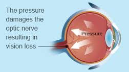
The Treatment of Glaucoma: The damage caused by glaucoma can’t be reversed. But treatment and regular checkups can help slow or prevent vision loss, especially in you catch the disease in its early stage. The goal of glaucoma treatment is to lower pressure in your eye (intraocular pressure). Depending on your situation, your options may include eyedrops, laser treatment or surgery. 

Remember The damage caused by glaucoma can’t be reversed. But treatment and regular checkups can help slow or prevent vision loss, especially in you catch the disease in its early stage. The goal of glaucoma treatment is to lower pressure in your eye (intraocular pressure). Depending on your situation, your options may include eyedrops, laser treatment or surgery.

Eyedrops

Glaucoma treatment often starts with prescription eyedrops. These can help decrease eye pressure by improving how fluid drains from your eye or by decreasing the amount of fluid your eye makes. Depending on how low your eye pressure needs to be, more than one of the eyedrops below may need to be prescribed.

Prescription eyedrop medications include:

  • Prostaglandins. These increase the outflow of the fluid in your eye (aqueous humor), thereby reducing your eye pressure. Medicines in this category include latanoprost (Xalatan), travoprost (Travatan Z), tafluprost (Zioptan), bimatoprost (Lumigan) and latanoprostene bunod (Vyzulta). Possible side effects include mild reddening and stinging of the eyes, darkening of the iris, darkening of the pigment of the eyelashes or eyelid skin, and blurred vision. This class of drug is prescribed for once-a-day use.
  • Beta blockers. These reduce the production of fluid in your eye, thereby lowering the pressure in your eye (intraocular pressure). Examples include timolol (Betimol, Istalol, Timoptic) and betaxolol (Betoptic). Possible side effects include difficulty breathing, slowed heart rate, lower blood pressure, impotence and fatigue. This class of drug can be prescribed for once- or twice-daily use depending on your condition.
  • Alpha-adrenergic agonists. These reduce the production of aqueous humor and increase outflow of the fluid in your eye. Examples include apraclonidine (Iopidine) and brimonidine (Alphagan P, Qoliana). Possible side effects include an irregular heart rate, high blood pressure, fatigue, red, itchy or swollen eyes, and dry mouth. This class of drug is usually prescribed for twice-daily use but sometimes can be prescribed for use three times a day.
  • Carbonic anhydrase inhibitors. These medicines reduce the production of fluid in your eye. Examples include dorzolamide (Trusopt) and brinzolamide (Azopt). Possible side effects include a metallic taste, frequent urination, and tingling in the fingers and toes. This class of drug is usually prescribed for twice-daily use but sometimes can be prescribed for use three times a day.
  • Rho kinase inhibitor. This medicine lowers eye pressure by suppressing the rho kinase enzymes responsible for fluid increase. It is available as netarsudil (Rhopressa) and is prescribed for once-a-day use. Possible side effects include eye redness, eye discomfort and deposits forming on the cornea.
  • Miotic or cholinergic agents. These increase the outflow of fluid from your eye. An example is pilocarpine (Isopto Carpine). Side effects include headache, eye ache, smaller pupils, possible blurred or dim vision, and nearsightedness. This class of medicine is usually prescribed to be used up to four times a day. Because of potential side effects and the need for frequent daily use, these medications are not prescribed very often anymore.

Because some of the eyedrop medicine is absorbed into your bloodstream, you may experience some side effects unrelated to your eyes. To minimize this absorption, close your eyes for one to two minutes after putting the drops in. You may also press lightly at the corner of your eyes near your nose to close the tear duct for one or two minutes. Wipe off any unused drops from your eyelid.

If you have been prescribed multiple eyedrops or you need to use artificial tears, space them out so that you are waiting at least five minutes in between types of drops.

Oral medications

If eyedrops alone don’t bring your eye pressure down to the desired level, your doctor may also prescribe an oral medication, usually a carbonic anhydrase inhibitor. Possible side effects include frequent urination, tingling in the fingers and toes, depression, stomach upset, and kidney stones.

Surgery and other therapies

Other treatment options include laser therapy and various surgical procedures. The following techniques are intended to improve the drainage of fluid within the eye, thereby lowering pressure:

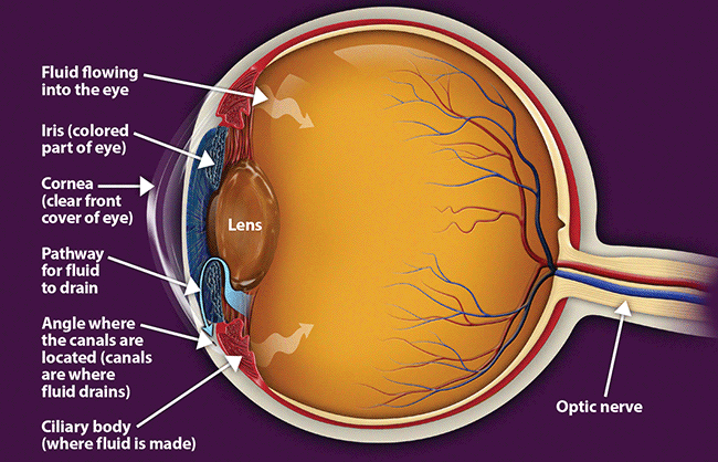
  • Laser therapy. Laser trabeculoplasty (truh-BEK-u-low-plas-tee) is an option if you have open-angle glaucoma. It’s done in your doctor’s office. Your doctor uses a small laser beam to open clogged channels in the trabecular meshwork. It may take a few weeks before the full effect of this procedure becomes apparent.
  • Filtering surgery. With a surgical procedure called a trabeculectomy (truh-bek-u-LEK-tuh-me), your surgeon creates an opening in the white of the eye (sclera) and removes part of the trabecular meshwork.
  • Drainage tubes. In this procedure, your eye surgeon inserts a small tube shunt in your eye to drain away excess fluid to lower your eye pressure.
  • Minimally invasive glaucoma surgery (MIGS). Your doctor may suggest a MIGS procedure to lower your eye pressure. These procedures generally require less immediate postoperative care and have less risk than trabeculectomy or installing a drainage device. They are often combined with cataract surgery. There are a number of MIGS techniques available, and your doctor will discuss which procedure may be right for you.

After your procedure, you’ll need to see your doctor for follow-up exams. And you may eventually need to undergo additional procedures if your eye pressure begins to rise again or other changes occur in your eye.


QUOTE FOR WEDNESDAY:

“More than 4 million people in the United States have glaucoma and about 35% have related vision loss. Both total glaucoma and vision-affecting glaucoma vary by demographic factors.

Glaucoma is called “the sneak thief of sight” since there are no symptoms and once vision is lost, it’s permanent. As much as 40% of vision can be lost without a person noticing.”

Glaucoma Research Foundation (January Is Glaucoma Awareness Month – Glaucoma Research Foundation)

Part II Glaucoma National Awareness: Secondary types of Glaucoma & The key to prevention of glaucoma.

glaucoma3   glaucoma2                                        Glaucoma-TableSecondary Types of Glaucoma:

Sometimes glaucoma is caused by another medical condition — this is called secondary glaucoma.

Neovascular glaucoma

Treatments: Medicines, laser treatment, surgery

Neovascular glaucoma happens when the eye makes extra blood vessels that cover the part of your eye where fluid would normally drain. It’s usually caused by another medical condition, like diabetes or high blood pressure.

If you have neovascular glaucoma, you may notice:

  • Pain or redness in your eye
  • Vision loss

This type of glaucoma can be hard to treat. Doctors need to treat the underlying cause (like diabetes or high blood pressure) and use glaucoma treatments to lower the eye pressure that results from it.

Pigmentary glaucoma

Treatments: Medicines, laser treatment, surgery

Pigment dispersion syndrome happens when the pigment (color) from your iris (the colored part of your eye) flakes off. The loose pigment may block fluid from draining out of your eye, which can increase your eye pressure and cause pigmentary glaucoma.

Young, white men who are near-sighted are more likely to have pigment dispersion syndrome than others. If you have this condition, you may have blurry vision or see rainbow-colored rings around lights, especially when you exercise.

Doctors can treat pigmentary glaucoma by lowering eye pressure, but there currently isn’t a way to prevent pigment from detaching from the iris.

Exfoliation glaucoma

Treatments: Medicines, laser treatment, surgery

Exfoliation glaucoma (sometimes called pseudoexfoliation) is a type of open-angle glaucoma that happens in some people with exfoliation syndrome, a condition that causes extra material to detach from parts of the eye and block fluid from draining.

Recent research shows that genetics may play a role in exfoliation glaucoma. You are at higher risk if someone else in your family has exfoliation glaucoma.

This type of glaucoma can progress faster than primary open-angle glaucoma, and often causes higher eye pressure. This means that it’s especially important for people who are at risk to get eye exams regularly.

Uveitic glaucoma

Treatments: Medicines, surgery

Uveitic glaucoma can happen in people who have uveitis, a condition that causes inflammation (irritation and swelling) in the eye. About 2 in 10 people with uveitis will develop uveitic glaucoma.

Experts aren’t sure how uveitis causes uveitic glaucoma, but they think that it may happen because uveitis can cause inflammation and scar tissue in the middle of the eye. This may damage or block the part of the eye where fluid drains out, causing high eye pressure and leading to uveitic glaucoma.

In some cases, the medicines that treat uveitis may also cause uveitic glaucoma, or make it worse. This is because corticosteroid medicines may cause increased eye pressure as a side effect.

Steroid-Induced Glaucoma

Treatments: The treatment for steroid-induced glaucoma involves stopping or reducing steroid use. Glaucoma medications can also help lower eye pressure. However, while the increase in eye pressure is treatable, damage to the optic nerve is irreversible.

Steroid-induced glaucoma is open-angle glaucoma caused by prolonged steroid use (longer than 3 weeks). It interferes with the outflow of aqueous humor, gradually increasing eye pressure.

Because the increase in eye pressure is slow, steroid-induced glaucoma is typically asymptomatic. Some people may notice blurry vision as an early warning sign. Eyebrow pain has also been associated with IOP. However, these symptoms only occur if IOP is very high.

Trauma Induced Glaucoma

Treatments:  After an eye injury, your doctor may recommend glaucoma medication, steroids, and antibiotics. These can reduce intraocular pressure -IOP, prevent inflammation, and reduce the risk of infection.

Trauma-induced glaucoma is caused by an eye injury that usually involves blunt trauma or penetrating eye injury. Penetrating and blunt trauma injuries can block your eye’s drainage system, gradually increasing IOP over time.

However, the symptoms of traumatic glaucoma depend on the nature of the injury and how fast IOP increases. For example, glaucoma from a penetrating eye injury may develop after the wound heals. Oftentimes, glaucoma symptoms won’t develop for months or years later.

The KEY is take the steps to help PREVENT Glaucoma:

These self-care steps can help you detect glaucoma in its early stages, which is important in preventing vision loss or slowing its progress.

  • Get regular dilated eye examinations. Regular comprehensive eye exams can help detect glaucoma in its early stages, before significant damage occurs. As a general rule, the American Academy of Ophthalmology recommends having a comprehensive eye exam every five to 10 years if you’re under 40 years old; every two to four years if you’re 40 to 54 years old; every one to three years if you’re 55 to 64 years old; and every one to two years if you’re older than 65. If you’re at risk of glaucoma, you’ll need more frequent screening. Ask your doctor to recommend the right screening schedule for you.
  • Know your family’s eye health history. Glaucoma tends to run in families. If you’re at increased risk, you may need more frequent screening.
  • Exercise safely. Regular, moderate exercise may help prevent glaucoma by reducing eye pressure. Talk with your doctor about an appropriate exercise program.
  • Take prescribed eyedrops regularly. Glaucoma eyedrops can significantly reduce the risk that high eye pressure will progress to glaucoma. To be effective, eyedrops prescribed by your doctor need to be used regularly even if you have no symptoms.
  • Wear eye protection. Serious eye injuries can lead to glaucoma. Wear eye protection when using power tools or playing high-speed racket sports in enclosed courts.


QUOTE FOR TUESDAY:

“Some types of glaucoma are caused by other medical conditions — but for other people, the doctor doesn’t find another condition that causes it. When the doctor doesn’t find another cause, it’s called primary glaucoma.

Sometimes glaucoma is caused by another medical condition — this is called secondary glaucoma.”

National Eye Institute – NIH (Types of Glaucoma | National Eye Institute)

Part I National Glaucoma Awareness: Definition of Glaucoma, the Primary Types of Glaucoma and their symptoms.

   glaucoma2                      

 Glaucoma-Table

A common eye condition in which the fluid pressure inside the eye rises to a level higher than healthy for that eye. If untreated, it may damage the optic nerve, causing the loss of vision or even blindness. The elderly, African-Americans, and people with family histories of the disease are at greatest risk.

Glaucoma is a multi-factorial, complex eye disease with specific characteristics such as optic nerve damage and visual field loss. While increased pressure inside the eye (called intraocular pressure or IOP) is usually present, even patients with normal range IOP can develop glaucoma.

There is no specific level of elevated eye pressure that definitely leads to glaucoma; conversely, there is no lower level of IOP that will absolutely eliminate a person’s risk of developing glaucoma. That is why early diagnosis and treatment of glaucoma is the key to preventing vision loss.

Eye pressure is measured in millimeters of mercury (mm Hg). Normal eye pressure ranges from 12-22 mm Hg, and eye pressure of greater than 22 mm Hg is considered higher than normal. When the IOP is higher than normal but the person does not show signs of glaucoma, this is referred to as ocular hypertension.

High eye pressure alone does not cause glaucoma. However, it is a significant risk factor. Individuals diagnosed with high eye pressure should have regular comprehensive eye examinations by an eyecare professional to check for signs of the onset of glaucoma.

A person with elevated IOP is referred to as a glaucoma suspect, because of the concern that the elevated eye pressure might lead to glaucoma. The term glaucoma suspect is also used to describe those who have other findings that could potentially, now or in the future, indicate glaucoma. For example, a suspicious optic nerve, or even a strong family history of glaucoma, could put someone in the category of a glaucoma suspect.

Vision loss from glaucoma occurs when the eye pressure is too high for the specific individual and damages the optic nerve. Any resultant damage cannot be reversed. The peripheral (side) vision is usually affected first. The changes in vision may be so gradual that they are not noticed until a lot of vision loss has already occurred.

In time, if the glaucoma is not treated, central vision will also be decreased and then lost; this is how visual impairment from glaucoma is most often noticed. The good news is that glaucoma can be managed if detected early, and with medical and/or surgical treatment, most people with glaucoma will not lose their sight.

Most common signs and symptoms of Glaucoma:

There are several forms of glaucoma; the two most common forms are primary open-angle glaucoma (POAG) and angle-closure glaucoma (ACG). Open-angle glaucoma is often called “the sneak thief of sight” because it has no symptoms until significant vision loss has occurred.

Types of Glaucoma:

1-Open Angle Glaucoma

2-Angle Closure Glaucoma

3-Normal Tension Glaucoma

1-Open-Angle Glaucoma

Open-angle glaucoma, the most common form of glaucoma, accounting for at least 90% of all glaucoma cases:

  • Is caused by the slow clogging of the drainage canals, resulting in increased eye pressure
  • Has a wide and open angle between the iris and cornea
  • Develops slowly and is a lifelong condition
  • Has symptoms and damage that are not noticed.

“Open-angle” means that the angle where the iris meets the cornea is as wide and open as it should be. Open-angle glaucoma is also called primary or chronic glaucoma. It is the most common type of glaucoma, affecting about three million Americans.

Symptoms of Open-Angle Glaucoma

There are typically no early warning signs or symptoms of open-angle glaucoma. It develops slowly and sometimes without noticeable sight loss for many years.

Most people who have open-angle glaucoma feel fine and do not notice a change in their vision at first because the initial loss of vision is of side or peripheral vision, and the visual acuity or sharpness of vision is maintained until late in the disease.

By the time a patient is aware of vision loss, the disease is usually quite advanced. Vision loss from glaucoma is not reversible with treatment, even with surgery.

Because open-angle glaucoma has few warning signs or symptoms before damage has occurred, it is important to see a doctor for regular eye examinations. If glaucoma is detected during an eye exam, your eye doctor can prescribe a preventative treatment to help protect your vision.

In open-angle glaucoma, the angle in your eye where the iris meets the cornea is as wide and open as it should be, but the eye’s drainage canals become clogged over time, causing an increase in internal eye pressure and subsequent damage to the optic nerve. It is the most common type of glaucoma, affecting about four million Americans, many of whom do not know they have the disease.

You are at increased risk of glaucoma if your parents or siblings have the disease, if you are African-American or Latino, and possibly if you are diabetic or have cardiovascular disease. The risk of glaucoma also increases with age.

2-Angle-Closure Glaucoma

Angle-closure glaucoma, a less common form of glaucoma:

  • Is caused by blocked drainage canals, resulting in a sudden rise in intraocular pressure
  • Has a closed or narrow angle between the iris and cornea
  • Develops very quickly
  • Has symptoms and damage that are usually very noticeable
  • Demands immediate medical attention.

It is also called acute glaucoma or narrow-angle glaucoma. Unlike open-angle glaucoma, angle-closure glaucoma is a result of the angle between the iris and cornea closing.

Symptoms of Angle-Closure Glaucoma

  • Hazy or blurred vision
  • The appearance of rainbow-colored circles around bright lights
  • Severe eye and head pain
  • Nausea or vomiting (accompanying severe eye pain)
  • Sudden sight loss

Angle-closure glaucoma is caused by blocked drainage canals in the eye, resulting in a sudden rise in intraocular pressure. This is a much more rare form of glaucoma, which develops very quickly and demands immediate medical attention

In contrast with open-angle glaucoma, symptoms of acute angle-closure glaucoma are very noticeable and damage occurs quickly. If you experience any of these symptoms, seek immediate care from an ophthalmologist.

If you are diagnosed with glaucoma, it is important to set a regular schedule of examinations with your eye doctor to monitor your condition and make sure that your prescribed treatment is effectively maintaining a safe eye pressure.

3-Normal-tension glaucoma

Normal-tension glaucoma is a type of open-angle glaucoma that happens in people with normal eye pressure.

You may be at higher risk for normal-tension glaucoma if you:

  • Are of Japanese ancestry
  • Have a family history of normal-tension glaucoma
  • Have had certain heart problems, like an irregular heartbeat
  • Have low blood pressure

Signs and symptoms of Normal-Tension Glaucoma:

  • Reduced vision
  • Loss of peripheral vision
  • Bulging or swollen cornea
  • Pupil dilation that doesn’t change with light adjustments
  • Redness in the white (sclera) of the eye

Experts don’t know what causes normal-tension glaucoma, but research shows that treatments that lower eye pressure can help slow the disease and stop vision loss.

For Treatments to ALL 3 primary types of Glaucoma listed above are:

Medicines, laser treatment, surgery.

Tomorrow stay tune for part II on glaucoma awareness giving other types of glaucoma, how its diagnosed & the risks.

QUOTE FOR MONDAY:

“In most children, the cause of tetralogy of Fallot isn’t known. It’s a common type of heart defect. It may be seen more commonly in children with Down syndrome or DiGeorge syndrome. Some children can have other heart defects along with tetralogy of Fallot.  Infants and young children with unrepaired tetralogy of Fallot are often blue (cyanotic-not enough oxygen getting to their tissues).”

AHA American Heart Association (www.heart.org/en/health-topics/congenital-heart-defects/about-congenital-heart-defects)

Part III Congenital Birth Defects Cardiac-Tetralogy of Fallot

What is it?

A heart defect that features four problems.

They are:

  • a hole between the lower chambers of the heart
  • an obstruction from the heart to the lungs
  • The aorta (blood vessel) lies over the hole in the lower chambers
  • The muscle surrounding the lower right chamber becomes overly thickened

More information for parents of children with Tetralogy of Fallot

What causes it?

In most children, the cause of tetralogy of Fallot isn’t known. It’s a common type of heart defect. It may be seen more commonly in children with Down syndrome or DiGeorge syndrome. Some children can have other heart defects along with tetralogy of Fallot.

How does it affect the heart?

Normally the left side of the heart only pumps blood to the body, and the heart’s right side only pumps blood to the lungs. In a child with tetralogy of Fallot, blood can travel across the hole (VSD) from the right pumping chamber (right ventricle) to the left pumping chamber (left ventricle) and out into the body artery (aorta). Obstruction in the pulmonary valve leading from the right ventricle to the lung artery prevents the normal amount of blood from being pumped to the lungs. Sometimes the pulmonary valve is completely obstructed (pulmonary atresia).

How does tetralogy of Fallot affect my child?

Infants and young children with unrepaired tetralogy of Fallot are often blue (cyanotic). The reason is that some oxygen-poor blood is pumped to the body through the hole in the wall between the right and left ventricle instead of being pumped to the lungs.

What can be done about tetralogy of Fallot?

Tetralogy of Fallot is treated surgically. A temporary operation may be done at first if the baby is small or if there are other problems. Complete repair comes later. Sometimes the first operation is a complete repair.

Tetralogy of Fallot patch diagramTetralogy of Fallot shunt diagram

Temporary Operation

In some infants, a shunt operation may be done first to provide adequate blood flow to the lungs. This is not open-heart surgery and doesn’t fix the inside of the heart. The shunt is usually a small tube of synthetic material sewn between a body artery (or the aorta) and the pulmonary artery. The shunt is closed when a complete repair is done later.

Complete Repair

Complete repair tends to be done early in life. The surgeon closes the ventricular septal defect with a patch and opens the right ventricular outflow tract by removing some thickened muscle below the pulmonary valve, repairing or removing the obstructed pulmonary valve and, if needed, enlarging the branch pulmonary arteries that go to each lung.

Sometimes a tube is placed between the right ventricle and the pulmonary artery. This is sometimes called a Rastelli repair. It’s similar to the type of repair used for some other heart defects.

Will my child’s activities be limited?

Your child may need to limit physical activity, particularly for competitive sports, if there is leftover obstruction or leak in the pulmonary valve, which is common after repair. Children with decreased heart function or rhythm disturbances may need to limit their activity more.

If the tetralogy has been repaired with surgery, and there’s no obstruction or leak in the pulmonary valve, your child may be able to participate in normal activities without much increased risk.

Your child’s pediatric cardiologist will help decide if your child needs limits on physical activity.

What will my child need in the future?

If your child has had tetralogy of Fallot repaired, he or she will need regular follow-up with a pediatric cardiologist. As an adult, your child will need lifelong regular follow-up with a cardiologist who’s had special training in congenital heart defects.

Some long-term problems can include leftover or worsening obstruction between the right pumping chamber and the lung arteries. Children with repaired tetralogy of Fallot have a higher risk of heart rhythm disturbances called arrhythmias. Sometimes these may cause dizziness or fainting.

Generally, the long-term outlook is good, but some children may need medicines, heart catheterization or even more surgery.

What about preventing endocarditis?

Children with tetralogy of Fallot are at increased risk for endocarditis. Some children, including those have had a valve replacement, still have a shunt or have leaks around surgical patches, and need to take antibiotics before certain dental procedures to help prevent endocarditis.

QUOTE FOR THE WEEKEND:

“The ductus arteriosus is a normal fetal artery connecting the aorta and the main lung artery (pulmonary artery). The ductus allows blood to detour away from the lungs before birth.

Every baby is born with a ductus arteriosus. After birth, the opening is no longer needed and it usually narrows and closes within the first few days.

Sometimes, the ductus doesn’t close after birth. Failure of the ductus to close is common in premature infants but rare in full-term babies. In most children, the cause of Patent Ductus Arteriosus-PDA isn’t known. Some children can have other heart defects along with the PDA.

Truncus arteriosus occurs when the two large arteries carrying blood away from the heart don’t form properly and one large artery is present instead. This artery (the truncus) sits over a large opening or hole in the wall between the two pumping chambers (ventricular septal defect). With only one artery, there is no specific path to the lungs for oxygen before returning to the heart to deliver oxygen to the body.”

American Heart Association-AHA   (www.heart.org/en/health-topics/congenital-heart-defects/about-congenital-heart-defects)

Part II Congenital Cardiac Defects Awareness Month-Patent ductus arteriosus & Truncus arteriosus

The duct should close in the first hours after birth. If it does not, the blood begins to shunt from the aorta into the pulmonary artery and hyperperfuse the lungs. The left side of the heart will have an increase in blood return and become volume overloaded. Too much blood is going to the lungs. RA – RV – Lungs _ LA – LV – Aorta now blood shunts backwards because pressure in L side higher than R so pressure in aorta in higher it backflows (it is already oxygenated) and prevents the blood that needs to be oxyenate doesn’t get there. Dispalces blood that needs to be oxygenated. Mixed blood in oxygenation. L sided heart failure. L to R shunt. THIS IS CALLED A LEFT-TO-RIGHT SHUNT.

-1 Patent ductus arteriosus (PDA).

Simply put, this is a hole in your baby’s aorta that doesn’t close.

Aorta is the largest artery in the body, the aorta arises from the left ventricle of the heart, goes up (ascends) a little ways, bends over (arches), then goes down (descends) through the chest and through the abdomen to where ends by dividing into two arteries called the common iliac arteries that go to the legs which is called the femoral artery than.

Anatomically, the aorta is traditionally divided into the ascending aorta, the aortic arch, and the descending aorta. The descending aorta is, in turn, subdivided into the thoracic aorta (that descends within the chest) and the abdominal aorta (that descends within the belly).

The aorta gives off branches that go to the head and neck, the arms, the major organs in the chest and abdomen, and the legs. It serves to supply them all with oxygenated blood. The aorta is the central conduit from the heart to the body. A hole in the aorta causes many problems.

During pregnancy, the hole allows your baby’s blood to bypass his lungs and get oxygen from your umbilical cord. After he’s born, he starts to get oxygen from his own lungs, and the hole has to close.

If it doesn’t, it’s called patent ductus arteriosus, or PDA. Small PDAs may get better on their own. A larger one could need surgery.

 2-Truncus arteriosus.

This is when your baby is born with one major artery instead of two that carry blood to the rest of his body. He will need surgery as an infant to repair the defect, and may need more procedures later in life.

I-transposition of the great arteries. This means that the right and left chambers of your baby’s heart are reversed. His blood still flows normally, but over time, his right ventricle doesn’t work as well because it must pump harder.

D-transposition of the great arteries. In this condition, the two main arteries of your baby’s heart are reversed. His blood doesn’t move through the lungs to get oxygen, and oxygen-rich blood doesn’t flow throughout his body. He will have to have surgery to repair this condition, usually within the first month of his life.

Single ventricle defects. Babies are sometimes born with a small lower chamber of the heart, or with one valve missing. Different types of single ventricle defects include:

  • Hypoplastic left heart syndrome: Your baby has an undeveloped aorta and lower left chamber, or ventricle.
  • Pulmonary atresia/intact ventricular septum: Your baby has no pulmonary valve, which controls blood flow from the heart to the lungs.
  • Tricuspid atresia: Your baby has no tricuspid valve, which should be between the upper and lower chambers of the right side of his heart.

Which Type?

In some cases, your doctor can spot congenital heart problems when your baby is still in the womb. But he can’t always diagnose the defect until after birth and until your baby shows signs of a problem.

Many mild congenital heart defects are diagnosed in childhood or even later because they don’t cause any obvious symptoms. Some people don’t find out they have them until they’re adults.

Whatever the type of congenital heart defect, rest assured that with advances in diagnostic tools and treatments, there’s a much greater chance of a long, normal life than ever before.

Heart transplants are recommended for children who have serious heart problems. These children are not able to live without having their heart replaced. Illnesses that affect the heart in this way include complex congenital heart disease, present at birth at times. They also include heart muscle disease (cardiomyopathy).


QUOTE FOR FRIDAY:

“Every 4.5 minutes a baby is born with a condition that affects the structure or function of their body. Collectively, these conditions are referred to as birth defects. They can vary widely in how and where they affect the body and include things from cleft lip to heart problems. While medical advancements have greatly improved health and survival, many of these conditions are lifelong and require lifelong care.

Achieving the best possible health will look different for different people. No two people living with these conditions are exactly alike. Everyone’s journey is unique, shaped by their specific condition, individual strengths, and the support system in place. However, a community of support can help people with these conditions no matter what health experiences they are navigating.”

Centers for Disease Control and Prevention – CDC

(Birth Defects Awareness Month | Birth Defects | CDC)