QUOTE FOR THE WEEKEND:

“Atrial fibrillation (AFib) is an irregular and often very rapid heart rhythm but it can also be a in slow rate for some. An irregular heart rhythm is called an arrhythmia and that is all AFIB patients who are diagnosed with it. AFib can lead to blood clots in the heart. The condition also increases the risk of stroke, heart failure and other heart-related complications.

During atrial fibrillation, the heart’s upper chambers — called the atria — beat chaotically and irregularly. They beat out of sync with the lower heart chambers, called the ventricles. For many people, AFib may have no symptoms. But AFib may cause a fast, pounding heartbeat, shortness of breath or light-headedness due to the rapid heart rate.

AFib can also be bradycardia (a slow heart beat rhythm).

You could have pauses  in that AFIB bradycardia that are called sick sinus syndrome or a longer pauses causing sinus arrest.  Each pt’s heart is unique on how it operates.”

MAYO CLINIC

Heart Rhythms, like A FIb, with a pause in it called Sick Sinus Syndrome-What it is, S/S, causes, tests, and treatment!

   TopBox-Normal Sinus Rhythm

Normally, the heartbeat starts in an area in the top chambers of the heart (atria). This area is the heart’s pacemaker. It is called the sinoatrial node, sinus node or SA node. Its role is to keep the heart beat steady and regular. This creates the normal rhythm of the human body call Normal Sinus Rhythm (listed above in pictures in 2nd box call NSR).

Sick sinus syndrome is a group of heart rhythm problems due to problems with the sinus node, such as:

  • The heartbeat rate is too slow, called sinus bradycardia
  • The heartbeat pauses or stops, called sinus pauses or sinus arrest
  • Episodes of a fast heart rate
  • Slow heart rhythms that alternate with fast heart rhythms, called bradycardia-tachycardia or “tachy-brady syndrome”  Tachy meaning fast and Brady meaning slow.

Alternative Names

Bradycardia-tachycardia syndrome; Sinus node dysfunction; Slow heart rate – sick sinus; Tachy-brady syndrome; Sinus pause – sick sinus; Sinus arrest – sick sinus

Causes

Sick sinus syndrome most often occurs in people older than age 50. It is often due to scar-like damage to electrical pathways in the heart muscle tissue.

In children, heart surgery on the upper chambers is a common cause of sick sinus syndrome.

Coronary heart disease, high blood pressure, and aortic and mitral valve diseases may occur with sick sinus syndrome. However, these diseases may have nothing to do with the syndrome.

Sick sinus syndrome is uncommon, but not rare. It is the most common reason people need to have an artificial pacemaker implanted. Sinus bradycardia occurs more often than the other types of the condition.

Tachycardias (rapid heart rhythms) that start in the upper chambers, when the SA node can’t work to intiate the person’s rhythm,  atrial rhythms start to take over as the pacemaker of the heart that may be part of the sick sinus syndrome for people who have it. These atrial rhythms include atrial fibrillation, atrial flutter, atrial tachycardia-all are types of upper chamber heart rhythms, where they intiate from. A period of fast heart rates is often followed by very slow heart rates. When there are periods of both slow and fast heart rates (rhythms) the condition often will be called tachy-brady syndrome; this is what makes up atrial fibrillation (a fib) irregular rhythm/slow and fast heart beats and due to this allows pooling of blood in the heart chamber putting it at a potential to clot and could be passed on from the heart into circulation if the clot breaks off and if it reaches the brain puts the patient at risk for stroke.  Or A Fib can cause heart failure or other cardiac problems.

Some medicines can make abnormal heart rhythms worse, especially when doses are high. These include digitalis, calcium channel blockers, beta-blockers, and antiarrhythmics.

Symptoms

Most of the time, there are no symptoms.

Symptoms that do occur may mimic those of other disorders.

Symptoms may include:

  • Chest pain or angina
  • Confusion or other changes in mental status
  • Fainting or near-fainting
  • Fatigue
  • Dizziness or lightheadedness
  • Sensation of feeling the heart beat (palpitations)
  • Shortness of breath, possibly only with physical activity like walking

Exams and Tests

The heart rate may be very slow at any time. Blood pressure may be normal or low.

Sick sinus syndrome may cause symptoms of heart failure to start or get worse. Sick sinus syndrome is diagnosed when the symptoms occur only during episodes of arrhythmia. However, the link is often hard to prove.

An ECG may show abnormal heart rhythms related to this syndrome.

Holter or longer term rhythm monitors are effective tools for diagnosing sick sinus syndrome. They may pick up very slow heart rates and long pauses, along with episodes of atrial tachycardias. Types of monitors include event monitors, loop recorders, and mobile telemetry.

An intracardiac electrophysiology study (EPS) is a very specific test for this disorder. However, it is not often needed and may not confirm the diagnosis.

In some cases, a person’s heart rate is observed when walking or exercising to see if it increases enough; a stress test.

Treatment

You may not need treatment if you do not have any symptoms. Your health care provider will review the medicines you take to make sure they are not making your condition worse. Do not stop taking any of your medicines unless your provider tells you to do so.

You may need a permanent implanted pacemaker if your symptoms are related to bradycardia (slow heart rate or with long pauses or rhythm arrest (completely stops).

QUOTE FOR THURSDAY:

“A direct blow to the head, or a blow to the body that suddenly rotates the head, can lead to a concussion, causing the brain to rapidly move back and forth inside the skull. “You don’t need impact to the head to have a concussion,” Dr. Sethi notes. For example, a football tackle or a fender bender may involve no head contact but could cause the head to whip around fast enough to cause a concussion.

Concussions typically do not damage the brain. “In the majority of cases, CAT-scans of concussion come back negative,” Dr. Sethi says. Yet, a concussion can temporarily stop the brain from working properly.

The danger is in not recognizing the signs of a concussion, especially since they are not always obvious, Dr. Sethi stresses. “Parents should know that concussion symptoms—a headache, dizziness, difficulty tolerating light or noise–could be very subtle,” he says. Athletes who have sustained a knock to the head commonly insist that they’re fine so that they can keep playing. “If you’re a coach on the sideline you may not notice anything wrong until you see that a player is off balance, looks confused or disoriented, or has a glassy look in his eyes,” he says.

Even severe head injuries, like hematomas, do not always produce immediate symptoms, Dr. Sethi says, recounting the late actor Natasha Richardson, who fell off her skis and hit the back of her head, insisted she felt fine, and refused medical care. “We call these injuries walking, talking, and dying. By the time she got to the hospital, her brain was hemorrhaging.”

Weill Cornell Medicine (https://weillcornell.org/news/national-winter-sports-traumatic-brain-injury-tbi-awareness)

Part I Total Brain Injury (TBI) in regards to Winter Sports.

BRAIN INJURY IMAGE   brain Lobe Regions

concussion 3a concussion 2


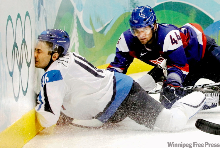
We are in winter now with the sports this season that can be very dangerous.  Football with hockey and boxing already started and yet they maybe very exciting they unfortunately can still can be severely dangerous.  One of the reasons when a football quaterback catches after hiked the ball or in a kick off to the other team the player who catches the ball and not running it out starts no longer at the 20 but the 25 yard line, is done all for more safety even though some of us football fanatics think how wimpy.

The NFL now even takes action.  Through Fall and Winter Sports TBI Awareness Month, The Johnny OTM Foundation (Johnny O) is hoping to raise awareness regarding the health risks athletes face when they participate in winter sports, specifically traumatic brain injuries and concussions. September is a great time to put a spotlight on fall/winter sports safety and preventive measures athlete can take to avoid TBIs and concussions.

The mission of Johnny O “is to educate the American public to the growing seriousness of Alzheimer’s, Dementia and Traumatic Brain Injuries in the American population by raising the necessary donations through strategic research initiatives and heightened public awareness to accomplish our objectives.”1 Fall and Winter Sports TBI Awareness Month is one of many initiatives Johnny O is undertaking to not only raise public awareness, but also improve safety and reduce TBIs in Americans of all ages.

 A concussion is a traumatic brain injury that alters the way your brain functions. Effects are usually temporary but can include headaches and problems with concentration, memory, balance and coordination.

Although concussions usually are caused by a blow to the head, they can also occur when the head and upper body are violently shaken. These injuries can cause a loss of consciousness, but most concussions do not. Because of this, some people have concussions and don’t realize it.

Concussions are common, particularly if you play a contact sport, such as football. But every concussion injures your brain to some extent. This injury needs time and rest to heal properly. Most concussive traumatic brain injuries are mild, and people usually recover fully.

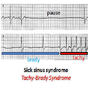
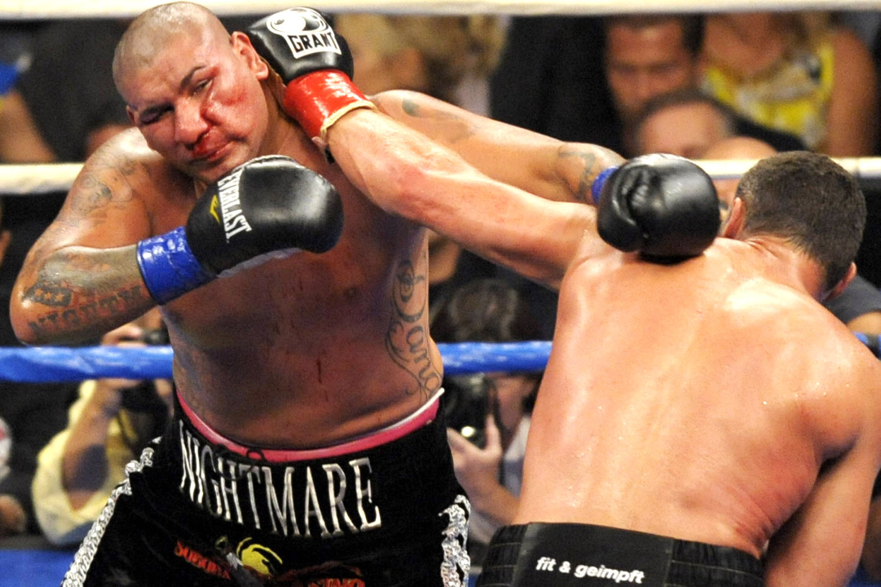
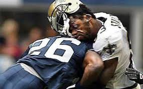
Remember the key to a brain concussion fully recovering is not to have impact to the head happening over and over again.  Based on the same concept if you get hit in the same spot over and over again anywhere in the body bruising to actual injury will happen whether it be muscle or bone.  Well get hit in the head over and over again like in sports especially boxing but now the big conversation with football even with a helmet on you will cause a permanent damage to the brain.   A perfect example of this is a boxer that gets hit over an over again to the head in a boxing ring.   The head is just another area of the body and no different than other areas of our body.

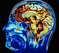
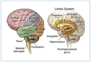
What actually happens is the concussion is most often caused by a sudden direct blow or bump to the head.  The brain is made of soft tissue. It’s cushioned by spinal fluid and encased in the protective shell of the skull. When you sustain a concussion, the impact can jolt your brain. Sometimes, it literally causes it to move around in your head. Traumatic brain injuries can cause bruising, damage to the blood vessels, and injury to the nerves.

concussion 5concussion 4

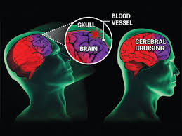
The result? Your brain doesn’t function normally. If you’ve suffered a concussion, vision may be disturbed, you may lose equilibrium, or you may fall unconscious. In short, the brain is confused. That’s why Bugs Bunny often saw stars after getting whacked in the head in his cartoon by some other character.

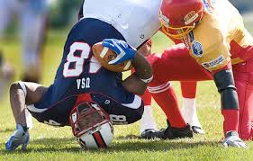
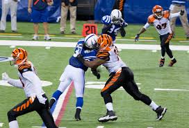
The new uptake with football is being concerned with players getting concussions from getting hit by their opponent players whether it be defense or offense while playing the game. Concussions have become big business in the football world. With 1,700 players in the NFL, 66,000 in the college game, 1.1 million in high school and 250,000 more in Pop Warner, athletes and families across the country are eager to find ways to cut the risks of brain injury, whose terrifying consequences regularly tear across the sports pages. And a wave of companies offering diagnostic tools and concussion treatments are just as eager to sell them peace of mind.

That’s actually a slogan for one company.   ImPACT, the maker of the world’s most popular concussion evaluation system, offers a 20-minute computerized test that players can take via software or online to measure verbal and visual memory, processing speed, reaction time and impulse control.  The idea behind ImPACT (Immediate Post-Concussion Assessment and Cognitive Testing) and similar batteries is that doctors or athletic trainers can give a baseline test to a healthy athlete, conduct follow-up tests after an injury and then compare the results to help figure out when it’s OK to return the athlete to play. Selling itself as “Valid. Reliable. Safe,” ImPACT dominates the testing market and has spread throughout the sports world: Most NFL clubs use the test, as do all MLB, MLS and NHL clubs, the national associations for boxing, hockey and soccer in the U.S., and nine auto racing circuits.

A total of 87 out of 91 former NFL players have tested positive for the brain disease at the center of the debate over concussions in football, according to new figures from the nation’s largest brain bank focused on the study of traumatic head injury.

Researchers with the Department of Veterans Affairs and Boston University have now identified the degenerative disease known as chronic traumatic encephalopathy, or CTE, in 96 percent of NFL players that they’ve examined and in 79 percent of all football players. The disease is widely believed to stem from repetitive trauma to the head, and can lead to conditions such as memory loss, depression and dementia.

In total, the lab has found CTE in the brain tissue in 131 out of 165 individuals who, before their deaths, played football either professionally, semi-professionally, in college or in high school.

Forty percent of those who tested positive were the offensive and defensive linemen who come into contact with one another on every play of a game, according to numbers shared by the brain bank with FRONTLINE. That finding supports past research suggesting that it’s the repeat, more minor head trauma that occurs regularly in football that may pose the greatest risk to players, as opposed to just the sometimes violent collisions that cause concussions.

But the figures come with several important caveats, as testing for the disease can be an imperfect process. Brain scans have been used to identify signs of CTE in living players, but the disease can only be definitively identified posthumously. As such, many of the players who have donated their brains for testing suspected that they had the disease while still alive, leaving researchers with a skewed population to work with.

 Even with those caveats, the latest numbers are “remarkably consistent” with past research from the center suggesting a link between football and long-term brain disease, said Dr. Ann McKee, the facility’s director and chief of neuropathology at the VA Boston Healthcare System.

“People think that we’re blowing this out of proportion, that this is a very rare disease and that we’re sensationalizing it,” said McKee, who runs the lab as part of a collaboration between the VA and BU. “My response is that where I sit, this is a very real disease. We have had no problem identifying it in hundreds of players.”

In a statement, a spokesman for the NFL said, “We are dedicated to making football safer and continue to take steps to protect players, including rule changes, advanced sideline technology, and expanded medical resources. We continue to make significant investments in independent research through our gifts to Boston University, the [National Institutes of Health] and other efforts to accelerate the science and understanding of these issues.”

The latest update from the brain bank, which in 2010 received a $1 million research grant from the NFL, comes at a time when the league is able to boast measurable progress in reducing head injuries. In its 2015 Health & Safety Report, the NFL said that concussions in regular season games fell 35 percent over the past two seasons, from 173 in 2012 to 112 last season. A separate analysis by FRONTLINE that factors in concussions reported by teams during the preseason and the playoffs shows a smaller decrease of 28 percent.


QUOTE FOR THE WEDNESDAY:

“Esophageal Atresia is a congenital disorder in which a baby’s esophagus does not form properly during pregnancy.  Symptoms include include white, frothy bubbles in the mouth, vomiting, and coughing/choking when feeding.  Treatment includes surgery”.

Yale Medicine (https://www.yalemedicine.org/conditions/tracheoesophageal-fistula-and-esophageal-atresia)

Congenital Defect Esophageal Atresia


Esophageal atresia is a birth defect in which part of a baby’s esophagus (the tube that connects the mouth to the stomach) does not develop properly.

Esophageal atresia is a birth defect of the swallowing tube (esophagus) that connects the mouth to the stomach. In a baby with esophageal atresia, the esophagus has two separate sections—the upper and lower esophagus—that do not connect. A baby with this birth defect is unable to pass food from the mouth to the stomach, and sometimes difficulty breathing.

Esophageal atresia often occurs with tracheoesophageal fistula, a birth defect in which part of the esophagus is connected to the trachea, or windpipe.

Types of Esophageal Atresia

There are four types of esophageal atresia: Type A, Type B, Type C and Type D.

  • Type A is when the upper and lower parts of the esophagus do not connect and have closed ends. In this type, no parts of the esophagus attach to the trachea.
  • Type B is very rare. In this type the upper part of the esophagus is attached to the trachea, but the lower part of the esophagus has a closed end.
  • Type C is the most common type. In this type the upper part of the esophagus has a closed end and the lower part of the esophagus is attached to the trachea, as is shown in the drawing.
  • Type D is the rarest and most severe. In this type the upper and lower parts of the esophagus are not connected to each other, but each is connected separately to the trachea.

How Many Babies are Born with Esophageal Atresia?

Researchers estimate that about 1 in every 4,100 babies is born with esophageal atresia in the United States.1 This birth defect can occur alone, but often occurs with other birth defects.

Causes

Like many families of children with a birth defect, CDC wants to find out what causes them. Understanding the factors that can increase the chance of having a baby with a birth defect will help us learn more about the causes. CDC funds the Centers for Birth Defects Research and Prevention, which collaborate on large studies such as the National Birth Defects Prevention Study (NBDPS; births 1997-2011) and the Birth Defects Study To Evaluate Pregnancy exposureS (BD-STEPS; began with births in 2014), to understand the causes of and risks for birth defects, including esophageal atresia.

The causes of esophageal atresia in most babies are unknown. Researchers believe that some instances of esophageal atresia may be caused by abnormalities in the baby’s genes. Nearly half of all babies born with esophageal atresia have one or more additional birth defects, such as other problems with the digestive system (intestines and anus), heart, kidneys, or the ribs or spinal column.

Recently, CDC reported on important findings about some factors that increase the risk of having a baby with esophageal atresia:

  • Paternal age – Older age of the father is related to an increased chance of having a baby born with esophageal atresia.
  • Assisted reproductive technology (ART) – Women who used ART to become pregnant have an increased risk of having a baby with esophageal atresia compared to women who did not use ART.

CDC continues to study birth defects, such as esophageal atresia, and how to prevent them. If you are pregnant or thinking about becoming pregnant, talk with your doctor about ways to increase your chances of having a healthy baby.

Diagnosis

Esophageal atresia is rarely diagnosed during pregnancy. Esophageal atresia is most commonly detected after birth when the baby first tries to feed and has choking or vomiting, or when a tube inserted in the baby’s nose or mouth cannot pass down into the stomach. An x-ray can confirm that the tube stops in the upper esophagus.

Treatment

Once a diagnosis has been made, surgery is needed to reconnect the two ends of the esophagus so that the baby can breathe and feed properly. Multiple surgeries and other procedures or medications may be needed, particularly if the baby’s repaired esophagus becomes too narrow for food to pass through it; if the muscles of the esophagus don’t work well enough to move food into the stomach; or if digested food in the stomach consistently moves back up into the esophagus.

QUOTE FOR TUESDAY:

“Radon is a naturally occurring radioactive gas emitted from the ground. Radon is odorless, tasteless and colorless, and can enter a home through cracks in floors, basement walls, foundations and other openings. Radon can be present at high levels inside homes, schools and other buildings. Radon in homes is more common than you think. Pennsylvania has one of the nation’s most serious radon problems. High levels of radioactive radon gas have been found in every county but most places in the Commonwealth remain undertested, so this isn’t something that should be taken lightly.”

American Lung Association (https://www.lung.org/media/press-releases/pa-radon-2024)

QUOTE FOR MONDAY:

“You can’t see, smell or taste radon, but it could be present at a dangerous level in your home. Radon is the leading cause of lung cancer deaths among nonsmokers in America and claims the lives of about 21,000 Americans each year. In fact, the EPA and the U.S. Surgeon General urge all Americans to protect their health by testing their homes, schools and other buildings for radon.

Exposure to radon is a preventable health risk and testing radon levels in your home can help prevent unnecessary exposure. If a high radon level is detected in your home, you can take steps to fix the problem to protect yourself and your family.”

United States Environmental Protection – EPA

(https://www.epa.gov/radon/national-radon-action-month-information)

QUOTE FOR THE WEEKEND:

“A few precautions that are recommended for all pregnant women include the following:

  • Limit your exposure to mercury by not eating bigeye tuna, king mackerel, marlin, orange roughy, shark, swordfish, or tilefish. Limit eating white (albacore) tuna to 6 ounces a week. You do not have to avoid all fish during pregnancy. In fact, fish and shellfish are nutritious foods with vital for a pregnant woman and her fetus. Be sure to eat at least 8–12 ounces of low-mercury fish and shellfish per week.
  • Avoid exposure to lead. Lead can be found in old paint, construction materials, alternative medicines, and items made in foreign countries, such as jewelry and pottery.
  • Avoid taking high levels of vitamin A. Very high levels of vitamin A have been linked to severe birth defects. You should consume no more than 10,000 international units of vitamin A a day.
  • Women who are obese (defined as having a of 30 or greater) when they get pregnant have an increased risk of having babies with certain birth defects than women who are a normal weight. Among the most common obesity-related birth defects are NTDs, heart defects, and . If you are planning a pregnancy, the best way to prevent problems caused by obesity is to be at a normal weight before you get pregnant.”

American College of Obstetricians and Gynecologists ACOG

(https://www.acog.org/womens-health/faqs/reducing-risks-of-birth-defects)

QUOTE FOR FRIDAY:

“The reported incidence of PDA in term neonates is only 1 in 2,000 births, accounting for 5%–10% of all congenital heart disease. The incidence of PDA in preterm neonates is far greater, with reports ranging from 20%–60% (depending on population and diagnostic criteria). The increased incidence of PDA in the preterm infant is attributable to the lack of normal closure mechanisms due to immaturity. Gestational age and weight are intimately linked to PDA in preterm neonates. Specifically, PDA is present in 80% of infants weighing less than 1,200 g at birth, compared to 40% of infants weighing less than 2,000 g at birth., Furthermore, symptomatic PDA is present in 48% of infants with a birth weight of less than 1,000 g.”

National Library of Medicine (https://www.ncbi.nlm.nih.gov/pmc/articles/PMC3462096/)