Archive | May 2025

Part II Lupus Awareness–on how it’s diagnosed, what’s the treatments & possible complications.


Diagnosing Lupus:

Diagnosing lupus is difficult because signs and symptoms vary considerably from person to person. Signs and symptoms of lupus may vary over time and overlap with those of many other disorders. No one test can diagnose lupus. The combination of blood and urine tests, signs and symptoms, and physical examination findings leads to the diagnosis.  Tests the MD will order:

Laboratory tests

Blood and urine tests may include:

  • Complete blood count. This test measures the number of red blood cells, white blood cells and platelets as well as the amount of hemoglobin, a protein in red blood cells. Results may indicate you have anemia, which commonly occurs in lupus. A low white blood cell or platelet count may occur in lupus as well.
  • Erythrocyte sedimentation rate. This blood test determines the rate at which red blood cells settle to the bottom of a tube in an hour. A faster than normal rate may indicate a systemic disease, such as lupus. The sedimentation rate isn’t specific for any one disease. It may be elevated if you have lupus, another inflammatory condition, cancer or an infection.
  • Kidney and liver assessment. Blood tests can assess how well your kidneys and liver are functioning. Lupus can affect these organs.
  • Urinalysis. An examination of a sample of your urine may show an increased protein level or red blood cells in the urine, which may occur if lupus has affected your kidneys.
  • Antinuclear antibody (ANA) test. A positive test for the presence of these antibodies — produced by your immune system — indicates a stimulated immune system. While most people with lupus have a positive ANA test, most people with a positive ANA do not have lupus. If you test positive for ANA, your doctor may advise more-specific antibody testing.If your doctor suspects that lupus is affecting your lungs or heart, he or she may suggest:
  • Chest X-ray. An image of your chest may reveal abnormal shadows that suggest fluid or inflammation in your lungs.

  • Ultrasound maybe used in where a M.D. orders a Echocardiogram. It’s a a noninvasive ultrasound test that evaluates heart function.This test uses sound waves to produce real-time images of your beating heart. It can check for problems with your valves and other portions of your heart.Lupus can harm your kidneys in many different ways, and treatments can vary, depending on the type of damage that occurs. In some cases, it’s necessary to test a small sample of kidney tissue to determine what the best treatment might be. The sample can be obtained with a needle or through a small incision.
  • First see a specialist for Lupus who is more out in knowing what to look for (just like a cardiologist for cardiac problems or endocrinologist for diabetes, etc…) So set up an appointment with a doctor to be examined!

Treatments for Lupus:

1. Biopsy

2. The M. D. will do a physical and determine whether your signs and symptoms your experiencing.  Treatment for lupus depends on your signs and symptoms and the cause for it so treatment varies. The symptoms should be treated as lupus and what medications to use requires a careful discussion of the benefits and risks with your doctor. As your signs and symptoms flare and subside, you and your doctor may find that you’ll need to change medications or dosages.

3.Medications, this could include the following:

a. Nonsteroidal anti-inflammatory drugs (NSAIDs).  This could include, over-the-counter NSAIDs, such as naproxen sodium (Aleve) and ibuprofen (Advil, Motrin IB, others), may be used to treat pain, swelling and fever associated with lupus. Stronger NSAIDs are available by prescription. Side effects of NSAIDs include stomach bleeding, kidney problems and an increased risk of heart problems.

b. Antimalarial drugs.  Medications commonly used to treat malaria, such as hydroxychloroquine (Plaquenil), also can help control lupus. Side effects can include stomach upset and, very rarely, damage to the retina of the eye.

c. Corticosteroids  This could include Prednisone and other types of corticosteroids can counter the inflammation of lupus but often produce long-term side effects — including weight gain, easy bruising, thinning bones (osteoporosis), high blood pressure, diabetes and increased risk of infection. The risk of side effects increases with higher doses and longer term therapy.

d. Immunosuppressants.  These drugs suppress the immune system which may be helpful in serious cases of lupus. Examples include azathioprine (Imuran, Azasan), mycophenolate (CellCept), leflunomide (Arava) and methotrexate (Trexall). Potential side effects may include an increased risk of infection, liver damage, decreased fertility and an increased risk of cancer. A newer medication, belimumab (Benlysta), also reduces lupus symptoms in some people. Side effects include nausea, diarrhea and fever.Take steps to care for your body if you have lupus. Simple measures can help you prevent lupus flares and, should they occur, better cope with the signs and symptoms you experience.

4. Other treatments could include:

  • See your doctor regularly. Having regular checkups instead of only seeing your doctor when your symptoms worsen may help your doctor prevent flare-ups, and can be useful in addressing routine health concerns, such as stress, diet and exercise that can be helpful in preventing lupus complications.
  • Get adequate rest. People with lupus often experience persistent fatigue that’s different from normal tiredness and that isn’t necessarily relieved by rest. For that reason, it can be hard to judge when you need to slow down. Get plenty of sleep at night and naps or breaks during the day as needed.
  • Be sun smart. Because ultraviolet light can trigger a flare, wear protective clothing — such as a hat, long-sleeved shirt and long pants — and use sunscreens with a sun protection factor (SPF) of at least 55 every time you go outside.
  • Get regular exercise. Exercise can help you recover from a flare, reduce your risk of heart attack, help fight depression and promote general well-being.
  • Don’t smoke. Smoking increases your risk of cardiovascular disease and can worsen the effects of lupus on your heart and blood vessels.
  • Eat a healthy diet. A healthy diet emphasizes fruits, vegetables and whole grains. Sometimes you may have dietary restrictions, especially if you have high blood pressure, kidney damage or gastrointestinal problems.

Possible complications to Lupus:

  • Kidneys. Lupus can cause serious kidney damage, and kidney failure is one of the leading causes of death among people with lupus. Signs and symptoms of kidney problems may include generalized itching, chest pain, nausea, vomiting and leg swelling (edema).
  • Brain and central nervous system. If your brain is affected by lupus, you may experience headaches, dizziness, behavior changes, hallucinations, and even strokes or seizures. Many people with lupus experience memory problems and may have difficulty expressing their thoughts.
  • Blood and blood vessels. Lupus may lead to blood problems, including anemia and increased risk of bleeding or blood clotting. It can also cause inflammation of the blood vessels (vasculitis).
  • Lungs. Having lupus increases your chances of developing an inflammation of the chest cavity lining (pleurisy), which can make breathing painful. You may also be more susceptible to pneumonia.
  • Heart. Lupus can cause inflammation of your heart muscle, your arteries or heart membrane (pericarditis). The risk of cardiovascular disease and heart attacks increases greatly.  Other areas of the body that can be effected by lupus causing problems include:  Infection, Cancer, Bone Tissue Death (avascular necrosis), and Pregnancy complications.



QUOTE FOR WEDNESDAY:

“Lupus is a complicated disease that affects different people in different ways. For some, lupus can be mild — for others, it can be life threatening.

Right now, there’s no cure for lupus. The good news is that with the support of your doctors and loved ones, you can learn to manage it.

Around 1.5 million people in the United States are living with lupus.”

LUPUS Foundation of America (https://www.lupus.org/understanding-lupus)

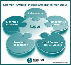
Part I Lupus Awareness Month-What it is, the facts of Lupus, the 4 major types, & the signs and symptoms!

Lupus Part1a Butterfly Rash

lupus part 1b


Lupus is an inflammatory disease that is caused when the auto, meaning “self”, immune system attacks its own tissues.

Lupus is a chronic, an autoimmune disease that can damage any part of the body (skin, joints, and/or organs inside the body). Chronic means that the signs and symptoms tend to last longer than six weeks and often for many years.

In lupus, something goes wrong with your immune system, which is the part of the body that fights off viruses, bacteria, and germs (“foreign invaders,” like the flu). Normally our immune system produces proteins called antibodies that protect the body from these invaders. Your autoimmune immune system cannot tell the difference between these foreign invaders and your body’s healthy tissues and creates auto-antibodies that attack and destroy healthy tissue. These auto-antibodies cause inflammation, pain, and damage in various parts of the body.

Lupus is also a disease of remissions (flare ups) whereby the symptoms worsen and you feel ill and remissions (the symptoms improve and you feel better) but the disease remains. So your goal is to keep the disease as best as possible in remissions.

FACTS TO KNOW IF YOU HAVE LUPUS:

  • Lupus is not contagious, not even through sexual contact. You cannot “catch” lupus from someone or “give” lupus to someone.
  • Lupus is not like or related to cancer. Cancer is a condition of malignant, abnormal tissues that grow rapidly and spread into surrounding tissues. Lupus is an autoimmune disease, as described above. However, some treatments for lupus may include immunosuppressant drugs that are also used in chemotherapy.
  • Lupus is not like or related to HIV (Human Immune Deficiency Virus) or AIDS (Acquired Immune Deficiency Syndrome) except that they both affect the immune system (but differently) in having remissions and exacerbations, that’s it. In HIV or AIDS the immune system is underactive; whereas in lupus, the immune system is overactive.
  • Lupus can range from mild to life-threatening and should always be treated by a doctor. With good medical care, most people with lupus can lead a full life.
  • Another factor in helping you reach remission is being compliant with the taking the meds ordered by your doctor.
  • Research estimates that at least 1.5 million Americans have a form of lupus. The actual number may be higher; however, there have been no large-scale studies to show the actual number of people in the U.S. living with lupus.
  • More than 16,000 new cases of lupus are reported annually across the country.
  • It is believed that 5 million people throughout the world have a form of lupus.
  • Lupus strikes mostly women of childbearing age (15-44). However, men, children, and teenagers develop lupus, too. Most people with lupus develop the disease between the ages of 15-44.
  • Women of color are two to three times more likely to develop lupus than Caucasians.
  • People of all races and ethnic groups can develop lupus.
  • Lupus effects 80% women.
  • Discoid lupus erythematosus—causes a skin rash that doesn’t go away
  • Subacute cutaneous lupus erythematosus—causes skin sores on parts of the body exposed to sun
  • Drug-induced lupus—can be caused by medications
  • Neonatal lupus—a rare type of lupus that affects newborns.      
  • Approximately 1.5 million Americans have a form of lupus.
  • Systemic lupus erythematosus cases: 70% of total lupus cases.
  • Of individuals diagnosed with lupus, 90% are women.
  • Eighty percent of people develop lupus between 15 and 45 years of age.
  • Lupus is two to three times more prevalent among people of color.
  • Chance of a parent or sibling having or developing lupus: 20%

There are 4 major kinds of lupus. Here are the types:

1-Systemic lupus erythematosus (SLE) is the most common and most serious type of lupus.

2-Cutaneous lupus erythematosus, which affects only the skin.

3-Drug-induced lupus, a short-term type of lupus caused by certain medicines.

4-Neonatal lupus, a rare type of lupus that affects newborn babies.

The most common type and serious type of lupus is systemic lupus erythematosus (SLE)

  • Systemic Lupus Erythematosus  or also known as (SLE):
  • The cause(s) of SLE are unknown, however, heredity, viruses, ultraviolet light, and drugs all may play some role.

Signs and Symptoms of Lupus:

1. *butterfly rash on the face – very common symptom with a flare up.

Including other possible symptoms:

2. appetite loss 3. hair loss 4. fever 5. fatigue 6. photosensitivity 7. Raynaud’s phenomenon 8. pleuritis and 9. pericarditis.

  • Up to 10% of people with lupus isolated to the skin will develop the systemic form of lupus (SLE).
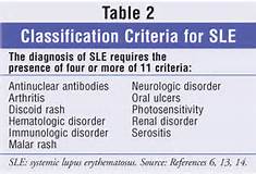
  • The criteria listed at the top in the picture help doctors diagnose SLE!

Stayed tune for part II tomorrow on how its diagnosed and the type of treatments!


QUOTE FOR TUESDAY:

“Buerger disease is a rare disease of the arteries and veins in the arms and legs. In Buerger disease — also called thromboangiitis obliterans — blood vessels become blocked. This reduces blood flow to the affected areas. Blood clots may form in the blood vessels.

Over time, the lack of blood flow damages or destroys skin tissue. The damage can lead to infection and death of body tissue, called gangrene. Buerger disease is usually first seen in the feet. It may eventually affect the blood vessels of the hand.”

MAYO Clinic

QUOTE FOR MONDAY:

In 2022, stroke accounted for approximately 1 of every 20 deaths in the United States.
• On average in 2022, someone died of stroke every 3 minutes 11 seconds in the United
States.
• Stroke caused 165 393 deaths in the United States in 2022.
• In 2022, the age-adjusted US stroke death rate as an underlying cause of death was 39.5
per 100 000, an increase of 7.0% from 36.9 per 100 000 in 2012, and the actual number
of stroke deaths increased 28.7% during the same time period.
• In 2021, there were 7.25 million deaths attributable to stroke worldwide (3.59 million
deaths from ischemic stroke, 3.31 million deaths from intracerebral hemorrhage, and
0.35 million from subarachnoid hemorrhage).
• Age-standardized mortality due to stroke amongst regions was highest for Oceania and
southeast Asia. Rates were lowest for Australasia and western Europe. Age-standardized
mortality due to ischemic stroke amongst regions was highest for eastern Europe,
followed by north Africa and the Middle East and central Asia. Mortality was lowest for
Australasia. Amongst regions, intracerebral hemorrhage mortality was highest for
Oceania, followed by southeast and east Asia and central and eastern sub-Saharan
Africa. Amongst regions, mortality estimated for subarachnoid hemorrhage was highest
for Oceania followed by southeast Asia and Andean Latin America.”

American Heart Association (American Stroke Month | American Stroke Association)

Part III Stroke Awareness – Rx and how to prevent one!

Part III stroke   stroke III

Part III stroke  Part III stroke

A.) TREATMENT OF STROKES

If you’re having a stroke, it’s critical that you get medical attention right away. Immediate treatment may minimize the long-term effects of a stroke and prevent death.

Ischemic Stroke Treatment

1.)tPA, the Gold Standard

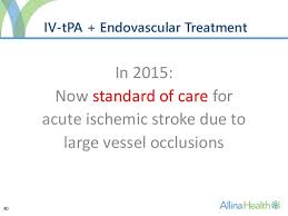
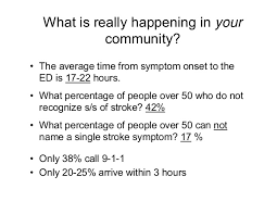
The only FDA approved treatment for ischemic strokes is tissue plasminogen activator (tPA, also known as IV rtPA, given through an IV in the arm). tPA works by dissolving the clot and improving blood flow to the part of the brain being deprived of blood flow. If administered within 3 hours(and up to 4.5 hours in certain eligible patients), tPA may improve the chances of recovering from a stroke. A significant number of stroke victims don’t get to the hospital in time for tPA treatment; this is why it’s so important to identify a stroke immediately.

2.)Endovascular Procedures

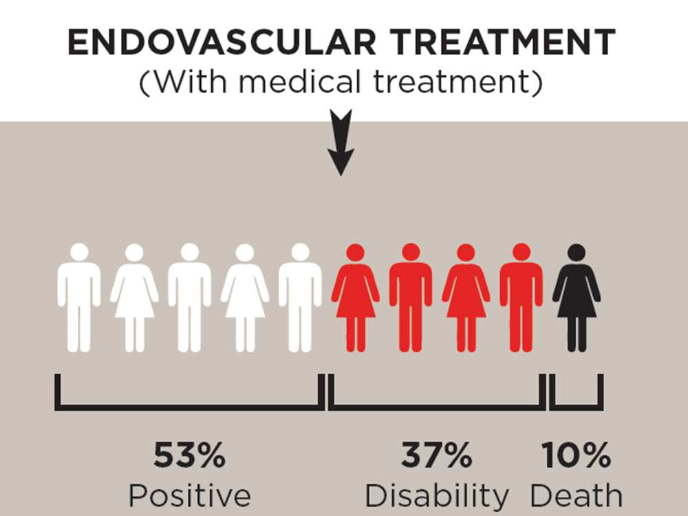
Another treatment option is an endovascular procedure* called mechanical thrombectomy, strongly recommended, in which  trained doctors try  removing a large blood clot by  sending a wired-caged device called a stent retriever, to the site of the blocked blood vessel in the brain. To remove the brain clot, doctors thread a catheter through an artery in the groin up to the blocked artery in the brain. The stent opens and grabs the clot, allowing doctors to remove the stent with the trapped clot. Special suction tubes may also be used. The procedure should be done within six hours of acute stroke symptoms, and only after a patient receives tPA.   *Note: Patients must meet certain criteria to be eligible for this procedure. Image courtesy of Medtronic

Hemorrhagic Stroke Treatment

1.)Endovascular Procedures Endovascular procedures may be used to treat certain hemorrhagic strokes similar to the way the procedure is used for treating an ischemic stroke. These procedures are less invasive than surgical treatments, and involve the use of a catheter introduced through a major artery in the leg or arm, then guided to the aneurysm or AVM; it then deposits a mechanical agent, such as a coil, to prevent rupture.

2.)Surgical Treatment For strokes caused by a bleed within the brain (hemorrhagic stroke), or by an abnormal tangle of blood vessels (AVM), surgical treatment may be done to stop the bleeding. If the bleed is caused by a ruptured aneurysm (swelling of the vessel that breaks), a metal clip may be placed surgically at the base of the aneurysm to secure it.

B.) How to prevent a stroke!

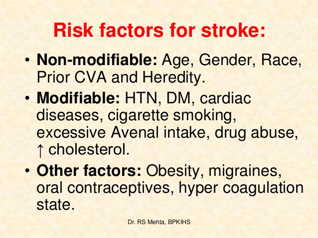
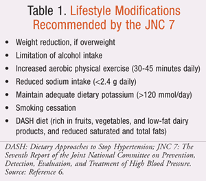
Treatment is also aimed at other factors that put you at risk, including high blood pressure, diabetes, and high cholesterol. But it takes more than just your doctor’s efforts. You also have an important role to play in preventing stroke. It’s up to you to make lifestyle changes that can lower your risk.

What you can do to prevent a stroke:

1-Control your blood pressure.

2-Lose Weight to the point that your in a healthy weight for your height. If you’re overweight, losing as little as 10 pounds can have a real impact on your stroke risk.      Try to eat no more than 1,500 to 2,000 calories a day (depending on your activity level and your current body mass index). Increase the amount of exercise you do with such activities as walking, golfing, or playing tennis, and by making activity part of every single day.

3-Exercise More-Exercise contributes to losing weight and lowering blood pressure, but it also stands on its own as an independent stroke reducer. Exercise at a moderate intensity 5x/wk and if you can’t do ½ hr as day spread it out into 2 15minute exercise moments for the day.

4- Drink-in moderation What you’ve heard is true. Drinking can make you less likely to have a stroke—up to a point. “Studies show that if you have about one drink per day, your risk may be lower. I am not saying drink one glass of liquor a day but if you have to limit it to one glass a day. Red wine your first choice, because it contains resveratrol, which is thought to protect the heart and brain.

5-Atrial Fibrillation-Atrial fibrillation is a form of irregular heartbeat that causes clots to form in the heart. Those clots can then travel to the brain, producing a stroke. “Atrial fibrillation carries almost a fivefold risk of stroke, and should be taken seriously; take your anticoagulant medication the MD orders to keep the blood thin to prevent clotting.

6-Treat diabetes –Having high blood sugar over time damages blood vessels, making clots more likely to form inside them putting the person at higher risk for a stroke. So simply keep your sugar under control.

7-QUIT Smoking-Along with a healthy diet and regular exercise, smoking cessation is one of the most powerful lifestyle changes that will help you reduce your stroke risk


QUOTE FOR THE WEEKEND:

“A stroke is a medical emergency that happens when something prevents your brain from getting enough blood flow=lack of 02 to the brain. A blocked blood vessel or bleeding in your brain can cause strokes.  They can be fatal and need immediate treatment. Call 911 or your local emergency services number right away if you think you or someone you’re with is having a stroke.  Strokes are the second leading cause of death worldwide and the fifth most common in the U.S.  The sooner someone is diagnosed and treated, the more likely it is they’ll survive a stroke. Every second counts!”

Cleveland Clinic (Stroke: What It Is, Causes, Symptoms & Treatment)

Stroke awareness – Symptoms of a Stroke and how its diagnosed!

stroke part II  stroke part IIb

Signs and Symptoms of a stroke happening:

Symptoms of stroke include trouble walking, speaking, and understanding, as well as paralysis or numbness of the face, arm, or leg.

People may experience the following:                                                                      

Muscular: difficulty walking, paralysis with weak muscles, problems with coordination, stiff muscles, overactive reflexes, or paralysis of one side of the body

Visual: blurred vision, double vision, sudden visual loss, or temporary loss of vision in one eye

Whole body: balance disorder, fatigue, or lightheadedness

Speech: difficulty speaking, slurred speech, or speech loss

Sensory: pins and needles or reduced sensation of touch

Facial: muscle weakness or numbness

Limbs: numbness or weakness

Also common: difficulty swallowing, headache, inability to understand, mental confusion, numbness, or rapid involuntary eye movement

What is done for a stroke regarding diagnostic tooling:

 To determine the most appropriate treatment for your stroke, your emergency team needs to evaluate the type of stroke you’re having and the areas of your brain affected by the stroke. They also need to rule out other possible causes of your symptoms, such as a brain tumor or a drug reaction. Your doctor may use several tests to determine your risk of stroke, including:

CT scan of brain tissue damaged by stroke

Cerebral angiogram A cerebral angiogram showing a carotid aneurysm due to a stroke.

Physical examination. Your doctor will ask you or a family member what symptoms you’ve been having, when they started and what you were doing when they began. Your doctor then will evaluate whether these symptoms are still present.

Your doctor will want to know what medications you take and whether you have experienced any head injuries. You’ll be asked about your personal and family history of heart disease, transient ischemic attack or stroke.

Your doctor will check your blood pressure and use a stethoscope to listen to your heart and to listen for a whooshing sound (bruit) over your neck (carotid) arteries, which may indicate atherosclerosis. Your doctor may also use an ophthalmoscope to check for signs of tiny cholesterol crystals or clots in the blood vessels at the back of your eyes.

Blood tests. You may have several blood tests, which tell your care team how fast your blood clots, whether your blood sugar is abnormally high or low, whether critical blood chemicals are out of balance, or whether you may have an infection. Managing your blood’s clotting time and levels of sugar and other key chemicals will be part of your stroke care.

Computerized tomography (CT) scan. A CT scan uses a series of X-rays to create a detailed image of your brain. A CT scan can show a hemorrhage, tumor, stroke and other conditions. Doctors may inject a dye into your bloodstream to view your blood vessels in your neck and brain in greater detail (computerized tomography angiography).  The goal is if the CT scan determined the stroke to be a ischemic stroke start rtpa a drug IV if the symptoms of the stroke started in the past 3 hrs if not treat it another way OR if the stroke is determined to be hemorrhagic than its the OR.  Will go into treatment in more detail in Part III tomorrow.

Magnetic resonance imaging (MRI). An MRI uses powerful radio waves and magnets to create a detailed view of your brain. An MRI can detect brain tissue damaged by an ischemic stroke and brain hemorrhages. Your doctor may inject a dye into a blood vessel to view the arteries and veins and highlight blood flow (magnetic resonance angiography, or magnetic resonance venography).

Carotid ultrasound. In this test, sound waves create detailed images of the inside of the carotid arteries in your neck. This test shows buildup of fatty deposits (plaques) and blood flow in your carotid arteries.

Cerebral angiogram. In this test, your doctor inserts a thin, flexible tube (catheter) through a small incision, usually in your groin, and guides it through your major arteries and into your carotid or vertebral artery. Then your doctor injects a dye into your blood vessels to make them visible under X-ray imaging. This procedure gives a detailed view of arteries in your brain and neck.

Echocardiogram. An echocardiogram uses sound waves to create detailed images of your heart. An echocardiogram can find a source of clots in your heart that may have traveled from your heart to your brain and caused your stroke.

You may have a transesophageal echocardiogram. In this test, your doctor inserts a flexible tube with a small device (transducer) attached into your throat and down into the tube that connects the back of your mouth to your stomach (esophagus). Because your esophagus is directly behind your heart, a transesophageal echocardiogram can create clear, detailed ultrasound images of your heart and any blood clots.

In reality going to an ER room if the pt comes suspected of a stroke and has symptoms or not than nationally in America the hospitals are to do the following:

-A neuro assessment should be done in 10 minutes  by the doctor.

-A CT SCAN ordered and pt sent off for the CT SCAN test and done within 25 minutes.

-The CT SCAN read and interpreted by the radiologist / neuro doctor within 45 minutes.                        At this point it tells the MD if the pt has a blockage or a hemmorage in the brain that caused the stroke. Remember a ischemic stroke and hemmoragic stroke are treated differently.

We’ll get into treatment tomorrow in Part III Treatment of a stroke.


QUOTE FOR FRIDAY:

“93,000 + Americans will be diagnosed with a primary brain tumor diagnosis in 2025 (An estimated 24,80o new primary malignant brain tumors (brain cancer) will be diagnosed in 2025 in this amount.); 35.7 percent is the five-year survival rate for patients with malignant brain tumors and 18, 330 Americans will die from a malignant brain tumor in 2025.  73% of all brain tumors are benign and approximately 27% are malignant.”

National Brain Tumor Society (NBTS) – Brain Tumor Awareness Month


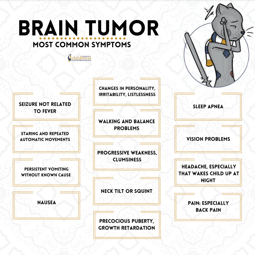
Brain Tumor Awareness-Types of brain tumors, brain tumor grading, basics of brain/CNS A&P and function to understand symptoms, statistics on brain tumors and risk factors!


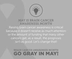
May is Brain Cancer and Brain Tumor Awareness Month (BTAM), a time to raise awareness about brain tumors and educate the community.

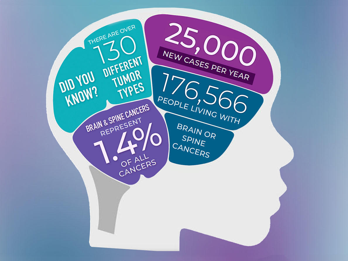
Doctors will diagnose cancers of the brain or central nervous system in about 25,400 people in the United States in 2024, according to the National Cancer Institute. All brain and spine tumors, collectively called central nervous system (CNS) tumors cover over 130 different CNS tumor types. These cancers make up a portion of the more than 94,000 brain tumors alone (including benign tumors) that will occur in this country in 2024.

It can be hard for people with CNS tumors to find accurate information, specialized support, and expert care. You can help by spreading awareness and sharing educational materials like through blogs live striveforgoodhealth.com and other sites in the internet.

There are many types of brain and spinal cord tumors. The tumors result from the abnormal growth of cells and may be either benign or malignant. Benign brain and spinal cord tumors grow and press on nearby areas of the brain. Normally, they rarely spread into other tissues; the brain tumors that are diagnosed malignant rapidly spread only in brain tissue and remember when your a fetus the brain develops that the spinal cord grows out of made of brain tissue so spreading can go in those 2 areas.  A brain tumor malignant can form in the brain or other parts of the central nervous system (CNS), being the spine or cranial nerves. So remember, Malignant tumors in the brain and spinal cord only grow quickly spreading only into the brain and (CNS) spinal cord tissue.  The positive note is the tumor stays in those areas but unfortunately it spreads rapidly for most brain tumors.  Survival in a brain tumor especially malignant is a survival rate of 5 years or less but there are those cases that have lasted longer but on average its 5 years or less and this would include a benign tumor not operable but it is suppose to grow slower than a malignant tumor.  Malignant brain tumors need to be treated as soon as possible to prolong life.

Tumor grading:

Tumor grade has long been a way to define the aggressiveness of a tumor, particularly for malignant brain tumors such as glioma but also for non-malignant (benign) brain tumors including meningioma.

Traditionally, tumors have been classified as grade 1 to 4 based on histology (cells as viewed under a microscope) and molecular markers. Grade 1 tumors occur primarily in children and represent a type separate from grade 2-4 (seen primarily in adults). Grade 2 tumors are considered low grade, but some can be aggressive. Grade 3 and 4 tumors are defined as high grade.

What are molecular markers?

Not all brain tumors are the same. Some tumors have differences in the genetic or molecular makeup of the cells. These differences are called molecular markers, or biomarkers. Molecular markers are becoming increasingly important for brain tumor diagnosis and treatment. For example, some molecular markers help determine how aggressive a tumor may be. Others determine how responsive a tumor will be to treatment.

Some common molecular markers include the following:

  • IDH1 and IDH2
  • MGMT
  • 1p/19q co-deletion
  • BRAF
  • EGFR
  • TP53
  • ATRX
  • TERT
  • PTEN
  • NTRK
  • FGFR

In 2016, the World Health Organization (WHO) included two molecular markers into the CNS tumor classification system that improved accuracy of glioma diagnosis. In 2021 WHO again updated CNS tumor classification, incorporating new knowledge gained from additional molecular markers and new diagnostic techniques. Tumors are now listed as “CNS grade 1-4” with presence or absence of IDH mutation, a key factor in glioma classification.

Basis Review of Brain & the CNS with how it functions:

The brain and spinal cord together form the central nervous system (CNS), like we said in knowing this the brain is a complex organ made up of nerves and connective tissue. Nerves in the brain and spinal cord transmit messages throughout the body. The CNS directs and regulates all of the body’s functions. The brain tumor can definitely mess up a lot of these functions depending on where the tumor is located since the brain is broken up in lobes to do different functions that is what causes the wide signs and symptoms of dysfunctions that occur in time with a brain tumor especially that is metastatic.

The CNS is the core of our existence. It controls:

> Personality: thoughts, memory, intelligence, speech, understanding and emotions

> Senses: vision, hearing, taste, smell and touch

> Basic body functions: breathing, heartbeat and blood pressure

> How we function in our environment: movement, balance and coordination

The brain is made up of multiple parts, and each part of the brain is responsible for different body functions. Therefore, brain tumor symptoms, and potential treatment options, depend a great deal on where the tumor is located.

Learning about the normal workings of the brain and spine will help you understand the symptoms of brain tumors, how they are diagnosed and how they are treated.

Major parts of the brain: There are three major parts of the brain:

1. Cerebrum: uses information from senses to tell our body how to respond. It controls reading, thinking, learning, movement, speech, vision, personality and emotions.

2. Cerebellum: controls balance for standing, walking and other motion.

3. Brain stem: connects the brain with the spinal cord and controls basic body functions such as breathing, sleeping, body temperature and blood pressure.

Lobes of the brain

Different lobes of the brain control different functions. The frontal lobe of the brain helps you think and reason. The temporal lobe contains the neural pathways for hearing and vision, as well as behavior and emotions. Having a tumor, or treatment, in one of these lobes could affect the lobe’s specific functions. Additionally, since the brain has areas that connect, it is possible for a brain tumor to impact a function of the brain where the tumor is not specifically located.

Other common brain tumor locations include the meninges (a layer of tissue that covers the brain and spinal cord), skull base (the bottom of the skull), spinal cord, pituitary tumor, and cranial nerves.

Brain tumor statistics:

Brain tumors are reported in people of all ages, races, ethnicities, and genders. Over 1.3 million Americans are living with a primary or secondary/metastatic brain tumor today. Primary tumors originate in the brain, and the most common types are meningiomas, pituitary tumors, and gliomas. Metastatic, or secondary brain tumors arise from outside the brain in another organ such as the breast or lung and spread to other areas of the brain. These are the most common brain tumors.

Unless otherwise specified, the follow statistics come from the Central Brain Tumor Registry of the United States Annual Report:

  • Approximately 90,000 people are diagnosed with a primary brain tumor every year.   
  • Brain and other CNS tumors are the fifth most common cancer. 
  • Over 1 million people are living with a diagnosis of a primary brain tumor. 
  • There are more than 100 different types of primary brain and CNS tumors. 
  • Nearly one-third (27.9 percent) of brain and central nervous system (CNS) tumors are malignant. 
  • Brain and CNS tumors are the most common cancer diagnosed in children aged 0-14. 
  • More than 28,000 children in the United States are currently diagnosed with a brain tumor. 
  • Approximately 3,400 children (aged 0-14) are diagnosed with a primary brain tumor each year. 
  • Approximately 12,800 adolescents and young adults (aged 15-39) are diagnosed with a primary brain tumor each year. 
  • The incidence rate for brain and CNS tumors is highest among those aged 85 years and older. 
  • Each year, approximately 17,200 people die from a malignant brain tumor. Survival after diagnosis with a primary brain tumor varies significantly by age, race, geographical location, tumor type, tumor location, and molecular markers. 

Risk Factors for Brain Tumors:

Genetic and hereditary risk factors

Inherited traits are carried in genes. Each individual has two copies of each gene, one from each parent. Genes often contain small changes. Sometimes these changes do not cause any problems, but sometimes these changes are more serious and can interfere with the way the gene is supposed to work.

There are a few rare, inherited genetic syndromes that are associated with brain tumors., including Neurofibromatosis 1 (NF1 gene), Neurofibromatosis 2 (NF2 gene), Turcot syndrome (APC gene), Gorlin syndrome (PTCH gene), Tuberous Sclerosis (TSC1 and TSC2 genes) and Li-Fraumeni syndrome (TP53 gene).

Although 5-10% of persons with brain tumors have a family history of a brain tumor, the vast majority of CNS tumors appear not to be a part of inherited genetic syndromes.  A number of studies have identified genetic variants that may be associated with an increased risk of certain brain tumors including glioma and meningioma.  Study results from 2017 show that while there are some hereditary similarities in glioma tumors between family members, there is not a statistically significant difference between families having tumors with similar hereditary features as compared to families with tumors having different hereditary features. Also, in families with more than one glioma, the tumors tend to have the same molecular markers. This study continues to collect and analyze data.

Environmental risk factors:

Other than family history, the most consistently identified risk factor associated with brain tumor development is therapeutic or high-dose ionizing radiation.  With regard to medical diagnostic radiation exposure, small increases in brain tumor risks have been reported.  Although certain brain scans and radiation therapy used to treat brain tumors use ionizing radiation, the risk of developing a new brain tumor due to these causes is very low. Occupational exposures among medical radiation workers have been associated with approximately twice the risk of brain cancer mortality, though data on the level of radiation exposure were not available.

With respect to the impact of non-ionizing radiation from cell phones, the association between this exposure and brain cancer has been the subject of much research. Radio frequency fields were classified by the World Health Organization’s International Agency for Research on Cancer in 2011 as a possible carcinogen following the observation of increased glioma risk among heavy cell phone users: the topic remains under study at present.

Industrial chemicals have long been suspected as a cause of glioma due to their ability to cross the blood–brain barrier.  The blood-brain barrier that the human brain has protects the brain from toxins and pathogens. Despite numerous chemical, environmental, and occupational exposures having been explored in epidemiological studies of glioma, results have been inconsistent for most factors. Although not precisely defined, an association between exogenous hormones (e.g., oral contraceptives, hormone replacement therapies) and meningioma risk is often reported and thus patients might discuss this topic with their health care providers.