Other perianal infections
Sometimes the skin glands near your anus become infected and need to be drained, like in this structural disease. Just behind the anus, abscesses can form that contain a small tuft of hair at the back of the pelvis (called a pilonidal cyst).
Sexually transmitted diseases that can affect the anus include anal warts, herpes, AIDS, chlamydia and gonorrhea.
Diverticular disease
The structural disease diverticulosis is the presence of small outpouchings (diverticula) in the muscular wall of your large intestine that form in weakened areas of the bowel. They usually occur in the sigmoid colon, the high-pressure area of the lower large intestine.
Diverticular disease is very common and occurs in 10% of people over age 40 and in 50% of people over age 60 in Western cultures. It is often caused by too little roughage (fiber) in the diet. Diverticulosis can sometimes develop/progress into diverticulitis
Complications of diverticular disease happen in about 10% of people with outpouchings. They include infection or inflammation (diverticulitis), bleeding and obstruction. Treatment of diverticulitis includes treating the constipation and sometimes antibiotics if really severe. Surgery is needed as last resort in those who have significant complications to remove the involved diseased segment of the colon.
Colon polyps and cancer
Each year, 130,000 Americans are diagnosed with colorectal cancer, the second most common form of cancer in the United States. Fortunately, with advances in early detection and treatment, colorectal cancer is one of the most curable forms of the disease. By using a variety of screening tests, it is possible to prevent, detect and treat the disease long before symptoms appear.
The importance of screening
Almost all colorectal cancers begin as polyps, benign (non-cancerous) growths in the tissues lining your colon and rectum. Cancer develops when these polyps grow and abnormal cells develop and start to invade surrounding tissue. Removal of polyps can prevent the development of colorectal cancer. Almost all precancerous polyps can be removed painlessly using a flexible lighted tube called a colonoscope. If not caught in the early stages, colorectal cancer can spread throughout the body. More advanced cancer requires more complicated surgical techniques.
Most early forms of colorectal cancer do not cause symptoms, which makes screening especially important. When symptoms do occur, the cancer might already be quite advanced. Symptoms include blood on or mixed in with the stool, a change in normal bowel habits, narrowing of the stool, abdominal pain, weight loss, or constant tiredness.
Most cases of colorectal cancer are detected in one of four ways:
- By screening people at average risk for colorectal cancer beginning at age 45.
- By screening people at higher risk for colorectal cancer (for example, those with a family history or a personal history of colon polyps or cancer).
- By investigating the bowel in patients with symptoms.
- A chance finding at a routine check-up.
Early detection is the best chance for a cure.
Colitis
There are several types of colitis, which are conditions that cause an inflammation of the bowel. These include:
- Infectious colitis.
- Ulcerative colitis (cause unknown).
- Crohn’s disease (cause unknown).
- Ischemic colitis (caused by not enough blood going to the colon).
- Radiation colitis (after radiotherapy).
Colitis causes diarrhea, rectal bleeding, abdominal cramps and urgency (frequent and immediate need to empty the bowels). Treatment depends on the diagnosis, which is made by colonoscopy and biopsy.
Prevention:
Can gastrointestinal diseases be prevented?
Many diseases of the colon and rectum can be prevented or minimized by maintaining a healthy lifestyle, practicing good bowel habits and getting screened for cancer.
A colonoscopy is recommended for average-risk patients at age 45. If you have a family history of colorectal cancer or polyps, a colonoscopy may be recommended at a younger age. Typically, a colonoscopy is recommended 10 years younger than the affected family member. (For example, if your brother was diagnosed with colorectal cancer or polyps at age 45, you should begin screening at age 35.)
If you have symptoms of colorectal cancer you should consult your healthcare provider right away. Common symptoms include:
- A change in normal bowel habits.
- Blood on or in the stool that is either bright or dark.
- Unusual abdominal or gas pains.
- Very narrow stool.
- A feeling that the bowel has not emptied completely after passing stool.
- Unexplained weight loss.
- Fatigue.
- Anemia (low blood count).
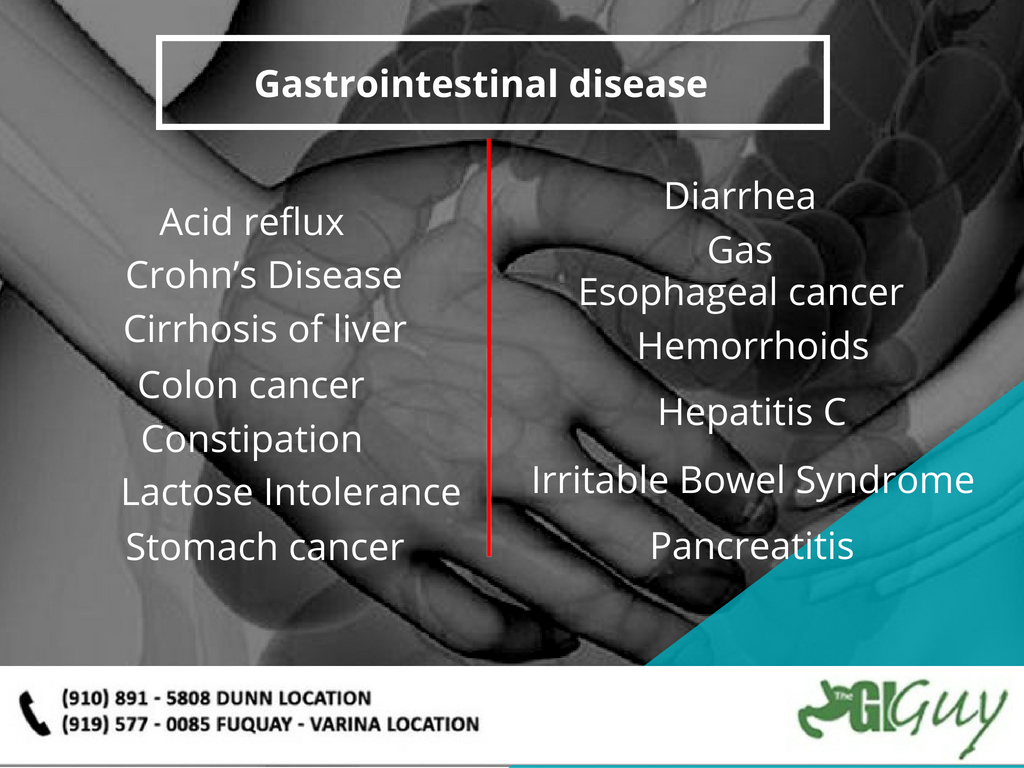
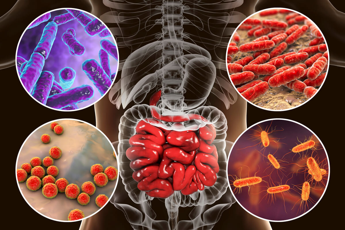
Other types of gastrointestinal diseases
There are many other gastrointestinal diseases. Some are discussed, but others are not covered here. Other functional and structural diseases include peptic ulcer disease, gastritis, gastroenteritis, celiac disease, Crohn’s disease, gallstones, fecal incontinence, lactose intolerance, Hirschsprung disease, abdominal adhesions, Barrett’s esophagus, appendicitis, indigestion (dyspepsia), intestinal pseudo-obstruction, pancreatitis, short bowel syndrome, Whipple’s disease, Zollinger-Ellison syndrome, malabsorption syndromes and hepatitis.