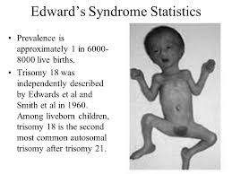
Trisomy 18, also known as Edwards syndrome, is a condition which is caused by a error in cell division, known as meiotic disjunction. When this happens, instead of the normal pair, an extra chromosome 18 results (a triple) in the developing baby and disrupts the normal pattern of development in significant ways that can be life-threatening, even before birth. A Trisomy 18 error occurs in about 1 out of every 2500 pregnancies in the United States and 1 in 6000 live births. The numbers of total births is much higher because it includes significant numbers of stillbirths that occur in the 2nd and 3rd trimesters of pregnancy.
Unlike Down syndrome, which also is caused by an extra chromosome, the developmental issues caused by Trisomy 18 are associated with more medical complications that are more potentially life-threatening in the early months and years of life. Studies have shown that only 50% of babies who are carried to term will be born alive, and baby girls will have higher rates of live birth than baby boys.
What are Related Conditions?
The most common trisomy is Trisomy 21, also known as Down syndrome, where a baby has three of the twenty-first chromosome. Trisomy 18 is the second most common trisomy and occurs when a baby has three of the eighteenth chromosome. This results in 47 chromosomes instead of the normal 46 in the affected cells. It is this extra genetic material that causes the problems associated with Trisomy 18. The third most common is Trisomy 13, also known as Patau syndrome.
While there are different types of Trisomy 18, this does not mean one is better for a child than another. With each type, there is a range of possibilities. Some children are medically fragile while others thrive; some children walk while others are confined to wheelchairs. It is hard to say how the extra chromosome will impact an individual child from the genetic diagnosis alone.
Types of Trisomy 18:
- Full Trisomy 18: The most common type of Trisomy 18 (occurring in about 95% of all cases) is full Trisomy. With full Trisomy, the extra chromosome occurs in every cell in the baby’s body. This type of trisomy is not hereditary. It is not due to anything the parents did or did not do—either before or during pregnancy. It is just an unfortunate error in nature.
- Partial Trisomy 18: Partial trisomies are very rare. They occur when only part of an extra chromosome is present. Some partial Trisomy 18 syndromes may be caused by hereditary factors. Very rarely, a piece of chromosome 18 becomes attached to another chromosome before or after conception. Affected people have two copies of chromosome 18, plus a “partial” piece of extra material from chromosome 18.
- Mosaic Trisomy 18: Mosaic trisomy is also very rare. It occurs when the extra chromosome is present in some (but not all) of the cells of the body. Like full Trisomy 18, mosaic Trisomy is not inherited and is a random occurrence that takes place during cell division.The extra genetic material from the extra eighteenth chromosome can cause a wide variety of problems (sometimes referred to as birth defects) in the developing child in the mother’s womb and after birth. Just as children with Down syndrome can range from mildly to severely affected, the same is true for children with Trisomy 18. This means that there is no hard and fast rule about what Trisomy 18 will mean for a specific child. Each child has their own unique profile of how Trisomy 18 is affecting their developing body and organs. However, all studies on survival rates show that there is a high mortality rate for children with Trisomy 18 before or shortly after birth.
- Common Problems associated with Trisomy 18 can include:
- Impact of Trisomy 18 on Baby?
- Heart defects:
- VSD (Ventricular Septal Defect): a hole between the lower chambers
- ASD (Atrial Septal Defect): a hole between the upper chambers
- Coarctation of the aorta: a narrowing of the exit vessel from the heart
- Kidney problems
- Part of the intestinal tract is outside the stomach (omphalocele)
- The esophagus doesn’t connect to the stomach (esophageal artesia)
- Excess amniotic fluid (polyhydramnios)
- Clenched hands
- Pocket of fluid on the brain (choroid plexus cysts)
- Rocker bottom feet
- Delayed growth
- Small jaw (mycrognathia)
- Small head (microcephaly)
- Low-set ears
- Strawberry-shaped head
- Severe developmental delays
- Umbilical or inguinal hernia
How is Trisomy 18 Diagnosed?
Most cases of Trisomy 18 are diagnosed prenatally in the United States. Regardless of whether the diagnosis is made prenatally or postnatally (after birth) the process is the same. A sample of the baby’s dna is extracted from a blood sample or other bodily cells or tissue and is cultured to examine a picture of the chromosomes called a karyotype. A karyotype is simply a picture of a person’s chromosomes. In order to get this picture, the chromosomes are isolated, stained, and examined under the microscope. Most often, this is done using the chromosomes in the white blood cells. A picture of the chromosomes is taken through the microscope. A visible extra 18th chromosome confirms a Trisomy 18 diagnosis.
Treatment varies
Treatment will depend on what conditions the child ends up with into adulthood and how it impacts the individual in adulthood.