Continuation of Types of skin cancer:
5-Akinetic Keratosis:
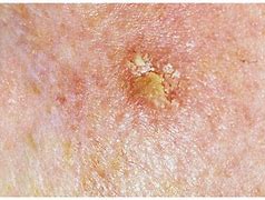
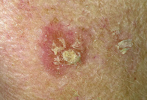
Actinic keratosis (AK) is a skin disorder that causes rough, scaly patches of skin. Another name for AK is solar keratosis. AK is a type of precancer, which means that if you don’t treat the condition, it could turn into cancer. Without treatment, AK can lead to a type of skin cancer called squamous cell carcinoma.
The risk factors of Akinetic Keratosis are:
–UV exposure from the sun or indoor tanning.
-History of skin cancer in particular history of actinic keratosis.
-Fair skin: People with fair skin including lighter color hair or eyes have an increased risk.
Warning Signs can help with early detection and treatment this can be successfully removed without complications. Look out for any new, changing or unusual skin growths, so you can spot skin cancers like BCC when they are easiest to treat and cure.
Treatments Akinetic Keratosis:
An actinic keratosis sometimes disappears on its own but might return after more sun exposure. It’s hard to tell which actinic keratoses will develop into skin cancer, so they’re usually removed as a precaution.
Medicines-
If you have several actinic keratoses, your health care provider might prescribe a medicated cream or gel to remove them, such as fluorouracil (Carac, Efudex others), imiquimod (Aldara, Zyclara) or diclofenac. These products might cause inflamed skin, scaling or a burning sensation for a few weeks.
Surgical and other procedures-
Many methods are used to remove actinic keratosis, including:
- Freezing (cryotherapy). Actinic keratoses can be removed by freezing them with liquid nitrogen. Your health care provider applies the substance to the affected skin, which causes blistering or peeling. As your skin heals, the damaged cells slough off, allowing new skin to appear. Cryotherapy is the most common treatment. It takes only a few minutes and can be done in your health care provider’s office. Side effects may include blisters, scarring, changes to skin texture, infection and changes in skin color of the affected area.
- Scraping (curettage). In this procedure, your health care provider uses a device called a curet to scrape off damaged cells. Scraping may be followed by electrosurgery, in which a pencil-shaped instrument is used to cut and destroy the affected tissue with an electric current. This procedure requires local anesthesia. Side effects may include infection, scarring and changes in skin color of the affected area.
- Laser therapy. This technique is increasingly used to treat actinic keratosis. Your health care provider uses an ablative laser device to destroy the patch, allowing new skin to appear. Side effects may include scarring and discoloration of the affected skin.
- Photodynamic therapy. Your health care provider might apply a light-sensitive chemical solution to the affected skin and then expose it to a special light that will destroy the actinic keratosis. Side effects may include inflamed skin, swelling and a burning sensation during therapy.
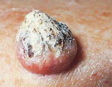
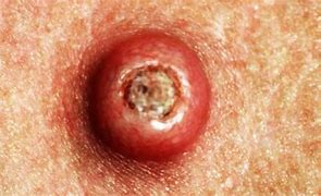
6-Keratocanthoma (KA)
The term “Keratoacanthoma” (KA) was coined by Freudenthal in the year 1936. It was first described way back in 1889 by Hutchinson and was called molluscum sebaceum and self-limiting epithelioma. KA is benign, self-limiting squamo-proliferative lesion.
It shows male preponderance and most commonly arises on the sun-exposed parts predominantly face, neck forearms, hands and legs. Cutaneous lesions arise from hair follicles whereas mucosal lesions originate from ectopic sebaceous glands. This is a slow growing cancer of the skin that looks like a dome or crater. This is common; more than 200,000 cases per year in US. Regarding treatment from medical professional is advised. This condition often requires lab test or imaging. Keratoacanthoma last several months. It is common for ages 60 and older and is more common in males.
The risk factors of Keratocanthoma (KA):
–UV exposure from the sun or indoor tanning.
-contact with chemical carcinogens, or cancer-causing chemicals
-Infection with some strains of a wart virus, such as papillomavirus
-History of skin cancer in particular history of Keratoacanthoma.
Warning Signs can help with early detection and treatment, this can be successfully removed without complications if caught early. Look out for any new, changing or unusual skin growths, so you can spot skin cancers like BCC when they are easiest to treat and cure.
Treatments:
If your medical professional suspects a keratoacanthoma, they will first want to establish the correct diagnosis by performing a biopsy. Than treatments could include the following:
- Removal (excision), in which a scalpel is used to cut away the keratoacanthoma and then place stitches to bring the wound edges together.
- Mohs micrographic surgery, in which tiny slivers of skin are removed until there are no more cancer cells. This technique is particularly useful for keratoacanthomas located on the nose, ears, lips, and hands.
- Electrodesiccation and curettage, also known as “scrape and burn.” After numbing the lesion, the medical professional uses a sharp instrument called a curette to scrape away the skin cancer cells, followed by an electric needle to burn (cauterize) the tissue. The electrodesiccation helps kill the cancer cells and stop bleeding at the site.
- Radiation treatment, where x-ray therapy is often useful for patients who may have difficulty with a surgical procedure due to other health issues.
Very rarely, keratoacanthomas are treated with medicine injected directly into the skin lesion (intralesional chemotherapy). In patients with more than one keratoacanthoma, the medical professional may suggest taking oral medication (ie, isotretinoin) to reduce their size and number.
Once the skin cancer has been removed, frequent follow-up appointments with a dermatologist or medical professional trained to examine the skin are essential to ensure that the keratoacanthoma has not returned and that no new skin cancer has developed elsewhere on your body. In addition, good sun protection habits (as noted in the Self-Care section) are vital to preventing further damage from UV light.