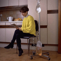
Peritoneal dialysis is a commonly used form of renal replacement therapy worldwide, although less frequently utilized in the United States (around 10% of prevalent dialysis patients). Two major variations of peritoneal dialysis are commonly used:
- Continuous ambulatory peritoneal dialysis (CAPD): The patient performs exchanges manually three to four times per day.
- Automated peritoneal dialysis (APD): An automated cycler performs multiple nighttime exchanges. At the end of this time the patient either instills fluid in the abdomen for a daytime dwell- continuous cycling peritoneal dialysis (CCPD) or leaves no dialysate in the abdomen for the daytime- nocturnal intermittent peritoneal dialysis (NIPD). NIPD is probably not suitable for patients with minimal residual kidney function (RKF).Important goals for a PD patient to achieve include: normalization of acid-base abnormalities, bone and mineral metabolism abnormalities, blood pressure, nutritional status, and functional status.Ultra-filtration failure is clinically recognized as the inability to maintain normal fluid homeostasis. While peripheral and pulmonary edema are specific findings of volume overload, more sensitive signs include hypertension and weight gain. In the evaluation of volume overload, the provider must assess contributing factors such as dietary sodium excess, non-compliance with medications or with the dialysis regimen, episodes of hyperglycemia (which decrease the osmotic stimulus for water removal), and decreased residual kidney function.
Difficulty with ultra-filtration may be associated with dialysate leaks. Leaks can occur into the pleural space (hydrothorax), abdominal wall (causing localized edema), or into a hernia. Due to sequestration of fluid and increased lymphatic absorption, ultrafiltration is decreased.
A rare condition, encapsulating peritoneal sclerosis (EPS) may present with ultrafiltration failure. The patient may also have symptoms of uremia (due to inadequate solute removal), nausea/vomiting, decreased appetite, and weight loss.
Soon after an episode of peritonitis, a decrease in drain volume can be detected in many patients. This may explain the correlation between peritonitis and a high rate of cardiovascular events.
Tests Performed:
Peritoneal equilibration test (PET)
After an overnight dwell, 2 liters of 2.5% dextrose solution is instilled and dwells for 4 hours. At time 0, 2 hrs, and 4 hours, samples of dialysate urea, glucose, sodium, and creatinine are measured along with serum values at 2 hours. One can then calculate the ratio of dialysate/plasma (D/P) creatinine and the ratio of dialysate glucose at 4 hrs to time 0 (D/Do glucose).
Patients who have rapid absorption of glucose and/or rapid removal of creatinine are classified as rapid (or high) transporters while patients who have slow equilibration of urea, creatinine, and dextrose are slow transporters. Using published nomograms, patients can be classified in one of four categories: High, High-Average, Low-Average, and Low transporters.
PET allows the provider to tailor a dialysis prescription to the patient’s peritoneal characteristics. In general, fast transporters will have better ultrafiltration with shorter exchanges and usually are treated with CCPD or NIPD. One should perform the first PET test 4 – 6 weeks after initiating dialysis as it may be inaccurate immediately after starting PD. Similarly, a PET should not be performed within one month of an episode of peritonitis. A PET only needs to be repeated for a clinical change. There is no role for routine monitoring of transport status.
Dialysate and urine collection for urea
- Based on a 24 hour collection of urine and sample of drained dialysate over 24 hours.
- Dialysis dose is typically quantified by the removal of urea (Kt/Vurea). The daily peritoneal Kt urea is calculated by multiplying D/P urea by 24 hour drain volume. To normalize for urea distribution volume (V), which is assumed to be total body water, Kt urea is divided by V which can be calculated through many methods (such as Watson method). Finally, to arrive at the weekly Kt/V (std Kt/V) the daily Kt/V is multiplied by 7. To calculate renal (residual) Kt/V, U/P urea is multiplied by 24 hour urine volume and divided by V. The renal Kt/V is multiplied by 7 as well. The renal Kt/V and peritoneal Kt/V can then be added together to give the total Kt/V result.
- In patients who rely on residual kidney function to achieve the minimum acceptable weekly Kt/V urea of 1.7, urine collection should be done every two months; dialysate collection and urine collection is otherwise typically done every four months.
- Observational studies suggested that higher Kt/V urea values are correlated with decreased mortality. However, the early studies did not separate renal urea removal from dialytic urea removal. Subsequent analysis of large cohorts, such as CANUSA, have demonstrated that the presence of residual kidney function is far more important to survival than peritoneal urea removal.
- Three randomized studies have compared different targets of solute removal in PD patients
- ADEMEX (Mexico): Achieved peritoneal Kt/V 2.2 vs 1.8. No difference in mortality or hospitalizations were seen although more patients withdrew from the low dose group due to uremia.
- Lo WK et al (Hong Kong): Patients randomized to three total (renal + peritoneal) Kt/V targets-1.5 to 1.7, 1.7 to 2.0, and > 2.0. This study demonstrated no difference in survival or hospitalizations but patients in the lowest dose group had worse anemia and higher erythropoietin requirements. Patients in that group were more likely to be removed from the study by their physician due to uremic symptoms.
- Mak et al (Hong Kong): Patients on CAPD randomized to extra exchange or not. Achieved peritoneal Kt/V was 1.56 vs 1.92. There was no difference in serum albumin but higher dose group had fewer hospitalizations.
- Most national and international guidelines recommend achieving total Kt/V urea of >=1.7
Abdominal X- ray/Peritoneography
- Plain abdominal X-rays are performed to evaluate for malposition of the peritoneal catheter.
- The normal position of catheter is in the pelvic gutter.
- In patients with normal inflow of dialysate but problems with outflow, the most common underlying cause is constipation. However, if symptoms do not improve after resumption of normal bowel movements, abdominal X-ray should be done to assess catheter position.
- For peritoneography, the initial X ray is taken, then 100-200 ml non-ionic contrast is mixed into a 2L dialysate bag and instilled in the patient. The patient changes positions to mix dialysate and a repeat X ray is taken.
- Can be used to diagnose an entrapped catheter or a peritoneal leak.
Abdominal computed tomography scan
- Can be used to evaluate the presence of dialysate leaks. Contrast injection as per peritoneography can help to delineate the leak.
- Abdominal computed tomography (CT) scan can also be used when EPS is suspected. Typical imaging findings include peritoneal calcifications, thick-walled “cocoon” encasing the intestines, and bowel dilatation.

