OSTEOPOROSIS what is it & what to do?

osteoporosis  osteoporosis skeletal areas affected

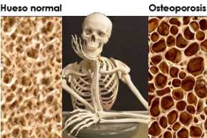
Decrease in bone mass and density and that leads to an increased risk of fracture.  In osteoporosis, the bone mineral density (BMD) is reduced, bone microarchitecture deteriorates, and the amount and variety of proteins in bone are altered.

Osteoporosis causes bones to become weak and brittle —- so brittle that a fall or even mild stresses like bending over or coughing can cause a fracture. Osteoporosis-related fractures most commonly occur in the hip, wrist or spine. Bone is living tissue, which is constantly being absorbed and replaced. Osteoporosis occurs when the creation of new bone doesn’t keep up with the removal of old bone. Osteoporosis affects men and women of all races; but white and Asian women–especially after menopause–are at highest risk. Medications, healthy diet and weight bearing exercise can help prevent bone loss or strengthen already weak bones.

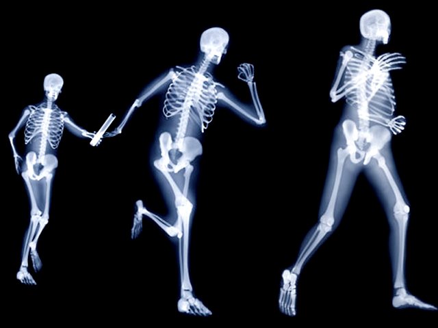
A weight bearing exercise is any exercise that has your legs and feet holding all of your weight. An example of this would be walking, yoga or even dancing.

The form of osteoporosis most common in women after menopause is referred to as primary type 1 or postmenopausal osteoporosis. Primary type 2 osteoporosis or senile osteoporosis occurs after age 75 and is seen in both females and males at a ratio of 2:1. Secondary osteoporosis may arise at any age and affect men and women equally. This form results from chronic predisposing medical problems or disease, or prolonged use of medications such as glucocorticoids, when the disease is called steroid- or glucocorticoid-induced osteoporosis.

The risk of osteoporosis fractures can be reduced with lifestyle changes and in those with previous osteoporosis related fractures medications. Lifestyle change includes diet, exercise, and preventing falls. The utility of calcium and vitamin D is questionable in most. Bisphosphonates are useful in those with previous fractures from osteoporosis but are of minimal benefit in those who have osteoporosis but no previous fractures. Osteoporosis is a component of the frailty syndrome.

Take the problem of Astronauts with osteoporosis:

Space travel has made it widely known that a stay outside the atmosphere – and thus outside the earth’s gravitational influence – disturbs the metabolism irreparably: the human body does not need any hard bones in zero-gravity, which leads to decalcification. A four year study of the ‘International Space Station’ showed that the bones of astronauts did not regenerate after even one year past their return to earth.

Anti-gravitational training is the key to osteoporosis if you can handle it (like jumping on a trampeline) Actual studies show that physical anti-gravitational activity helps the effected patients to regain their mobility and lessen the risk of bone fractures .

Osteoporosis in the U.S.A.

The National Osteoporosis Foundation (NOF) today released updated prevalence data estimating that a total of 54 million U.S. adults age 50 and older are affected by osteoporosis and low bone mass. Recently published online by the Journal of Bone and Mineral Research, the study, “The Recent Prevalence of Osteoporosis and Low Bone Mass in the United States Based on Bone Mineral Density at the Femoral Neck or Lumbar Spine,” includes the number of adults age 50 and over from the institutionalized and non-institutionalized population affected by osteoporosis and low bone mass and is an update to the prevalence data NOF released in 2013. Revealing that 10.2 million adults have osteoporosis and another 43.4 million have low bone mass, more than one-half of the total U.S. adult population is currently affected.

Assuming osteoporosis and low bone mass prevalence remain unchanged, the study projects that by 2020, the number of adults over age 50 with osteoporosis or low bone mass will grow from approximately 54 million to 64.4 million and by 2030, the number will increase to 71.2 million (a 29% increase from 2010); and it is anticipated that the number of fractures will grow proportionally.

“This disease causes an estimated two million broken bones each year and often results in immobility, pain, placement in a nursing home, isolation and other health problems, said Amy Porter, executive director and CEO of NOF. “Medicare (our tax dollars) pays for the cost for repair of 80 percent of broken bones that occur because of osteoporosis; these costs make osteoporosis the 10th ranked major illness among the top 5% highest cost Medicare beneficiaries (12% of all beneficiaries and 18% of high costs beneficiaries). We have to continue our efforts to eradicate this disease.”

The data is one of the first to look at the burden of osteoporosis using NOF’s criteria for diagnosing osteoporosis based on bone mineral density (BMD) at the hip or spine. Prior to 2005, the National Health and Nutrition Examination Survey (NHANES) only measured BMD at the hip. Spine BMD was added in 2005, providing the opportunity to estimate the burden of osteoporosis using BMD at either the hip or spine. The study estimates that among adults age 50 years and older, 10.2 million have osteoporosis at the femoral neck or lumbar spine and an additional 43.4 million have low bone mass at either skeletal site, placing them at increased risk for osteoporosis and broken bones.

Benefits of exercise

Women who have been physically active throughout their lives generally have stronger bones than do women who have led more sedentary lives. But it’s never too late to start exercising. For postmenopausal women, regular physical activity can:

  • Increase your muscle strength
  • Improve your balance
  • Make you better able to carry out daily tasks and activities
  • Maintain or improve your posture
  • Relieve or decrease pain
  • Improve your sense of well-being Before you start having osteoporosis.
  • Consult your doctor before starting any exercise program for osteoporosis. You may need some tests first, including:
  • Exercising if you have osteoporosis means finding the safest, most enjoyable activities for you given your overall health and amount of bone loss. There’s no one-size-fits-all prescription.
  • Bone density measurement
  • Fitness assessment

In the meantime, think about what kind of activities you enjoy most. If you choose an exercise you like your more likely to stick to it.


QUOTE FOR THE WEEKEND:

“WM is a cancer that starts in B cells. The cancer cells in people with WM are similar to those of 2 other types of cancer: multiple myeloma and non-Hodgkin lymphoma. Multiple myeloma is considered a cancer of plasma cells, and non-Hodgkin lymphoma is a cancer of lymphocytes. WM cells have features of both plasma cells and lymphocytes and are called lymphoplasmacytoid. The buildup of this M protein in the body can lead to many of the symptoms of WM, including excess bleeding, problems with vision, and nervous system problems.”

American Cancer Society

QUOTE FOR FRIDAY:

“Waldenstrom’s macroglobulinemia (WM) is a lymphoma, or cancer of the lymphatic system. It is sometimes referred to as a lymphoplasmacytic lymphoma (LPL) with an associated monoclonal IgM paraprotein. The disease occurs in a type of white blood cell called a B-lymphocyte or B-cell, which normally matures into a plasma cell whose job is to manufacture immunoglobulins (antibodies) to help the body fight infection.”

IWMF International Waldenstrom’s Macroglobulemia Foundation (https://www.iwmf.com/about-wm/newly-diagnosed)

QUOTE FOR THURSDAY:

“Overall, the most common risk factors for stroke are:high blood pressure, high cholesterol, smoking, diabetes, and increasing age.”

American Stroke Association

QUOTE FOR TUESDAY:

“A stroke occurs when the blood supply to part of your brain is interrupted or reduced, depriving brain tissue of oxygen and nutrients. Within minutes, brain cells begin to die.  A stroke is a medical emergency. Prompt treatment is crucial. Early action can minimize brain damage and potential complications.”

MAYO Clinic

QUOTE FOR MONDAY:

A-fib is the most common rhythm disorder of the heart. In the United States, it affects more than 2.7 million people.  In people with A-fib, the heart beats irregularly and often too quickly, getting out of sync with the two lower chambers.  This irregularity can cause clotting of the blood in the heart chambers putting the individual at risk for heart attack or stroke or pulmonary thrombus if the clot breaks loose”.

American Heart Association

Atrial Fib with Rapid Ventricular Rate!

afibafib RVR

                                  afib

Atrial fibrillation, or AFib, is the most common type of arrhythmia.

A heart arrhythmia is an abnormal rate or rhythm in your heartbeat. This can mean your heartbeat is too slow, too fast, or has an irregular rhythm.

Most arrhythmias are harmless, and may not cause symptoms. Some types, however, can have serious consequences and require treatment. Dangerous arrhythmias may cause heart failure, stroke, or low blood flow that results in organ damage. Most people with arrhythmias, even serious ones with treatment, live normal and healthy lives.

Working of the heart:

To easily identify atrial fibrillation with RVR=Rapid Ventricular Rate, it is vital to understand the working of the heart. The atrium or atria (plural) is the upper chamber of the heart, bigger in size compared to the lower chambers known as the ventricles. The atria function by gathering blood as it flows into the heart and shrinking to forward the blood into the ventricles. At the very moment, the smaller ventricle must shrink to forward the blood to all parts of the body. This rhythm of blood flow creates a heart signature voice referred to as the Sinus rhythm. It is important that the sinus rhythm is synchronized so that the atrium does not send blood into the ventricle out of cue. To achieve this, an electric signal is generated to ensure the atrium contracts. When this signal short circuits (bypasses) the atrium, atrial fibrillation with RVR occurs, and the atrium is seen to vibrate just like jelly on a flat surface.

Atrial fib with RVR refers to atrial fibrillation with rapid ventricular rate. Usually the heart is like clockwork, the top (collecting) chambers beat then the bottom (main pumping) chambers sense this and also beat, and so on, in a nice regular fashion just like a clock ticking second after second. Usually the heart beats at about 60-80 beats per minute.

In atrial fibrillation the top chamber basically goes crazy often firing off over 400 beats per minute! Atrial fibrillation with RVR (Rapid Ventricular Response) is a heart condition caused by irregular electrical activity that results in irregular contractions of the 2 top heart chambers fibrillating. This means the heart (atriums), shakes with a rapid tremulous movement or makes fine irregular twitching movements, generally referred to as fibrillating causing little control in the heart output of blood by the heart but the lower chambers called the ventricles take over.

These bottom chambers don’t allow all those impulses through but it does let every second or third one through. This can give a heart rate of 100-180 beats per minute at rest, still too many beats, known as Afib with RVR, leading to symptoms and problems with heart function. Afib does not necessarily lead to Afib with RVR however, Afib can be rate controlled, sometimes naturally, sometimes using medications and sometimes requiring procedures as discussed below.

In most people with AFib although symptoms can sometimes be unpleasant it is generally not harmful as long as the afib is controlled, meaning the heart in the afib rhythm with the pulse under 100. The main concern is stroke, but that can be treated with the use of blood thinning medications in people at risk. In Afib with RVR, basically the heart is beating too fast. Of course palpitations are the most common symptom. Other symptoms of AFib with RVR may include dizziness, lack of energy, exercise intolerance and shortness of breath. If Afib with RVR goes on for too long then this may result in heart failure and of course worsening of existing heart failure. Control of the heart rate in patients with Afib with RVR often causes these symptoms to improve, again meaning the HR is under 100 with the heart rhythm in afib.

A major indication of atrial fibrillation with RVR is a very rapid heartbeat rate, although some patients are known to have the condition without showing symptoms. Atrial fibrillation with RVR may occur when cardiac muscle cells overcome their intrinsic pacemaker’s signals and fire rapidly differently from their normal pattern spreading the abnormal activity to the ventricles. The rapid heart rate can strain the heart, developing a situation referred to as Tachycardia (meaning a pulse greater than 100). Atrial fibrillation with RVR can be detected from the various symptoms though it is important to remember that some patients have experienced the condition without symptoms.

Symptoms:Some of the symptoms of this disease include heart palpitations (described as unnoticed skipped beats or skipped beats noticed from experienced dizziness or difficulty in breathing), shortness of breath when lying flat (orthopnea), shortness of breath (dyspnea after exertion) sudden onset of short breath during the night (also called paroxysmal nocturnal dyspnea) and gradual swelling of lower extremities. As a result of inadequate blood flow, some patients complain of light headedness and may feel like they are about to faint, a condition referred to as presyncope and may actually lose consciousness (syncope). Some patients experience respiratory distress that results in them appearing blue. A close examination of jugular veins usually reveals elevated pressure in some patients (jugular venous distention). When some patients are subjected to lung examinations, crackles and rales may be observed pointing to possible lung edema.

Importance of proper diagnosis:

A good diagnosis of the symptoms shown by patients is important to ascertain that the patient is suffering from atrial fibrillation with RVR.  This is because some forms or irregular and rapid heart rates, tachyarrhythmia, are dangerous and must be ruled out as they are life threatening – such as ventricular tachycardia. Some patients are usually placed on continuous cardio respiratory monitoring, but an electrocardiogram ECG is vital for correct diagnosis.

 How is it diagnosed?

 Simple, a typical 12 lead electrocardiogram (ECG). This test shows cardiac rhythms which atrial fibrillation is. Rhythms are made up of types of waves that the ECG shows which are P waves, QRS waves, T waves and U waves.

The QRS complexes should be narrow, to signify that they are being initiated by normal conduction of atrial electrical activity through the Intra-ventricular conduction system, or heart conduction system. Wide QRS complexes could point to ventricular tachycardia, although wide complexes may also be an indication of disease processes in the Intra-ventricular conduction system. The R-R internal will also likely be irregular. Meaning measuring from each R section of the QRS rhythm. It is also important to find out if there are triggering causes for the tachycardia which include dehydration, Hypovolemia – a decrease in blood volume, and more specifically decrease in blood plasma volume. You can go ahead to eliminate Acute coronary syndrome – which refers to any diseases that are directly attributed to the obstruction of coronary arteries.

WHAT IS THE TREATMENT:

The cornerstones of atrial fibrillation (AF) management are rate control and anticoagulation  and rhythm control for those symptomatically limited by AF.  The clinical decision to use a rhythm-control or rate-control strategy requires an integrated consideration of several factors, including degree of symptoms, likelihood of successful cardioversion, presence of comorbidities, and candidacy for AF ablation (eg, catheter-based pulmonary vein electric isolation or surgical ablation.)

A Shock

This is known as cardioversion and is used typically either when an immediate result is required or used when the Afib is of relatively recent onset or only intermittent, and so has more chance of staying in normal rhythm. In cardioversion a small shock is given using defibrillation pads in sync mode. It is done under light anesthesia therefore it doesn’t hurt; pt is sedated if allowable. The Afib may or may not return however.  Highier odds the afibrillation could turn back to normal sinus rhythm if this afibrillation is newly diagnosed not chronic.

Rate Control Drugs

The biggest problem in Afib with RVR is too fast a heart rate. In a rhythm control strategy we use drugs such as beta-blockers to slow the heart rate down. These drugs typically will leave the patient in AF. For many people with AF it turns out that a rate control strategy is preferred as it is considered less risky than the rhythm control drugs used to get rid of the AF while being just as effective. In Afib with RVR rate control drugs can often slow the heart rate down fairly quickly and improve symptoms.

In the ER with newly diagnosed with pt stable the MD will order medication to decrease the rate

Rhythm Control Drugs

These medications are generally more powerful than the rate control drugs and attempt to convert the Afib back in to a normal rhythm. They are often given after a shock treatment to try and help the heart stay in normal rhythm. These drugs are also commonly used in hospitalized Afib with RVR patients. The problem with these drugs is that they may have side effects and associated risks. Many patients simply cannot tolerate Afib even if the rate is controlled and therefore require rhythm control drugs. They may be safe and effective however if used in selected patients. In cases of Afib with RVR these medications may need to be used if patients cannot tolerate other rate control medications.

Ablation Procedures

Ablation is a minimally invasive procedures typically done through the groin. They are typically used in patients that have tried, or cannot tolerate medicines for control of AFib. Ablation is typically not used as an emergency treatment of Afib with RVR, rather it is used for stable patients in AF, or those with intermittent AFib that wish to remain in normal rhythm. In patients that have had persistent Afib for a long time these procedures are not likely to be successful in the long term.

Pacemaker

This is typically the last throw of the dice for AF control. In some patients, drugs can either not control the rate in AFib with RVR, or the drugs can simply not be tolerated. In these patients who have no other choice, and in whom it is determined the Afib is causing harmful effects, a procedure called AV node ablation and pacemaker is done. In a relatively minor procedure, a small burn is made to the connection that connects the top and bottom chambers of the heart. A pacemaker is then inserted. This prevents Afib with RVR as although the top chambers continue to fire at a fast rate, the pacemaker now controls the bottom chamber, in a nice regular way. The downside of course is that now although the patient cannot have Afib with RVR, they have a pacemaker rather than the SA Node, our natural pacemaker in the heart in the R upper atrium.

Acute afib RVR patients are more likely to be converted to Normal Sinus Rhythm (the best rhythm you could be in) as opposed to patients with chronic afib. There are complete resolutions for both kind of afib but atrial fibrillation in RVR the heart can handle for only so long and remembering the engine of our body is the heart so take good care of it for if you don’t it could allow you to die.


QUOTE FOR THE WEEKEND:

“Achalasia is a rare disorder that makes it difficult for food and liquid to pass into your stomach. Achalasia occurs when nerves in the tube connecting your mouth and stomach (esophagus) become damaged.”

MAYO Clinic

Achalasia

Achalasia is a motility disorder in which the esophagus (food tube) empties slowly. The delay results from poor opening of the lower esophageal sphincter (valve) in association with the loss of the normal, orderly muscle activity (peristalsis) that propels foods and liquids along the esophagus into the stomach.

Achalasia results from the nerves in the esophagus and lower esophageal valve being damaged or destroyed. Most cases are idiopathic, meaning the cause is unknown. The origin may possibly be viral or autoimmune, and rarely it may be due to a cancer.

Men and women are equally affected. The average age of presentation is approximately 50 years, but it may also be diagnosed in children or the elderly.

Symptoms of Achalasia

The symptoms of achalasia most often occur during and after a meal. All individuals experience the sensation of solids, and usually liquids, hanging up and passing slowly into the stomach. This may occur several times a week or at every meal.

Effortless regurgitation of bland, undigested food or whitish foam (saliva) is common and may be associated with coughing and choking at night.

Some people experience chest pain, and heartburn is common. This heartburn is not related to acid reflux into the esophagus, but rather due to retained acidic food or the fermentation of food in the esophagus.

Approximately 50% of people with achalasia will lose weight.

Tests for Achalasia

The diagnosis of achalasia is suspected by barium x-rays and confirmed by esophageal manometry.

Barium studies of the esophagus (esophagram) show a distinctive narrowing of the lower esophageal valve, esophageal dilation with retained barium in the upright position, poor esophageal emptying, and to-and-fro movement due to the loss of orderly peristalsis (coordinated wave-like muscle contractions).

Esophageal manometry involves placing a small tube with pressure sensors into the stomach, and slowly withdrawing while measuring lower esophageal valve pressure and peristalsis. All achalasia patients have abnormal relaxation (opening) of the lower esophageal valve combined with a lack of peristalsis (orderly wave progression) in the esophageal body. Sometimes the lower esophageal valve pressure will also be abnormally high.

All achalasia patients should also have an upper GI endoscopy to exclude the uncommon case of a cancer (usually adenocarcinoma of the stomach) causing a pseudo-achalasia picture. [Endoscopy is a procedure that uses a thin, flexible tube with a light and a lens on the end to look into the esophagus and stomach.]

Treatment of Achalasia

The goal of treating achalasia is to disrupt and open the lower esophageal valve to improve esophageal emptying and relieve symptoms. Unfortunately, no treatment is available to promote the return of peristalsis.

The best treatments for healthy patients are pneumatic dilatation or laparoscopic Heller myotomy. Frail or elderly patients may do well with botulinum toxin (Botox) injections.

Treatment markedly relieves symptoms, but is rarely curative. Overall, the success of both the pneumatic dilation and heller myotomy procedures is 80%-90% and dependent on the skills of the operator. Retreatment may be required and alternative treatments may be needed.

  • Pneumatic dilatation – This procedure involves upper GI endoscopy with the passage of various size balloons to tear the esophagus from within, opening the valve. The procedure is done with conscious sedation and takes about 30 minutes, with a loss of one day of activity. The major complication, esophageal perforation (hole in the esophagus), is rare (less than 5%), but will require major surgery.
  • Heller myotomy – Surgery for achalasia involves cutting the muscle (myotomy) from the outside via small laparoscopic sites on the abdomen. The procedure requires general anesthesia, hospitalization for up to two days, and limited activity for two weeks. The major side effect is cutting the muscle too much, causing bad acid reflux.
  • Botulinum toxin – Botox can be injected into the esophagus and lower esophageal valve, via a needle passed through an upper endoscope. The toxin relaxes the sphincter, and thereby relieves symptoms. In young patients, symptoms relief is generally for only 3-6 months, while older patients may have relief for one year or longer. Adverse events are rare.

Medications may also be tried to relax the lower esophageal sphincter when surgery or pneumatic dilation is not an option, or Botox therapy has failed. Calcium channel blockers and long-acting nitrates are commonly used.