Archive | July 2015

QUOTE FOR THURSDAY:

“For diabetes in particular, we know there’s a relationship between lack of glucose regulation and complications like blindness and kidney failure. So if you were diabetic and you knew that you could get your glucose in a tight, normal range just by adjusting your lifestyle, wouldn’t that be great?”

Eric Topol   MD, (born 1954 who is an American cardiologist, geneticist, and digital medicine researcher.)

Acute Renal Failure versus Chronic Renal Failure

chronic-kidney-disease-image

Acute Renal Failure:

Kidney failure occurs when the kidneys lose their ability to function. To treat kidney failure effectively, it is important to know whether kidney disease has developed suddenly (acute) or over the long term (chronic). Many conditions, diseases, and medicines can create situations that lead to acute and chronic kidney disease. Acute kidney injury, also called acute renal failure, is more commonly reversible than chronic kidney failure since the chronic condition has lasted longer in the body affecting systems for several months to years (some decades). Acute Renal Failure is new to the body as opposed to chronic; making it higher odds this can be treated and cured.

When acute kidney injury occurs, the kidneys are unable to remove waste products and excess fluids, which then build up in the body and upset the body’s normal chemical balance.*

The most common causes of acute kidney injury are:

-dehydration

-blood loss from major surgery or injury

-medicines such as nonsteroidal anti-inflammatory drugs (NSAIDs), antibiotics, or the dyes (contrast agents) used in X-ray tests.

Symptoms depend on the cause of the problem of acute renal failure and can include:

  • -Little or no urine output.
  • -Dizziness upon standing.
  • -Swelling, especially of the legs and feet.
  • -Loss of appetite, nausea, and vomiting.
  • -Feeling confused, anxious and restless, or sleepy.
  • -Pain in the flank, which is felt just below the rib cage and above the waist on one or both sides of the back.*                                                                                                                                                                                                                                                                                Most cases of acute kidney injury occur in people who are already in the hospital for other reasons. In these people, acute kidney injury is usually diagnosed when routine tests show a sudden increase in creatinine and blood urea nitrogen (BUN) levels. A buildup of these waste products in the blood points to a loss of kidney function.

Further your doctor will do the following to diagnose the condition other than blood tests:

  • -Your doctor will compare these levels to previous tests to find out if kidney disease is acute or chronic.
  • -Also commonly done is an ultrasound of the kidneys which may help determine whether kidney problems are acute or chronic. Normal-sized kidneys may be present in either condition, but when both kidneys are smaller than normal, chronic kidney disease is usually the problem.  This helps rule out acute from chronic.             

The treatment of acute kidney injury includes:

correcting the cause and supporting the kidneys with dialysis until proper functioning is restored. Most people who develop acute kidney injury are already in the hospital.

Chronic Renal Failure:

In giving a short and easily understandable definition Chronic kidney disease happens when your kidneys no longer filter your blood the way they should, so wastes (toxins, usually end products of an acid) build up in your blood. This has probably been going on for years, and it may keep getting worse over time. Just like a car engine damaged but still using the car without getting the engine repaired sooner or later in time the engine no longer functions the same with any organ of the body getting damaged by some long term condition. If your disease gets worse and worse over time, you could have kidney failure for some multi organ failure, depending on the condition causing this.*

The most common causes of Chronic Renal Failure are:

-Diabetes (uncontrolled diabetes (Type 1 or 2) for many years.

*-High blood pressure for many years.

These are the top 2 causes of most chronic kidney disease. Controlling these diseases can help slow or stop the damage to the individual’s kidneys who has one of these, if not both.

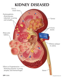
Other causes that can lead to chronic kidney disease include:   -Kidney diseases and infections, such as polycystic kidney disease, pyelonephritis, and glomerulonephritis, or a kidney problem you were born with.

-A narrowed or blocked renal artery. A renal artery carries blood to the kidneys.

-Long-term use of medicines that can damage the kidneys. Examples include nonsteroidal anti-inflammatory drugs (NSAIDs), such as celecoxib and ibuprofen.

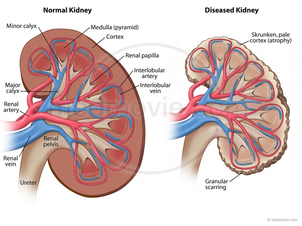
Know this for starters, each of your kidneys has about a million tiny filters, called nephrons.The nephron is the tiny filtering structure in your kidneys. Each of your kidneys contain more than a million tiny filtering nephrons that help clean your blood removing toxins dumping them into your urinary bladder so you can evacuate them though urine (urea, urine; get it). Your nephrons play a vital role to our essential daily living. They help all humans do the following if there kidneys or one kidney is functioning properly. They:

  • -Remove excess water, wastes (like urea, ammonia, etc.) & other substances from your blood.
  • -Return substances like sodium, potassium or phosphorus whenever any of these substances run low in your body.
  • If nephrons are damaged by the high sugar content or high blood pressure in the kidneys, they stop working. For a while, healthy nephrons can take on the extra work or overload. But if the damage continues, more and more nephrons shut down. After a certain point, the nephrons that are left cannot filter your blood well enough to keep you’re blood filtered properly to keep you healthy. Just like running from a bear in the street chancing you. We can run only so long but sooner or later we will run out of energy and not be able to run anymore, same concept for the kidney nephrons when they run out of enough not properly working.

How well your kidneys work is called kidney function. As your kidney function gets worse, you may show these symptoms:

-Urinate less than normal.

-Have swelling and weight gain from fluid buildup in your tissues. This is called edema.

-Feel very tired or sleepy.

-Not feel hungry, or you may lose weight without trying.

-Often feel sick to your stomach (nauseated) or vomit.

-Have trouble sleeping.

-Have headaches or trouble thinking clearly.

To diagnose chronic renal failure is the same listed above on acute renal failure plus:

One way to measure how well your kidneys are working is to figure out your glomelular filtration rate (GFR). The GFR is usually calculated using results from your blood creatinine test. Then the stage of kidney disease is figured out using the GFR. There are five stages of kidney disease, from kidney damage with normal GFR to kidney failure.

There are things you can do to slow or stop the damage to your kidneys. Taking medicines and making some lifestyle changes can help you manage your disease and feel better.

Chronic kidney disease is also called chronic renal failure or chronic renal insufficiency.

Chronic kidney disease may seem to have come on suddenly. But it has been happening bit by bit for many years as a result of damage to your kidneys.

There are five stages of kidney disease, from kidney damage with normal GFR to kidney failure.

Chronic kidney disease is also called chronic renal failure or chronic renal insufficiency.

Chronic kidney disease is caused by damage of the kidneys whether the cause of it be primary a Renal or Kidney problem or a secondary, another disease or disorder that affects the kidneys in doing their job, like hyperglycemia related to a individual with uncontrolled diabetes, for instance.

Your doctor will do blood and urine tests to help find out how well your kidneys are working. These tests can show signs of kidney disease and anemia. (You can get anemia from having damaged kidneys.) You may have other tests to help rule out other problems that could cause your symptoms.

Your doctor will do tests that measure the amount of urea (BUN) and creatinine in your blood. These tests can help measure how well your kidneys are filtering your blood. As your kidney function gets worse, the amount of nitrogen (shown by the BUN test) and creatinine in your blood increases. The level of creatinine in your blood is used to find out the glomerular filtration rate (GFR). The GFR is used to show how much kidney function you still have. The GFR is also used to find out the stage of your kidney disease your in if you have it and its to guide decisions about treatment.

Your doctor will ask questions about any past kidney problems. He or she will also ask whether you have a family history of kidney disease and what medicines you take, both prescription and over-the-counter drugs.

You may have a test that lets your doctor look at a picture of your kidneys, such as an ultrasound or CT (Cat Scan of the kidneys). These tests can help your doctor measure the size of your kidneys, estimate blood flow to the kidneys, and see if urine flow is blocked. In some cases, your doctor may take a tiny sample of kidney tissue (biopsy) to help find out what caused your kidney disease.

 Treatment for Chronic Kidney Failure:

There are things you can do to slow or stop the damage to your kidneys. Taking medicines and making some lifestyle changes can help you manage your disease, prevent further damage to the kidneys, if their functioning at all and make you possibly feel better.

Very hard, never a complete 100 % resolution. It is like emphysema done by smokers the damage is done or like a heart attack the area of the infarction is heart muscle now scared and the damage is done, so its get the organ to its optimal level of functioning.

Kidney disease is a complex problem. You will probably need to take a number of medicines and have many tests. To stay as healthy as possible, take your medicines just the way your doctor says to and work closely with your doctor. Go to all your appointments for the MD to see a increase in function or decrease in function of your kidney or kidneys you have still functioning to a level. To do that you can’t just go every 6 months especially when first diagnosed with it or with a collapse of an exacerbation of kidney failure in a worse level that brought on new symptoms that brought you to the ER. Lifestyle changes are an important part of your treatment. Taking these steps can help slow down kidney disease and reduce your symptoms. These steps may also help with high blood pressure, diabetes, and other problems that make kidney disease worse or made the kidney disease happen with the secondary diagnosis you had originally for years (ex. Hypertension or Diabetes if not both especially is uncontrolled).

  • Follow a diet that is easy on your kidneys. A dietitian can help you make an eating plan with the right amounts of salt (sodium) and protein. You may also need to watch how much fluid you drink each day.
  • Make exercise a routine part of your life. Work with your doctor to design an exercise program that is right for you.
  • Do not smoke or use tobacco.
  • Do not drink alcohol. When kidney function falls below a certain point, it is called Kidney failure. Kidney failure affects your whole body. It can cause serious heart, bone, and brain problems and make you feel very ill. Untreated kidney failure will be life-threatening at some point.
  • When you have kidney failure, you will probably have two choices: start dialysis or get a new kidney (transplant). Both of these treatments have risks and benefits. Talk with your doctor to decide which would be best for you.
  • Always talk to your doctor before you take any new medicine, including over-the-counter remedies, prescription drugs, vitamins, or herbs. Some of these can hurt your kidneys further.
  • In complete renal failure you have 2 choices for Rx.
  • 1.)**Dialysis is a process that filters your blood when your kidneys no longer can. It is not a cure, but it can help you feel better and live longer.
  • 2.)**Kidney transplant may be the best choice if you are otherwise healthy. With a new kidney, you will feel much better and will be able to live a more normal life. But you may have to wait for a kidney that is a good match for your blood and tissue type. And you will have to take medicine for the rest of your life to keep your body from rejecting the new kidney.    
  • Making treatment decisions when you are very ill is hard. It is normal to be worried and afraid. Discuss your concerns with your loved ones and your doctor. It may help to visit a dialysis center or transplant center and talk to others who have made these choices.

3260_KidneyPathologyCard-1


Kidneys anatomy and functions to understand what happens with kidney failure!

npml_0003_normal_versus_diseased-1030x772

The kidneys are important organs with many functions in the body, including producing hormones, absorbing minerals, and filtering blood and producing urine. While they are important and kidney failure can be fatal, a human only needs one healthy kidney to survive.

The kidneys are two bean-shaped organs that extract waste from blood, balance body fluids, form urine, and aid in other important functions of the body.

They reside against the back muscles in the upper abdominal cavity. They sit opposite each other on either side of the spine. The right kidney sits a little bit lower than the left to accommodate the liver.

When it comes to components of the urinary system, the kidneys are multi-functional powerhouses of activity, for if the kidneys aren’t working, meaning they don’t filter toxic wastes out of our blood stream (with other functions it does) than the waste products don’t get dumped into the urinary bladder from the renal tubes, called right and left ureters. In human anatomy, the ureters are tubes made of smooth muscle fibers that propel urine from the kidneys to the urinary bladder. If the kidneys are not working they are not filtering our blood (same principle as filtering beer to make it to perfection, the kidneys do it for our blood to be able to have the cells do their function to the optimal levels with keeping toxins out of the body in preventing many blood problems with more due to acidosis (toxin build up). In the adult, the ureters are usually 25–30 cm (10–12 in) long and ~3–4 mm in diameter.

The kidneys have multiple functions.                                                       Some of the core actions of a healthy kidney or kidneys of a human body include:

  • Waste excretion: There are many things your body doesn’t want inside of it. The kidneys filter out toxins, excess salts, and urea (a toxin), a nitrogen-based waste created by cell metabolism.                                                                                                                               * Urea is an organic chemical compound and is essentially the waste produced by the body after metabolizing protein. Naturally the compound urea is produced when the liver breaks down protein or amino acids, and ammonia, the kidneys then transfer the urea from the blood to the urine, when they do filtering of the blood.                                                                                                            * Urea is a byproduct of protein metabolism, the ending result.                                                                                                  *Extra nitrogen is expelled from the body through urea because it is extremely soluble (solid); it is a very efficient process. The average person excretes about 30 grams of urea a day, mostly through urine but a small amount is also secreted in perspiration. Synthetic versions of the chemical compound can be created in liquid or solid form and is often an ingredient found in fertilizers, animal food, and diuretics, just to name a few . Urea is what gives our urine the color yellow.                                                     *In the gastrointestinal tract, blood proteins are broken down into ammonia (could be due to high protein eating to drugs with actual conditions); and goes to the liver converting it to Urea. It is then released into the blood stream where the kidney’s take it up and eliminate it. Urea is then eliminated by the kidney’s, but not produced by it.                                                                                                                                                                                   *Urea is synthesized in the liver and transported through the blood to the kidneys for removal.
  • A Healthy Kidney or Kidneys functions in the human body:
  • Water level balancing: As the kidneys are key in the chemical breakdown of urine, they react to changes in the body’s water level throughout the day. As water intake decreases, the kidneys adjust accordingly and leave water in the body instead of helping excrete it which aides in electrolyte balancing in the blood with keeping the body hydrated properly.
  • Blood pressure regulation: The kidneys need constant pressure to filter the blood. When it drops too low, the kidneys increase the pressure. One way is by producing a blood vessel-constricting protein (angiotensin) that also signals the body to retain sodium and water. Both the constriction and retention help restore normal blood pressure.
  • Red blood cell regulation: When the kidneys don’t get enough oxygen, they send out a distress call in the form of erythropoietin, a hormone that stimulates the bone marrow to produce more oxygen-carrying red blood cells.
  • Acid regulation: As cells metabolize, they produce acids. Foods we eat can either increase the acid in our body or neutralize it. If the body is to function properly, it needs to keep a healthy balance of these chemicals. The kidneys do that, too.Because of all of the vital functions the kidneys perform and the toxins they encounter, the kidneys are susceptible to various problems.
  • Acute kidney failure is a condition in which the kidneys suddenly lose their ability to function properly. This can occur for many reasons, including:
  • Most people are born with two kidneys, but many people can live on just one. Kidney transplant surgeries with live donors are common medical procedures today.
  • Infection
  • Blood-clotting disorders
  • Decreased blood flow caused by low blood pressure
  • Autoimmune kidney disorders
  • Urinary tract infections
  • Complications from pregnancy
  • DehydrationDiseases and conditions that commonly cause chronic kidney disease include:
  • Chronic kidney failure – same as acute in that the kidney (s) loses its function.
  • Type 1 or type 2 diabetes
  • High blood pressure
  • Glomerulonephritis (gloe-mer-u-lo-nuh-FRY-tis), an inflammation of the kidney’s filtering units (glomeruli)
  • Interstitial nephritis, an inflammation of the kidney’s tubules and surrounding structures
  • Polycystic kidney disease
  • Prolonged obstruction of the urinary tract, from conditions such as enlarged prostate, kidney stones and some cancers
  • Vesicoureteral (ves-ih-koe-yoo-REE-ter-ul) reflux, a condition that causes urine to back up into your kidneys
  • Recurrent kidney infection, also called pyelonephritis (pie-uh-lo-nuh-FRY-tis)
  • A chronic condition causes the failure to happen, what we call a secondary diagnosis.  *                                                                                                                                 Learn more tomorrow, on Thursday, about Acute and Chronic kidney failure.


QUOTE FOR TUESDAY:

“I would not have traded two minutes of the joy and the grief with that man for two decades of anything with another.”

 
– Lou Gehrig’s wife, Eleanor

Lou Gehrigs Disease=Amyotrophic lateral sclerosis

ALS, or amyotrophic lateral sclerosis, is a progressive neurodegenerative disease that affects nerve cells in the brain and the spinal cord. A-myo-trophic comes from the Greek language. “A” means no. “Myo” refers to muscle, and “Trophic” means nourishment – “No muscle nourishment.” When a muscle has no nourishment, it “atrophies” or wastes away. “Lateral” identifies the areas in a person’s spinal cord where portions of the nerve cells that signal and control the muscles are located. As this area degenerates it leads to scarring or hardening (“sclerosis”) in the region.

Motor neurons reach from the brain to the spinal cord and from the spinal cord to the muscles throughout the body. The progressive degeneration of the motor neurons in ALS eventually leads to their demise. When the motor neurons die, the ability of the brain to initiate and control muscle movement is lost. With voluntary muscle action progressively affected, people may lose the ability to speak, eat, move and breathe. The motor nerves that are affected when you have ALS are the motor neurons that provide voluntary movements and muscle control. Examples of voluntary movements are making the effort to reach for a smart phone or step off a curb. These actions are controlled by the muscles in the arms and legs. Motor neurons are nerve cells located in the brain, brain stem, and spinal cord that serve as controlling units and vital communication links between the nervous system and the voluntary muscles of the body. Messages from motor neurons in the brain called upper motor neurons are transmitted to motor neurons in the spinal cord which are lower motor neurons and from them to particular muscles. The problem with ACL, both the upper motor niurons and the ower motor niurons degenerate or die which causes stoppage of sending messages to muscles. Unable to function, the muscles gradually weaken, waste away (atrophy), and have very fine twitches (called fasciculations). Eventually, the ability of the brain to start voluntary movement with messages is unable to work anymore.

Symptoms:

The onset of ALS may be so subtle that the symptoms are overlooked. The earliest symptoms may include fasciculations, cramps, tight and stiff muscles (spasticity), muscle weakness affecting an arm or a leg, slurred and nasal speech, or difficulty chewing or swallowing. These general complaints then develop into more obvious weakness or atrophy that may cause a physician to suspect ALS. Regardless of the part of the body first affected by the disease, muscle weakness and atrophy spread to other parts of the body as the disease progresses.

The parts of the body showing early symptoms of ALS depend on which muscles in the body are affected. Many individuals first see the effects of the disease in a hand or arm as they experience difficulty with simple tasks requiring manual dexterity such as buttoning a shirt, writing, or turning a key in a lock. In other cases, symptoms initially affect one of the legs, and people experience awkwardness when walking or running or they notice that they are tripping or stumbling more often.

There are two different types of ALS, sporadic and familial. Sporadic which is the most common form of the disease in the U.S., is 90 – 95 percent of all cases. It may affect anyone, anywhere. Familial ALS (FALS) accounts for 5 to 10 percent of all cases in the U.S. Familial ALS means the disease is inherited. In those families, there is a 50% chance each offspring will inherit the gene mutation and may develop the disease. French neurologist Jean-Martin Charcot discovered the disease in 1869.

The cause of ALS is not known, and scientists do not yet know why ALS strikes some people and not others. An important step toward answering this question was made in 1993 when scientists supported by the National Institute of Neurological Disorders and Stroke (NINDS) discovered that mutations in the gene that produces the SOD1 enzyme were associated with some cases of familial ALS.

Recent years have brought a wealth of new scientific understanding regarding the physiology of this disease. There is currently one FDA approved drug, riluzole, that modestly slows the progression of ALS in some people. Although there is not yet a cure or treatment that halts or reverses ALS, scientists have made significant progress in learning more about this disease. In addition, people with ALS may experience a better quality of life in living with the disease by participating in support groups and attending an ALS Association Certified Treatment Center of Excellence or a Recognized Treatment Center. Such Centers provide a national standard of best-practice multidisciplinary care to help manage the symptoms of the disease and assist people living with ALS to maintain as much independence as possible for as long as possible. According to the American Academy of Neurology’s Practice Paramater Update, studies have shown that participation in a multidisciplinary ALS clinic may prolong survival and improve quality of life.  To find a Center near you, visit http://www.alsa.org/community/certified-centers/.

ALS usually strikes people between the ages of 40 and 70, and approximately 20,000 Americans can have the disease at any given time (although this number fluctuates). For unknown reasons, military veterans are approximately twice as likely to be diagnosed with the disease than the general public.  Notable individuals who have been diagnosed with ALS include baseball great Lou Gehrig, Hall of Fame pitcher Jim “Catfish” Hunter, Toto bassist Mike Porcaro, Senator Jacob Javits, actor David Niven, “Sesame Street” creator Jon Stone, boxing champion Ezzard Charles, NBA Hall of Fame basketball player George Yardley, golf caddie Bruce Edwards, , musician Lead Belly (Huddie Ledbetter), photographer Eddie Adams, entertainer Dennis Day, jazz musician Charles Mingus, former vice president of the United States Henry A. Wallace, U.S. Army General Maxwell Taylor, and NFL football players Steve Gleason, O.J. Brigance and Tim Shaw.

Who gets ALS?

More than 12,000 people in the U.S. have a definite diagnosis of ALS, for a prevalence of 3.9 cases per 100,000 persons in the U.S. general population, according to a report on data from the National ALS Registry. ALS is one of the most common neuromuscular diseases worldwide, and people of all races and ethnic backgrounds are affected. ALS is more common among white males, non-Hispanics, and persons aged 60–69 years, but younger and older people also can develop the disease. Men are affected more often than women.

In 90 to 95 percent of all ALS cases, the disease occurs apparently at random with no clearly associated risk factors. Individuals with this sporadic form of the disease do not have a family history of ALS, and their family members are not considered to be at increased risk for developing it.

About 5 to 10 percent of all ALS cases are inherited. The familial form of ALS usually results from a pattern of inheritance that requires only one parent to carry the gene responsible for the disease. Mutations in more than a dozen genes have been found to cause familial ALS.

About one-third of all familial cases (and a small percentage of sporadic cases) result from a defect in a gene known as “chromosome 9 open reading frame 72,” or C9orf72. The function of this gene is still unknown. Another 20 percent of familial cases result from mutations in the gene that encodes the enzyme copper-zinc superoxide dismutase 1 (SOD1).

Treatment:

No cure has yet been found for ALS. However, the Food and Drug Administration (FDA) approved the first drug treatment for the disease—riluzole (Rilutek)—in 1995. Riluzole is believed to reduce damage to motor neurons by decreasing the release of glutamate. Clinical trials with ALS patients showed that riluzole prolongs survival by several months, mainly in those with difficulty swallowing. The drug also extends the time before an individual needs ventilation support. Riluzole does not reverse the damage already done to motor neurons, and persons taking the drug must be monitored for liver damage and other possible side effects. However, this first disease-specific therapy offers hope that the progression of ALS may one day be slowed by new medications or combinations of drugs.


QUOTE FOR MONDAY:

A Japanese legend says that if you can’t sleep at night its because you are awake in someone else’s dream.  (I must be in a lot of people’s dreams).

Insomnia

Insomnia is a sleeping disorder that is characterized by difficulty falling and/or staying asleep. People with Insomnia have one or more of the following symptoms:

  • Difficulty falling asleep
  • Waking up often during the night and having trouble going back to sleep
  • Waking up too early in the morning
  • Feeling tired upon waking *                                                                                                                                    There are noted to be 2 types: Primary and Secondary
  • Primary insomnia: Primary insomnia means that a person is having sleep problems that are not directly associated with any other health condition or problem.
  • Secondary insomnia: Secondary insomnia means that a person is having sleep problems because of something else, such as a health condition (like asthma, depression, arthritis, cancer or heartburn). Other conditions could be pain, medication they are taking or a substance they are using (like alcohol).                            *AcuteInsomnia                                                                                                                                                                                                                                                                Insomnia also varies in how long it lasts and how often it occurs. It can be short-term (acute insomnia) or can last a long time (chronic insomnia). It can also come and go, with periods of time when a person has no sleep problems. Acute insomnia can last from one night to a few weeks. Insomnia is called chronic when a person has insomnia at least three nights a week for a month or longer.
  • Significant life stress (job loss or change, death of a loved one, divorce, moving)
  • Illness
  • Emotional or physical discomfort
  • Environmental factors like noise, light, or extreme temperatures (hot or cold) that interfere with sleep
  • Some medications (for example those used to treat colds, allergies, depression, high blood pressure and asthma) which may interfere with sleep
  • Interferences in normal sleep schedule ( jet lag or switching from a day to night shift, for example)

ChronicInsomnia caused by:

  • Depression and/or anxiety
  • Chronic stress
  • Pain or discomfort at night

*Symptoms of insomnia can include:

  • Sleepiness during the day
  • General tiredness
  • Irritability
  • Problems with concentration or memory*                                    How Insomnia can be diagnosed: If you think you have insomnia, talk to your health care provider or a doctor who majors in Insomnia. An evaluation may include a physical, a medical history, and a sleep history. You may be asked to keep a sleep diary for a week or two, keeping track of your sleep patterns and how you feel during the day. Your health care provider may want to interview your bed partner about the quantity and quality of your sleep. In some cases, you may be referred to a sleep center for special tests.*                                                                     Treatment of InsomniaYou should seek help if your insomnia has become a pattern, or if you often feel fatigued or unrefreshed during the day and it interferes with your daily life. Many people have brief periods of difficulty sleeping (for example, a few days after starting a new job), but if insomnia lasts longer or has become a regular occurrence, you should ask for help.If you don’t feel satisfied after your conversation with your primary care physician, ask for a referral to a doctor who specializes in sleep medicine or consult other available resources. It’s important to find a doctor who has the proper knowledge and training to treat your insomnia.
  • Non-Medical (Cognitive & Behavioral) Treatments for InsomniaSome of these techniques can be self-taught, while for others it’s better to enlist the help of a therapist or sleep specialist.
  • Stimulus control helps to build an association between the bedroom and sleep by limiting the type of activities allowed in the bedroom. An example of stimulus control is going to bed only when you are sleepy, and getting out of bed if you’ve been awake for 20 minutes or more. This helps to break an unhealthy association between the bedroom and wakefulness. Sleep restriction involves a strict schedule of bedtimes and wake times and limits time in bed to only when a person is sleeping.
  • Medical Treatments for InsomniaDetermining which medication may be right for you depends on your insomnia symptoms and many different health factors. This is why it’s important to consult with a doctor before taking a sleep aid.
  • Alternative Medicine
  • Major classes of prescription insomnia medications include benzodiazepine hypnotics, non-benzodiazepine hypnotics, and melatonin receptor agonists.
  • There are many different types of sleep aids for insomnia, including over-the-counter (non-prescription) and prescription medications.
  • Cognitive behavioral therapy (CBT) includes behavioral changes (such as keeping a regular bedtime and wake up time, getting out of bed after being awake for 20 minutes or so, and eliminating afternoon naps) but it adds a cognitive or “thinking” component. CBT works to challenge unhealthy beliefs and fears around sleep and teach rational, positive thinking. There is a good amount of research supporting the use of CBT for insomnia. For example, in one study, patients with insomnia attended one CBT session via the internet per week for 6 weeks. After the treatment, these people had improved sleep quality.
  • Relaxation training, or progressive muscle relaxation, teaches the person to systematically tense and relax muscles in different areas of the body. This helps to calm the body and induce sleep. Other relaxation techniques that help many people sleep involve breathing exercises, mindfulness, meditation techniques, and guided imagery.
  • Many people listen to audio recordings to guide them in learning these techniques. They can work to help you fall asleep and also return to sleep in the middle of the night.
  • There are psychological and behavioral techniques that can be helpful for treating insomnia. Relaxation training, stimulus control, sleep restriction, and cognitive behavioral therapy are some examples.
  • Many cities also have sleep centers and clinics (sometimes connected to a hospital) that offer assessments, testing, and treatment. An Internet search will help you locate the nearest center.
  • Start by calling your primary care physician or bringing up the topic of sleep at your next well visit if you have one scheduled. If your doctor is knowledgeable about sleep disorders, he or she will guide you through the next steps, which may involve an assessment and further testing, or a referral to a sleep specialist. Your doctor may also start by giving you some basic information and resources about healthy sleep habits—these behavioral tips may help certain people with insomnia—or discussing potential medical treatment options to consider. Your doctor could refer you to a psychotherapist if your sleep struggles seem connected to anxiety, depression, or a major life adjustment.                         There are alternative medicines that may help certain people sleep. It’s important to know that these products are not required to pass through the same safety tests as medications, so their side effects and effectiveness are not as well understood.

QUOTE FOR THURSDAY:

“A Baker’s cyst, also called a popliteal (pop-luh-TEE-ul) cyst, is usually the result of a problem with your knee joint, such as arthritis or a cartilage tear.”

MAYO CLINIC

What is a Baker cyst?

bakers cyst 2

A Baker cyst is swelling caused by fluid from the knee joint protruding to the back of the knee. The back of the knee is also referred to as the popliteal area of the knee. A Baker cyst is sometimes referred to as a popliteal cyst. When an excess of knee joint fluid is compressed by the body weight between the bones of the knee joint, it can become trapped and separate from the joint to form the fluid-filled sac of a Baker cyst. The name of the cyst is in memory of the physician who originally described the condition, the British surgeon William Morrant Baker (1839-1896).

  • A Baker cyst is swelling caused by fluid from the knee joint protruding to the back of the knee.
  • Baker cysts are common and can be caused by virtually any cause of joint swelling (arthritis).
  • A Baker cyst may not cause symptoms or be associated with knee pain and/or tightness behind the knee, especially when the knee is extended or fully flexed.
  • Baker cysts can rupture and become complicated by spread of fluid down the leg between the muscles of the calf (dissection).
  • Baker cysts can be treated with medications, joint aspiration and cortisone injection, and surgical operation, usually arthroscopic surgery.SIGNS & SYMPTOMS OF BAKER CYSTS:Baker cysts can become complicated by spread of fluid down the leg between the muscles of the calf (dissection). The cyst can rupture, leaking fluid down the inner leg to sometimes cause the appearance of a painless bruise under the inner ankle. Baker cyst dissection and rupture are frequently associated with swelling of the leg and can mimic phlebitis of the leg. A ruptured Baker cyst typically causes rapid-onset swelling of the leg.How is a Baker cyst treated? Baker cysts often resolve with aspiration (removal) of excess knee fluid in conjunction with cortisone injection. Medications are sometimes given to relieve pain and inflammation.
  • DIAGNOSING BAKER CYSTS: Baker cysts can be diagnosed by the doctor’s examination and confirmed by imaging tests (either ultrasound, injection of contrast dye into the knee followed by imaging, called an arthrogram, or MRI scan) if necessary.
  • A Baker cyst may cause no symptoms or be associated with knee pain and/or tightness behind the knee, especially when the knee is extended or fully flexed. Baker cysts are usually visible as a bulge behind the knee that is particularly noticeable on standing and when compared to the opposite uninvolved knee. They are generally soft and minimally tender.
  • Baker cysts are not uncommon and can be caused by virtually any cause of joint swelling (arthritis). The excess joint fluid (synovial fluid) bulges to the back of the knee to form the Baker cyst. The most common type of arthritis associated with Baker cysts is osteoarthritis, also called degenerative arthritis. Baker cysts can occur in children with juvenile arthritis of the knee. Baker cysts also can result from cartilage tears (such as a torn meniscus), rheumatoid arthritis, and other knee problems.

When cartilage tears or other internal knee problems are associated, physical therapy or surgery can be the best treatment option. During a surgical operation, the surgeon can remove the swollen tissue (synovium) that leads to the cyst formation. This is most commonly done with arthroscopic surgery.