Archive | April 2020

QUOTE FOR THURSDAY:


“Lymphoma affects the body’s lymph system (also known as the lymphatic system). The lymph system is part of the immune system, which helps fight infections and some other diseases. It also helps fluids move through the body.”

American Cancer Society


Hodgkin’s Lymphoma versus Non-Hodgkin’s Lymphoma

A particular cell known as the Reed-Sternberg cell is found in the biopsies. This cell is not usually found in other lymphomas, therefore they are called non Hodgkins lymphoma. This may not seem a very big difference, but it is important because the treatment for Hodgkins and non Hodgkins lymphomas can be very different.

Although the diseases may sound similar, there is a lot of difference between Hodgkin and non-Hodgkin lymphoma.

Both Hodgkin and non-Hodgkin lymphoma are malignancies of a family of white blood cells known as lymphocytes, which help the body fight off infections and other diseases.

Both Hodgkin’s lymphoma and non-Hodgkin’s lymphoma are lymphomas — a type of cancer that begins in a subset of white blood cells and these are called lymphocytes. Lymphocytes are an integral part of your immune system, which protects you from germs.

The main difference between Hodgkin’s lymphoma and non-Hodgkin’s lymphoma is in the specific lymphocyte each involves but also it includes:

Hodgkin lymphoma Non-Hodgkin lymphoma
Thirty-two percent of patients diagnosed with Hodgkin lymphoma are 20-34 years old. The median age of a patient diagnosed with the disease is 39. Seventy-five percent of patients diagnosed with Hodgkin lymphoma 55 or older. The median age of a patient diagnosed with the disease is 66.
Hodgkin lymphoma is rare, accounting for about .5 percent of all new cancers diagnosed. An estimated 8,500 cases were diagnosed in 2016. Non-Hodgkin lymphoma is the seventh most diagnosed cancer, accounting for an estimated 72,500 cases in 2016.
More than 86 percent of patients diagnosed with Hodgkin lymphoma survive five years or more. About 70 percent of patients diagnosed with non-Hodgkin lymphoma survive five years or more.
There are six varieties of Hodgkin lymphoma. The most common forms are nodular sclerosis classical Hodgkin lymphoma and mixed cellularity classical Hodgkin lymphoma. They account for about 90 percent of all cases. There are more than 61 types and subtypes of non-Hodgkin lymphoma. B-cell lymphomas account for 85 percent of all cases. Diffuse large B-cell lymphoma is the most common form on non-Hodgkin lymphoma.

Sources: National Cancer Institute and Lymphoma Research Foundation 2016

Hodgkin lymphoma is marked by the presence of Reed-Sternberg cells, which are mature B cells that have become malignant, are unusually large, and carry more than one nucleus. The first sign of the disease is often the appearance of enlarged lymph nodes. Non-Hodgkin lymphoma, by contrast, can be derived from B cells or T cells and can arise in the lymph nodes as well as other organs. (B cells and T cells play different roles in the body’s immune response to disease.)

In 2017 by the Mayo Clinic it states both diseases are relatively rare, but non-Hodgkin lymphoma is more common in the United States, with more than 70,000 new cases diagnosed each year, compared to about 8,000 for Hodgkin lymphoma. The median age of patients with non-Hodgkin lymphoma is 60, but it occurs in all age groups. Hodgkin lymphoma most often occurs in people ages 15 to 24 and in people over 60. There are more than 60 distinct types of non-Hodgkin lymphoma, whereas Hodgkin lymphoma is a more homogeneous disease.

The two forms of lymphoma are marked by a painless swelling of the lymph nodes. Hodgkin lymphomas are more likely to arise in the upper portion of the body (the neck, underarms, or chest). Non-Hodgkin lymphoma can arise in lymph nodes throughout the body, but can also arise in normal organs. Patients with either type can have symptoms such as weight loss, fevers, and night sweats.

The diseases often follow different courses of progression. Hodgkin lymphoma tends to progress in an orderly fashion, moving from one group of lymph nodes to the next, and is often diagnosed before it reaches an advanced stage. Most patients with non-Hodgkin lymphoma are diagnosed at a more advanced stage.

Treatments for lymphoma vary depending on the type of disease, its aggressiveness, and location, along with the age and general health of the patient. As a general rule, however, Hodgkin lymphoma is considered one of the most treatable cancers, with more than 90 percent of patients surviving more than five years. Survival rates for patients with non-Hodgkin lymphoma tend to be lower, but for certain types of the disease, the survival rates are similar to those of patients with Hodgkin lymphoma.

A doctor can tell the difference between Hodgkin’s lymphoma and non-Hodgkin’s lymphoma by examining the cancer cells under a microscope. If in examining the cells, the doctor detects the presence of a specific type of abnormal cell called a Reed-Sternberg cell, the lymphoma is classified as Hodgkin’s. If the Reed-Sternberg cell is not present, the lymphoma is classified as non-Hodgkin’s.

Many subtypes of lymphoma exist, and your doctor will use laboratory tests to examine a sample of your lymphoma cells to determine your specific subtype. Expect to wait a few days to receive results from these specialized tests.

Your type of lymphoma helps your doctor determine your prognosis and your treatment options. The types of lymphoma have very different disease courses and treatment choices, so an accurate diagnosis is an integral part of getting the care you need.

How both Hodgkin’s and Non-Hodgkin’s Disease are diagnosed:

  • Physical exam. Your doctor checks for swollen lymph nodes, including in your neck, underarm and groin, as well as for a swollen spleen or liver.
  • Blood and urine tests. Blood and urine tests may help rule out an infection or other disease.  A sample of your blood is examined in a lab to see if anything in your blood indicates the possibility of cancer (in both of these diseases particularly the WBCs).
  • Imaging tests. Your doctor may recommend imaging tests to look for tumors in your body. Tests may include X-ray, CT, MRI and positron emission tomography (PET).
  • Lymph node test. Your doctor may recommend a lymph node biopsy procedure to remove all or part of a lymph node for laboratory testing. Analyzing lymph node tissue in a lab may reveal whether you have non-Hodgkin’s lymphoma and, if so, which type.  Your doctor may recommend a lymph node biopsy procedure to remove a lymph node for laboratory testing. He or she will diagnose classical Hodgkin’s lymphoma if abnormal cells called Reed-Sternberg cells are found within the lymph node.
  • Bone marrow test. A bone marrow biopsy and aspiration procedure involves inserting a needle into your hipbone to remove a sample of bone marrow. The sample is analyzed to look for non-Hodgkin’s lymphoma cells to look for non-Hodgkin’s lymphoma cells or to look for non-Hodgkin’s lymphoma cells.  This test is done on numerous types of cancer patients in helping to diagnose the cancer they have.

QUOTE FOR WEDNESDAY:

“An estimated 12 million people ages of 5 and 22 years suffer a sport-related injury annually, which leads to 20 million lost days of school2 and approximately $33 billion in healthcare costs.3 Yet, some of these injuries could be prevented with protective gear. “A properly fitted mouth guard is an essential piece of any athlete’s protective equipment,  says Dr. Paul Nativi, DMD, FASD, and past president of the Academy for Sports Dentistry. Talk with your dentist about what kinds of activities your family enjoys and ask about ways to make sure their teeth and face stay protected.”

The Academy of Sports Dentistry (Established 1983)


National Facial Protection Month

April is National Facial Protection Month, founded by the AAOMS to promote the use of protective gear — such as mouth guards and helmets — to prevent dental facial injuries resulting from sports and recreational activities.

Five of the nation’s top dental associations want to remind young athletes to play it safe by wearing a mouth guard during recreational and organized sports this spring.  Research estimates that about 2 percent of all children or adolescents who participate in sports eventually will suffer a facial injury severe enough to require medical attention.

“A properly fitted mouth guard is an essential piece of any athlete’s protective equipment,” says Dr. Paul Nativi, DMD, FASD, and past president of the Academy for Sports Dentistry.  “Mouth guards protect the teeth from being knocked out, broken and displaced.  Mouth guards prevent injuries to the bone and tissues around the teeth. They also help prevent injuries to the mandible (lower jaw) and temporomandibular joint in the jaw. Tooth loss incurs a tremendous financial, emotional, and psychological expense.  Protect what you have – wear a properly fitted mouth guard.”

The Academy for Sports Dentistry (ASD), American Academy of Pediatric Dentistry (AAPD), American Association of Oral and Maxillofacial Surgeons (AAOMS), American Association of Orthodontists (AAO), and the American Dental Association (ADA) are collaborating to promote National Facial Protection Month in April.  National Facial Protection Month strives to raise public awareness and remind parents/caregivers, coaches and athletes to play it safe while playing sports.

According to the American Academy of Pediatric Dentistry’s Policy on Prevention of Sports-related Orofacial  Injuries, sports accidents reportedly account for 10 to 39% of all dental injuries in children and are most often caused by direct hits with a hard object, such as a puck or ball, and player-to-player contact.

The dental associations offer the following five tips to help prevent facial injury:

  1. Wear a mouth guard when playing contact sports: Mouth guards are significantly less expensive than the cost to repair an injury, and dentists and dental specialists can make customized mouth guards that hold teeth in place and allow for normal speech and breathing.
  2. Wear a helmet: Helmets absorb the energy of an impact and help prevent damage to the head.
  3. Wear protective eyewear: Eyes are extremely vulnerable to damage, especially when playing sports.
  4. Wear a face shield to avoid scratched or bruised skin: Hockey pucks, basketballs and racquetballs can cause severe facial damage at any age.
  5. Make protective gear mandatory for all sports: Athletes who participate in football, hockey and boxing are required to wear mouth guards.  If  mouth guards have been proven to significantly decrease the risk of oral injuries, why is it not mandatory in every sport for kids to be required to wear them, particularly when participating in:


QUOTE FOR TUESDAY:

“Sarcoidosis is a rare disease. The Foundation for Sarcoidosis Research estimates that there are fewer than 200,000 cases per year in the United States. African-Americans are three times more likely to be diagnosed with sarcoidosis than Caucasians and tend to have more severe disease. This disease causes clusters of immune cells called granulomas to infiltrate your organs and lymph nodes. Sarcoidosis most commonly affects your lungs, but it can affect any organ in your body.”
 
American Lung Association

Part III Sarcoidosis Awareness Month-Diagnosis, Staging, & Rx.

2020

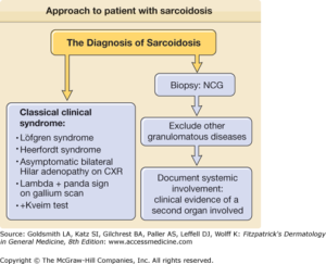
How Sarcoidosis can be diagnosed:

  • biopsy of the lungs, liver, skin, or other affected organs to check for granulomas
  • Blood tests, including complete blood counts, to check hormone levels and to test for other conditions that may cause sarcoidosis
  • Bronchoscopy, which may include rinsing an area of the lung to get cells or using a needle to take cells from the lymph nodes in the chest
  • Chest X-ray to look for granulomas in the lungs and heart and determine the stage of the disease. Often, sarcoidosis is found because a chest X-ray is performed for another reason.
  • Neurological tests, such as electromyography, evoked potentials, spinal taps, or nerve conduction tests, to detect problems with the nervous system caused by sarcoidosis
  • Eye exam to look for eye damage, which can occur without symptoms in a person with sarcoidosis
  • Gallium scan external link , which uses a radioactive material called gallium to look for inflammation, usually in the eyes or lymph nodes.
  • High-resolution computed tomography (CT) scan to look for granulomas
  • Magnetic resonance imaging (MRI) to help find granulomas. Learn more in our Chest MRI Health Topic.
  • Positron electron tomography (PET) scan, a type of imaging that can help find granulomas
  • Pulmonary function tests to check whether you have breathing problems
  • Ultrasound to look for granulomas

There is a staging to Sarcoidosis:

Doctors use stages to describe the various imaging findings of sarcoidosis of the lung or lymph nodes of the chest. There are four stages of sarcoidosis, and they indicate where the granulomas are located. In each of the first three stages, sarcoidosis can range from mild to severe. This helps the doctor in deciding treatment for the patient.  Stage IV is the most severe and indicates permanent scarring in the lungs.

  • Stage I: Granulomas are located only in the lymph nodes.
  • Stage II: Granulomas are located in the lungs and lymph nodes.
  • Stage III: Granulomas are located in the lungs only.
  • Stage IV: Pulmonary fibrosis.

Treatment for Sarcoidosis:

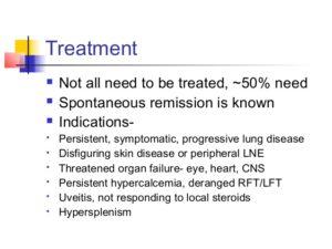
Remember there is no cure.  The goal of treatment is remission, a state in which the condition is not causing problems. Not everyone who is diagnosed with sarcoidosis needs treatment. Sometimes the condition goes away on its own. Whether you need treatment—and what type you need—will depend on your signs and symptoms, which organs are affected, and whether those organs are working well; this is also where staging of the disease comes into play. Some people do not respond to treatment.


QUOTE FOR MONDAY:

“Sarcoidosis can occur in any organ, but it most commonly affects the lungs, lymph nodes and heart. Signs and symptoms of sarcoidosis vary depending on which organs are affected. Doctors don’t know the exact cause of sarcoidosis.  While anyone can develop sarcoidosis, factors that may increase your risk include: age, sex, race and family history. Sometimes sarcoidosis causes long-term problems.  Complications depend on the organ effected.”

MAYO CLINIC

QUOTE FOR THE WEEKEND:

“In 2008, Congress declared April as National Sarcoidosis Awareness Month to bring more attention to this rare, multisystem disease. Five years later, we are slowly but surely making progress, bothS in public awareness and in research of sarcoidosis. Purple is the color of the ribbon for sarcoidosis awareness.”

The Bernie Mac Foundation

Part I Sarcoidosis Awareness Month

Sarcoidosis (pronounced SAR-COY-DOE-SIS) is an inflammatory disease characterized by the formation of granulomas, tiny clumps of inflammatory cells, in one or more organs of the body. When the immune system goes into overdrive and too many of these clumps form, they can interfere with an organ’s structure and function. When left unchecked, chronic inflammation can lead to fibrosis, which is permanent thickening or scarring of organ tissue.

This disorder can affect almost any organ in the body, including the heart, skin, liver, kidneys, brain, sinuses, eyes, muscles, bones, and other areas. Sarcoidosis most commonly targets the lungs and the lymph nodes, which are an important part of the immune system. When it affects the lungs, it is called pulmonary sarcoidosis. Ninety percent or more of people diagnosed with the disease have lung involvement.

Doctors believe sarcoidosis results from the body’s immune system responding to an unknown substance, most likely something inhaled from the air.

There is no cure for sarcoidosis, but most people do very well with little or only modest treatment. In half of cases, sarcoidosis goes away on its own. In a few cases, however, sarcoidosis may last for years and may cause organ damage.

General symptoms

For many people, sarcoidosis begins with these symptoms:

  • Fatigue
  • Fever
  • Swollen lymph nodes
  • Weight loss

Lung symptoms

Many patients with sarcoidosis experience lung problems, which may include:

  • Persistent dry cough
  • Shortness of breath
  • Wheezing
  • Chest pain

Skin symptoms

Some people who have sarcoidosis develop skin problems, which may include:

  • A rash of red or reddish-purple bumps, usually located on the shins or ankles, which may be warm and tender to the touch
  • Disfiguring sores (lesions) on the nose, cheeks and ears
  • Areas of skin that are darker or lighter in color
  • Growths under the skin (nodules), particularly around scars or tattoos

Eye symptoms

Sarcoidosis can affect the eyes without causing any symptoms, so it’s important to have your eyes checked. When eye symptoms do occur, they may include:

  • Blurred vision
  • Eye pain
  • Severe redness
  • Sensitivity to light

Heart symptoms

Cardiac sarcoidosis: A heart under attack

Signs and symptoms related to cardiac sarcoidosis may include:

  • Chest pain
  • Shortness of breath (dyspnea)
  • Fainting (syncope)
  • Fatigue
  • Irregular heartbeats (arrhythmias)
  • Rapid or fluttering heart beat (palpitations)
  • Swelling caused by excess fluid (edema)

Stay tune for Part II tomorrow on Sardcoidosis Awareness Month!

QUOTE FOR FRIDAY:

Dogs relieve stress. According to a study conducted in the 1980s by Aaron Katcher of the University of Pennsylvania and Alan Beck of Purdue, playing with a dog or even just petting a familiar dog lowers blood pressure and heart rate, slows breathing, and relaxes muscle tension almost immediately. Dogs are good for your heart. Literally. The “American Journal of Cardiology” published and researched. Dogs make us happy. In fact, even just looking at a dog can make you happier. A study in Japan in 2009 found that just staring into your dog’s eyes raises your level of oxytocin, also known as the “love hormone. Dogs make us want to take care of them. There must be a reason why so many people become virtually addicted to dog videos, and why puppies are so irresistible. ”  

American Kennel Club AKC.org