
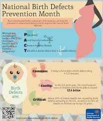
Health care providers are encouraged to provide women to plan for pregnancy; avoid harmful substances, like tobacco (2) and alcohol (3); and choose a healthy lifestyle, like eating a healthy diet (4), to increase their chances of a healthy pregnancy. Health care providers also discuss with women any medications they might be taking, both prescription and over-the-counter, to ensure they are taking only what is necessary. If yours is not maybe you need a new one. Prevention is the key to giving highier odds the baby will be healthier when born. Re-enforcement is a great tool and that’s where the medical profession comes into play with pregnant women who is their clientele.
Know that not all birth defects can be prevented. But, we also know that women can increase their chances of having a healthy baby by managing health conditions and adopting healthy behaviors before becoming pregnant. Make a commitment to yourself, to get healthy before and during pregnancy by actively trying to plan ahead, avoid harmful substances, choose a healthy lifestyle, and talk with your healthcare provider. There are some that can be prevented.
1.Plan ahead.
Get 400 micrograms (mcg) of folic acid every day.
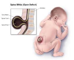
Folic acid is a B vitamin. If a woman has enough folic acid in her body at least one month before and during pregnancy, it can help prevent major birth defects of the developing brain and spine defects like anencephaly or spina bifida. Anencephaly is a serious birth defect in which a baby is born without parts of the brain and skull. It is a type of neural tube defect (NTD). As the neural tube forms and closes, it helps form the baby’s brain and skull (upper part of the neural tube), spinal cord, and back bones (lower part of the neural tube). Spina bifida is a condition that affects the spine and is usually apparent at birth. It is a type of neural tube defect (NTD). Spina bifida can happen anywhere along the spine if the neural tube does not close all the way. The backbone that protects the spinal cord does not form and close as it should. This often results in damage to the spinal cord and nerves. Spina bifida might cause physical and intellectual disabilities that range from mild to severe. The severity depends on:
- The size and location of the opening in the spine.
- Whether part of the spinal cord and nerves are affected.
- There are 3 types and they are: A-Myelomeningocele, B-Meningocele, and C-Spina Bifida Occulta.
- A-Myelomeningocele is the most serious type of spina bifida. With this condition, a sac of fluid comes through an opening in the baby’s back. Part of the spinal cord and nerves are in this sac and are damaged. This type of spina bifida causes moderate to severe disabilities, such as problems affecting how the person goes to the bathroom, loss of feeling in the person’s legs or feet, and not being able to move the legs.
- B-Meninocele is a sac of fluid comes through an opening in the baby’s back. But, the spinal cord is not in this sac. There is usually little or no nerve damage. This type of spina bifida can cause minor disabilities.
- C-Spina bifida occulta is the mildest type of spina bifida. It is sometimes called “hidden” spina bifida. With it, there is a small gap in the spine, but no opening or sac on the back. The spinal cord and the nerves usually are normal. Many times, spina bifida occulta is not discovered until late childhood or adulthood. This type of spina bifida usually does not cause any disabilities.
- Women can get folic acid from fortified foods or supplements, or a combination of the two, in addition to a varied diet rich in folate.
- See a healthcare professional regularly.
A woman should be sure to see her doctor when planning a pregnancy and start prenatal care as soon as she thinks that she is pregnant. It is important to see the doctor regularly throughout pregnancy, so a woman should keep all her prenatal care appointments. If you are trying to have a baby or are just thinking about it, it is not too early to start getting ready for pregnancy. Use these checklists to help you write down your goals and have them in a place that you reinforce yourself to maintain them as best as possible for your child’s sake and your own sake as well.
2.Avoid harmful substances.
- Avoid alcohol at any time during pregnancy. Alcohol in a woman’s bloodstream passes to the developing baby through the umbilical cord. There is no known safe amount of alcohol use during pregnancy or while trying to get pregnant. There is also no safe time during pregnancy to drink. All types of alcohol are equally harmful, including wine and beer. Drinking alcohol during pregnancy can cause miscarriage, stillbirth, and a range of lifelong physical, behavioral, and These disabilities in the child, which occur because the mother drank alcohol during the pregnancy, are known as fetal alcohol spectrum disorders (FASDs). The best advice for women is to stop drinking alcohol when trying to get pregnant.
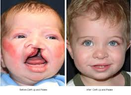
- Avoid smoking cigarettes. The dangers of smoking during pregnancy include preterm birth, certain birth defects from cleft lip or cleft palate to even infant death and more diseases inherited by mom through smoking. Even being around tobacco smoke puts a woman and her pregnancy at risk for problems. Quitting smoking before getting pregnant is best. For a woman who is already pregnant, quitting as early as possible can still help protect against some health problems for the baby, such as low birth weight. It’s never too late to quit smoking.
- Avoid marijuana and other “street drugs”. A woman who uses marijuana or other “street” drugs during pregnancy can have a baby who is born preterm, of low birth weight, or has other health problems, such as birth defects. Marijuana is the illicit drug most commonly used during pregnancy. Since we know of no safe level of marijuana use during pregnancy, women who are pregnant, or considering becoming pregnant, should not use marijuana, even in states where marijuana is legal. Women using marijuana for medical reasons should speak with their doctor about an alternative therapy with pregnancy-specific safety data.
- Prevent infections. Some infections that a woman can get during pregnancy can be harmful to the developing baby and can even cause birth defects. Some easy steps to prevent infections include frequent hand-washing, cooking meat until its well done, and staying away from people who have an infection.
3.Choose a healthy lifestyle.
- Keep diabetes under control.
Poor control of diabetes during pregnancy increases the chances for birth defects and other problems for the pregnancy. It can also cause serious complications for the woman. Proper healthcare before and during pregnancy can help prevent birth defects and other poor outcomes.
- Strive to reach and maintain a healthy weight.
Do you know …Your body mass index (BMI)? Calculate it. Where? Just look it up on the internet anywhere for free.
A woman who is obese (a Body Mass Index of 30 or higher) before pregnancy is at a higher risk for complications during pregnancy. Obesity also increases a pregnant woman’s risk of several serious birth defects. Even if a woman is not actively planning a pregnancy, getting healthy can help boost her health and her mood. If a woman is overweight or obese, she should talk with her doctor about ways to reach a healthy weight before she gets pregnant.
4.Talk with your healthcare provider.
- Talk to a healthcare provider about taking any medications.
We know that certain medications can cause serious birth defects if they are taken during pregnancy. For many medications taken by pregnant women, the safety has been difficult to determine. Despite the limited safety data, some medications are needed to treat serious conditions. If a woman is pregnant or planning a pregnancy, she should not stop taking medications she needs or begin taking new medications without first talking with her healthcare provider. This includes prescription and over-the-counter medications and dietary or herbal products.
- Talk to a healthcare provider about vaccinations (shots).
Most vaccinations are safe during pregnancy and some vaccinations, such as the flu vaccine and the Tdap vaccine (adult tetanus, diphtheria and acellular pertussis vaccine), are specifically recommended during pregnancy. Some vaccines protect women against infections that can cause birth defects. Having the right vaccinations at the right time can help keep a woman and her baby healthy. She should talk to her doctor about which vaccines are recommended for her during pregnancy.


