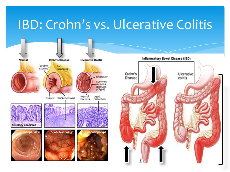
IBD refers to both Crohn’s disease and ulcerative colitis, however they can be distinguished from one another by their symptoms, GI involvement, biopsy, and antibody testing.
Your doctor will likely diagnose ulcerative colitis after ruling out other possible causes for your signs and symptoms. To help confirm the diagnosis the MD may have one or more of the following tests and procedures.
Diagnostic Tests for Ulcerative Colitis:
To help confirm a diagnosis of ulcerative colitis, you may have one or more of the following tests and procedures:
Lab tests
- Blood tests. Your provider may suggest blood tests to check for anemia — a condition in which there aren’t enough red blood cells to carry adequate oxygen to your tissues — or to check for signs of infection or inflammation.
- Stool studies. White blood cells or certain proteins in your stool can indicate ulcerative colitis. A stool sample also can help rule out other disorders, such as infections caused by bacteria, viruses and parasites.
Endoscopic procedures
- Colonoscopy. This exam allows your provider to view your entire colon using a thin, flexible, lighted tube with a camera on the end. During the procedure, tissue samples are taken for laboratory analysis. This is known as a tissue biopsy. A tissue sample is necessary to make the diagnosis.
- Flexible sigmoidoscopy. Your provider uses a slender, flexible, lighted tube to examine the rectum and sigmoid colon — the lower end of your colon. If your colon is severely inflamed, this test may be preferred instead of a full colonoscopy.
Imaging procedures
- X-ray. If you have severe symptoms, your provider may use a standard X-ray of your abdominal area to rule out serious complications, such as a megacolon or a perforated colon.
- CT scan. A CT scan of your abdomen or pelvis may be performed if a complication from ulcerative colitis is suspected. A CT scan may also reveal how much of the colon is inflamed.
- Computerized tomography (CT) enterography and magnetic resonance (MR) enterography. These types of noninvasive tests may be recommended to exclude any inflammation in the small intestine. These tests are more sensitive for finding inflammation in the bowel than are conventional imaging tests. MR enterography is a radiation-free alternative.
Diagnostic tests for Chron’s Disease:
Blood tests
-
Lab tests
- Blood tests. Your provider may suggest blood tests to check for anemia — a condition in which there aren’t enough red blood cells to carry adequate oxygen to your tissues — or to check for signs of infection or inflammation.
- Tests for anemia or infection. Your doctor may suggest blood tests to check for anemia — a condition in which there aren’t enough red blood cells to carry adequate oxygen to your tissues — or to check for signs of infection. Expert guidelines do not currently recommend antibody or genetic testing for Crohn’s disease.
- Fecal occult blood test. You may need to provide a stool sample so that your doctor can test for hidden (occult) blood in your stool. Red blood cells would be determined.
- Also further Stool studies. White blood cells or certain proteins in your stool can indicate ulcerative colitis. A stool sample also can help rule out other disorders, such as infections caused by bacteria, viruses and parasites.
Diagnostic Procedures
- Colonoscopy. This test allows your doctor to view your entire colon and the very end of your ileum (terminal ileum) using a thin, flexible, lighted tube with an attached camera. During the procedure, your doctor can also take small samples of tissue (biopsy) for laboratory analysis, which may help confirm a diagnosis. Clusters of inflammatory cells called granulomas, if present, help confirm the diagnosis of Crohn’s.
- Computerized tomography (CT). You may have a CT scan — a special X-ray technique that provides more detail than a standard X-ray does. This test looks at the entire bowel as well as at tissues outside the bowel. CT enterography is a special CT scan that provides better images of the small bowel. This test has replaced barium X-rays in many medical centers.
- Magnetic resonance imaging (MRI). An MRI scanner uses a magnetic field and radio waves to create detailed images of organs and tissues. MRI is particularly useful for evaluating a fistula around the anal area (pelvic MRI) or the small intestine (MR enterography).
- Capsule endoscopy. For this test, you swallow a capsule that has a camera in it. The camera takes pictures of your small intestine, which are transmitted to a recorder you wear on your belt. The images are then downloaded to a computer, displayed on a monitor and checked for signs of Crohn’s disease. The camera exits your body painlessly in your stool. You may still need endoscopy with biopsy to confirm the diagnosis of Crohn’s disease.
- Balloon-assisted enteroscopy. For this test, a scope is used in conjunction with a device called an overtube. This enables the doctor to look further into the small bowel where standard endoscopes don’t reach. This technique is useful when capsule endoscopy shows abnormalities, but the diagnosis is still in question.