Part III Skin Cancer Awareness – Akinetic Keratosis & Keratoacanthoma covering risk factors, warning signs, and treatments.

Continuation of Types of skin cancer:


5-Akinetic Keratosis:

Actinic keratosis (AK) is a skin disorder that causes rough, scaly patches of skin. Another name for AK is solar keratosis. AK is a type of precancer, which means that if you don’t treat the condition, it could turn into cancer. Without treatment, AK can lead to a type of skin cancer called squamous cell carcinoma.

A condition which causes scaly patches on the skin from exposure to the sun over the years. It is commonly found on face, lips, ears, neck, back of the hand and forearms.  Very common (More than 3 million cases per year in US)
Rarely requires lab test or imaging.  Treatable by a medical professional.  Can last several months or years.
Knowing the causes, risk factors and warning signs can help you detect them early, when they are easiest to treat and cure.

The risk factors of Akinetic Keratosis are:

UV exposure from the sun or indoor tanning.

-History of skin cancer in particular history of actinic keratosis.

– Age over 40.

-Fair skin: People with fair skin including lighter color hair or eyes have an increased risk.

Warning Signs can help with early detection and treatment this can be successfully removed without complications. Look out for any new, changing or unusual skin growths, so you can spot skin cancers like BCC when they are easiest to treat and cure.

Treatments Akinetic Keratosis:

An actinic keratosis sometimes disappears on its own but might return after more sun exposure. It’s hard to tell which actinic keratoses will develop into skin cancer, so they’re usually removed as a precaution.

Medicines-

If you have several actinic keratoses, your health care provider might prescribe a medicated cream or gel to remove them, such as fluorouracil (Carac, Efudex others), imiquimod (Aldara, Zyclara) or diclofenac. These products might cause inflamed skin, scaling or a burning sensation for a few weeks.

Surgical and other procedures-

Many methods are used to remove actinic keratosis, including:

  • Freezing (cryotherapy). Actinic keratoses can be removed by freezing them with liquid nitrogen. Your health care provider applies the substance to the affected skin, which causes blistering or peeling. As your skin heals, the damaged cells slough off, allowing new skin to appear. Cryotherapy is the most common treatment. It takes only a few minutes and can be done in your health care provider’s office. Side effects may include blisters, scarring, changes to skin texture, infection and changes in skin color of the affected area.
  • Scraping (curettage). In this procedure, your health care provider uses a device called a curet to scrape off damaged cells. Scraping may be followed by electrosurgery, in which a pencil-shaped instrument is used to cut and destroy the affected tissue with an electric current. This procedure requires local anesthesia. Side effects may include infection, scarring and changes in skin color of the affected area.
  • Laser therapy. This technique is increasingly used to treat actinic keratosis. Your health care provider uses an ablative laser device to destroy the patch, allowing new skin to appear. Side effects may include scarring and discoloration of the affected skin.
  • Photodynamic therapy. Your health care provider might apply a light-sensitive chemical solution to the affected skin and then expose it to a special light that will destroy the actinic keratosis. Side effects may include inflamed skin, swelling and a burning sensation during therapy.


6-Keratocanthoma (KA)

The term “Keratoacanthoma” (KA) was coined by Freudenthal in the year 1936. It was first described way back in 1889 by Hutchinson and was called molluscum sebaceum and self-limiting epithelioma. KA is benign, self-limiting squamo-proliferative lesion.

It shows male preponderance and most commonly arises on the sun-exposed parts predominantly face, neck forearms, hands and legs. Cutaneous lesions arise from hair follicles whereas mucosal lesions originate from ectopic sebaceous glands. This is a slow growing cancer of the skin that looks like a dome or crater.  This is common; more than 200,000 cases per year in US.  Regarding treatment from medical professional is advised.  This condition often requires lab test or imaging.  Keratoacanthoma last several months.  It is common for ages 60 and older and is more common in males.

KA is benign despite its similarities to squamous cell carcinoma (SCC), or the abnormal growth of cancerous cells on the skin’s most outer layer.

The risk factors of Keratocanthoma (KA):

UV exposure from the sun or indoor tanning.

-contact with chemical carcinogens, or cancer-causing chemicals

 -trauma 

-Infection with some strains of a wart virus, such as papillomavirus

-History of skin cancer in particular history of Keratoacanthoma.

Age over 60.

-People with fair skin.

Warning Signs can help with early detection and treatment, this can be successfully removed without complications if caught early. Look out for any new, changing or unusual skin growths, so you can spot skin cancers like BCC when they are easiest to treat and cure.

Treatments:

If your medical professional suspects a keratoacanthoma, they will first want to establish the correct diagnosis by performing a biopsy.  Than treatments could include the following:

  • Removal (excision), in which a scalpel is used to cut away the keratoacanthoma and then place stitches to bring the wound edges together.
  • Mohs micrographic surgery, in which tiny slivers of skin are removed until there are no more cancer cells. This technique is particularly useful for keratoacanthomas located on the nose, ears, lips, and hands.
  • Electrodesiccation and curettage, also known as “scrape and burn.” After numbing the lesion, the medical professional uses a sharp instrument called a curette to scrape away the skin cancer cells, followed by an electric needle to burn (cauterize) the tissue. The electrodesiccation helps kill the cancer cells and stop bleeding at the site.
  • Radiation treatment, where x-ray therapy is often useful for patients who may have difficulty with a surgical procedure due to other health issues.

Very rarely, keratoacanthomas are treated with medicine injected directly into the skin lesion (intralesional chemotherapy). In patients with more than one keratoacanthoma, the medical professional may suggest taking oral medication (ie, isotretinoin) to reduce their size and number.

Once the skin cancer has been removed, frequent follow-up appointments with a dermatologist or medical professional trained to examine the skin are essential to ensure that the keratoacanthoma has not returned and that no new skin cancer has developed elsewhere on your body. In addition, good sun protection habits (as noted in the Self-Care section) are vital to preventing further damage from UV light.


QUOTE FOR MONDAY:

“Squamous cells are flat cells of epithelial origin forming the outermost and uppermost layer of skin and even epidermis. These are keratin containing cells which perform the function of protecting the body against traumas and abrasions by maintaining its continuity.

The factors responsible for both cancers are nearly the same. Prolonged exposure to the sunlight, in particular, the ultraviolet rays is the risk and etiological factor for the skin cancers.”

ePainAssist (Merkel Cell Carcinoma Vs. Squamous Cell Carcinoma)

Part II Skin Cancer Awareness – Squamous Cell Carcinoma & Merkel Cell Carcinoma including risk factors and treatment.

Continuation of Types of Skin Cancer:


3-Squamous cell carcinoma – SCC

Squamous cell carcinoma is the second most common form of skin cancer in the United States. It accounts for about 15 percent of all skin cancers.  It is caused due to over production of skin cells. Squamous cell carcinoma of the skin is caused by DNA damage that leads to abnormal changes (mutations) in the squamous cells in the outermost layer of skin. This cancer is common (More than 200,000 cases per year in US).  The majority of squamous cell skin cancers are easily and successfully treated with current therapies.

Knowing the causes, risk factors and warning signs can help you detect them early, when they are easiest to treat and cure.

The risk factors of SCC:

UV exposure from the sun or indoor tanning.

-History of skin cancer, including squamous cell carcinoma (SCC) or melanoma

– Age over 50: Most BCCs appear in people over age 50.

-Fair skin: People with fair skin have an increased risk.

Warning Signs can help with early detection and treatment this can be successfully removed without complications. Look out for any new, changing or unusual skin growths, so you can spot skin cancers like BCC when they are easiest to treat and cure.

IT’S A FACT, Squamous cell carcinoma is considered more aggressive than basal cell carcinoma.  If squamous cell carcinoma does spread to internal organs it can be life threatening. The quicker the treatment when SCC is in the one layer of skin only the better the results.  The primary symptom to look out for with SCC is a growing bump or lesion on the skin which has a rough scaly surface or flat red patches.

Treatments for SCC:

If the skin cancer is small, not deep into the skin, called superficial, and has a low risk of spreading, less-invasive treatment choices include:

  • Curettage and electrodessication. This treatment involves removing the top of the skin cancer with a scraping tool called a curet. Then an electric needle is used to sear the base of the cancer.
  • Laser therapy. This treatment uses an intense beam of light to destroy growths. There’s usually little damage to nearby tissue. And there’s a reduced risk of bleeding, swelling and scarring.
  • Freezing. This treatment, called cryosurgery, involves freezing cancer cells with liquid nitrogen. Freezing might be done after using a scraping tool, called a curet, to remove the surface of the skin cancer.
  • Photodynamic therapy. During photodynamic therapy, a liquid medicine that makes the cancer cells sensitive to light is applied to the skin. Later, a light that destroys the skin cancer cells is shined on the area. This treatment might be used with surgery or other treatments.

Treatments for larger skin cancers:

More-invasive treatments might be recommended for larger squamous cell carcinomas and those that go deeper into the skin. Options might include:

  • Simple excision. This involves cutting out the cancer and a margin of healthy skin around it. Sometimes more skin around the tumor is removed, called a wide excision.
  • Mohs surgery. Mohs surgery involves removing the cancer layer by layer and looking at each layer under the microscope until no cancer cells are left. This allows the surgeon to remove the whole growth without taking too much of the healthy skin around it.
  • Radiation therapy. Radiation therapy uses powerful energy beams to kill cancer cells. Radiation therapy is sometimes used after surgery when there is an increased risk that the cancer might return. It also might be an option for people who can’t have or don’t want surgery.

Treatments for skin cancer that spreads past the skin:

When squamous cell carcinoma spreads to other parts of the body, medicines might be recommended, including:

  • Chemotherapy. Chemotherapy uses strong medicines to kill cancer cells. If squamous cell carcinoma spreads to the lymph nodes or other parts of the body, chemotherapy can be used alone or with other treatments, such as targeted therapy and radiation therapy.
  • Targeted therapy. Targeted therapy uses medicines that attack specific chemicals in the cancer cells. By blocking these chemicals, targeted treatments can cause cancer cells to die. Targeted therapy is usually used with chemotherapy.
  • Immunotherapy. Immunotherapy is a treatment with medicine that helps the body’s immune system kill cancer cells. The immune system fights off diseases by attacking germs and other cells that shouldn’t be in the body. Cancer cells survive by hiding from the immune system. Immunotherapy helps the immune system cells find and kill the cancer cells.For squamous cell carcinoma of the skin, immunotherapy might be considered when the cancer is advanced and other treatments aren’t an option.


4-Merkel Cell Carcinoma

This is a type of skin cancer characterized by flesh-colored nodule that occurs on the face, head or neck. It begins in the cells at the base of the uppermost layer of the skin (epidermis).  A normal Merkel cell is a cross between a nerve cell and an endocrine (or hormone-producing) cell located on or just below the skin in the underlying tissue, and functions predominantly as a touch receptor. Merkel cell carcinoma occurs when these cells begin to grow uncontrollably.

Merkel cell tumors typically arise on, but are not limited to, sun-exposed parts of the body such as the face and neck. Their shape and color are less distinctive than other skin cancers, and they can often appear as an innocent pink pearly nodule. As a result, it is usually only the speed with which they grow that attracts the attention of patients and their doctors.

With early detection and treatment, Merkel cell carcinoma can be well contained and even cured. Treatment becomes more difficult as the tumor grows and spreads, but aggressive therapy can still lead to high rates of survival.

Again, Warning Signs can help with early detection and treatment this can be successfully removed without complications. Look out for any new, changing or unusual skin growths, get yourself to the doctor immediately so you can spot skin cancers like BCC when they are easiest to treat and cure.

Risk Factors of Merkel Cell Carcinoma:

UV exposure from the sun or indoor tanning.

-History of skin cancer, including squamous cell carcinoma (SCC) or melanoma

– Age over 50: Most BCCs appear in people over age 50.

-Fair skin: People with fair skin have an increased risk.

-Male gender: Men are more likely to develop Basal Cell Carcinoma.

-Chronic infections and skin inflammation from burns, scars and other conditions-weakens the immune system.

-Merkel Cell Virus.  Recently, researchers have linked a virus to many cases of Merkel cell carcinoma. However, it remains to be determined if the Merkel cell polyomarvirus causes the disease, and if it might help guide future treatment. If so, the virus could offer promising new targets for immunotherapy.

IT’S A FACT, Memorial Sloan Kettering Hospital in NYC states  “Merkel cell carcinoma, also called neuroendocrine cancer of the skin, is an aggressive type of skin cancer that affects only about 400 people in the United States each year. But like other skin cancers, that number is growing.”.

Treatments:

Treatment of stage I and stage II Merkel cell carcinoma:

  • Surgery to remove the tumor, such as wide local excision with or without lymph node dissection.
  • Radiation therapy after surgery.

Treatment of stage III:

  • Wide local excision with or without lymph node dissection.
  • Radiation therapy.
  • Immunotherapy (immune checkpoint inhibitor therapy using pembrolizumab), for tumors that cannot be removed by surgery.
  • A clinical trial of chemotherapy.
  • A clinical trial of immunotherapy (nivolumab).

Treatment of Stage IV Merkel Cell Carcinoma

  • Immunotherapy (immune checkpoint inhibitor therapy using avelumab or pembrolizumab).
  • Chemotherapy, surgery or radiation therapy as palliative treatment to relieve symptoms and improve quality of life.
  • A clinical trial of immunotherapy (nivolumab and ipilimumab).


QUOTE FOR THE WEEKEND:

“With over 5 million cases diagnosed in the United States each year, skin cancer is America’s most common cancer. Fortunately, skin cancer is also one of the most preventable cancers. By sharing facts about the dangers of unprotected sun exposure and encouraging people to check their skin for warning signs, we can and will save lives.”

Skin Cancer Foundation (Skin Cancer Awareness Month)

Part I Skin Cancer Awareness-An Overview of the A&P of skin including Basal Cell Carcinoma and Melanoma and the risk factors & treatments of both.

  BASAL CELL CARCINONA

The skin is the largest organ of your body. It acts as a barrier between invaders (pathogens) and your body. Skin forms a waterproof mechanical barrier. Microorganisms that live all over your skin can’t get through your skin unless it’s broken.  The skin and mucous membranes act as a physical barrier preventing penetration by microbes. If the skin is cut then the blood produces a clot which seals the wound and prevents microbes from entering.


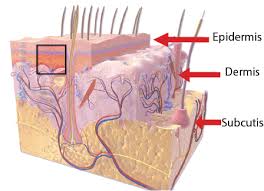
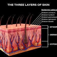
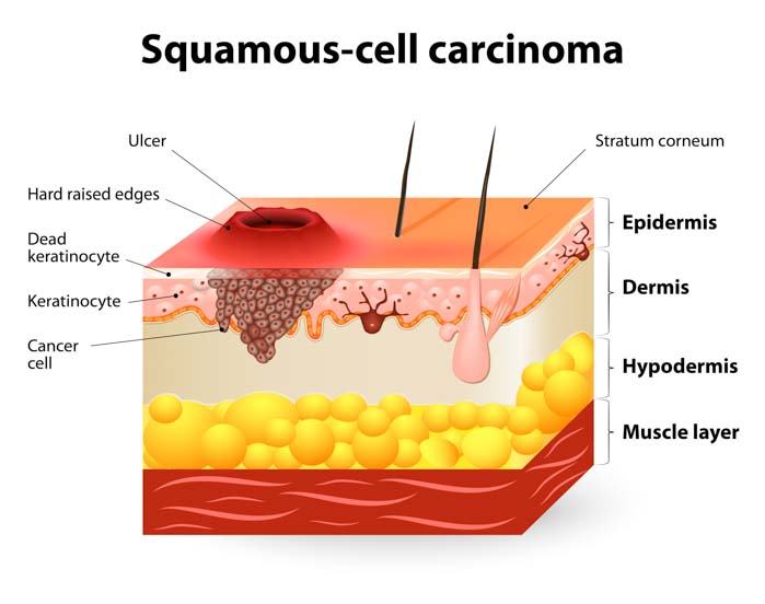
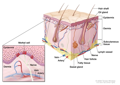
There are layers of skin and the first five layers form the epidermis, which is the outermost, thick layer of the skin and is listed above in the pictures.  Notice in the second picture on the Rt. shows all blood vessels below epidermis.

All seven layers vary significantly in their anatomy and function.

It is made up of three main layers, the epidermis, dermis, and the hypodermis, all three varying significantly in their anatomy and function. The skin’s structure is made up of an intricate network which serves as many functions for the body’s initial barrier against pathogens, UV light, and chemicals, and mechanical injury. It also maintains body temperature and prevents water loss from the body.

Of all the organs in the human body, few take the pounding your skin does. Yes, your skin is an organ, your body’s largest, in fact, and among your most important, considering you cannot live without it.

Your skin is a biological marvel capable of performing remarkable functions every day. It protects your muscles and organs from outside threats. It endures bumps and bruises, cuts and scratches, the sun’s burning rays and the grime left by dirt and dust. It moves and stretches when you do and mostly bounces back to form when you’re still.

Even when your body is at rest, your skin is a bustle of cellular activity. Basal cells change shape as they move to the surface to replace dying squamous cells. Merkel cells help your nerves sense the touch of another. Melanocytes produce melanin, the skin-darkening pigment that protects your skin from the sun.

And like other organs, your skin may develop cancer.

Skin cancer:

Skin cancer is the abnormal growth of skin cells — most often develops on skin exposed to the sun. But this common form of cancer can also occur on areas of your skin not ordinarily exposed to sunlight.

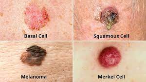
There are three major types of skin cancer — basal cell carcinoma, squamous cell carcinoma and melanoma.

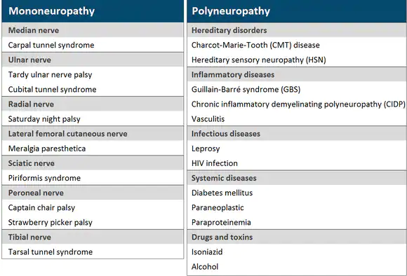
You can reduce your risk of skin cancer by limiting or avoiding exposure to ultraviolet (UV) radiation. Checking your skin for suspicious changes can help detect skin cancer at its earliest stages. Early detection of skin cancer gives you the greatest chance for successful skin cancer treatment.

The first 2 types of skin cancer, which are:

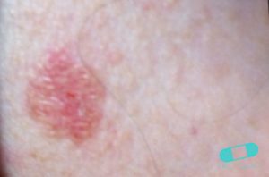
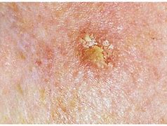
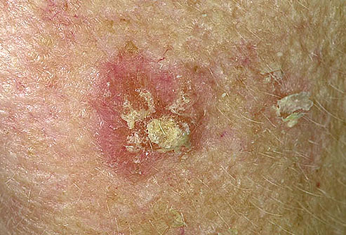
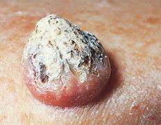
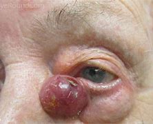
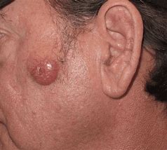
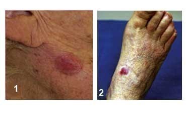
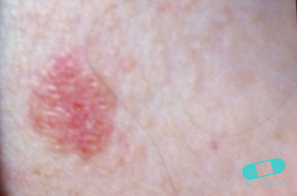
1.Basal Cell Carcinoma  

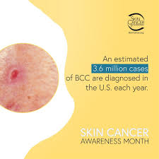
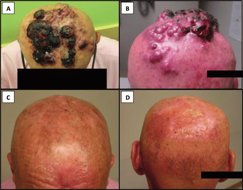
Basal cell carcinoma (BCC) is the most common form of skin cancer and the most frequently occurring form of all cancers. In the U.S. alone, an estimated 3.6 million cases are diagnosed each year. BCCs arise from abnormal, uncontrolled growth of basal cells.  Basal Cell Carcinoma grows slowly, most are curable and cause minimal damage when caught and treated early.

Knowing the causes, risk factors and warning signs can help you detect them early, when they are easiest to treat and cure.

The risk factors of BCC are:

UV exposure from the sun or indoor tanning.

-History of skin cancer, including squamous cell carcinoma (SCC) or melanoma

– Age over 50: Most BCCs appear in people over age 50.

-Fair skin: People with fair skin have an increased risk.

-Male gender: Men are more likely to develop BCC.

-Chronic infections and skin inflammation from burns, scars and other conditions.

Warning Signs can help with early detection and treatment, almost all basal cell carcinomas (BCCs) can be successfully removed without complications. Look out for any new, changing or unusual skin growths, so you can spot skin cancers like BCC when they are easiest to treat and cure.

IT’S A FACT 90% of nonmelanoma skin cancers (mainly BCCs and SCCs) are associated with exposure to UV radiation from the sun.

Treatments for BCC:

When detected early, most basal cell carcinomas (BCCs) can be treated and cured. Prompt treatment is vital, because as the tumor grows, it becomes more dangerous and potentially disfiguring, requiring more extensive treatment. Certain rare, aggressive forms can be fatal if not treated promptly.

If you’ve been diagnosed with a small or early BCC, a number of effective treatments can usually be performed on an outpatient basis, using a local anesthetic with minimal pain. Afterwards, most wounds can heal naturally, leaving minimal scarring.

Options include:

  • Curettage and electrodesiccation (electrosurgery)
  • Mohs surgery
  • Excisional surgery
  • Radiation therapy
  • Photodynamic therapy
  • Cryosurgery
  • Laser surgery
  • Topical medications
  • Medications for advanced BCC

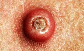
2. Melanoma – worst cancer the deepest in skin  

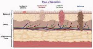
Melanoma is a type of cancer that usually begins in the skin. Specifically, it begins in cells called melanocytes. These are cells that produce melanin. Melanin is the pigment that gives skin, hair, and eyes their color.

Melanoma is among the most serious forms of skin cancer. 

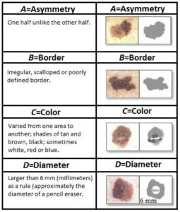
Melanoma is the deadliest type of skin cancer. It can be “in situ” which means that the cancer is confined to the top layer of skin, thus being highly curable. It can also be “malignant” which means that the cancer can spread to other parts of the body which significantly decreases the survivability rate. Melanoma in situ can grow to be malignant melanoma if not treated. The key to surviving melanoma is early detection, and especially before it becomes malignant. Melanoma caught in the early stages of its development is highly curable with a 97% survival rate.

Risk Factors of Melanoma are:

-Ultraviolet light exposure

-Moles

-Fair skin, freckling, light hair

-Family history of melanoma

-Personal history of melanoma or skin cancers

-Having a weakened immune response

-Being older

-Being male

-Xeroderma pigmentosum (XP): This is a rare, inherited condition that affects skin cells’ ability to repair damage to their DNA. People with XP have a high risk of developing melanoma and other skin cancers when they are young, especially on sun-exposed areas of their skin.

Again warning signs can count help with early detection and treatment this can be successfully removed without complications. Look out for any new, changing or unusual skin growths, so you can spot skin cancers like BCC when they are easiest to treat and cure.

IT’S A FACT Only 20-30% of melanomas are found in existing moles.  While 70-80% arise on normal-looking skin.

Treatments vary depending on the stage its in:

Stage I melanoma:

Stage I melanomas have grown into deeper layers of the skin, but they haven’t grown beyond the area where they started.

These cancers are typically treated by wide excision (surgery to remove the tumor as well as a margin of normal skin around it). The width of the margin depends on the thickness and location of the melanoma. Most often, no other treatment is needed.

Some doctors may recommend a sentinel lymph node biopsy (SLNB) to look for cancer in nearby lymph nodes, especially if the melanoma is stage IB or has other traits that make it more likely to have spread. You and your doctor should discuss this option.

If the SLNB does not find cancer cells in the lymph nodes, then no further treatment is needed, although close follow-up is still important.

If cancer cells are found on the SLNB (which changes the cancer stage to stage III – see below), a lymph node dissection (removal of all lymph nodes near the cancer) might be recommended. Another option might be to watch the lymph nodes closely by getting an imaging test such as ultrasound of the nodes every few months.

If the SLNB found cancer, adjuvant (additional) treatment with immune checkpoint inhibitors or targeted therapy drugs (if the melanoma has a BRAF gene mutation) might be recommended to try to lower the chance the melanoma will come back. Other drugs or perhaps vaccines might also be options as part of a clinical trial.

Stage II melanoma:

This stage II skin melanoma have grown deeper into the skin than stage I melanomas, but they still haven’t grown beyond the area in the skin where they started.

Wide excision (surgery to remove the melanoma and a margin of normal skin around it) is the standard treatment for these cancers. The width of the margin depends on the thickness and location of the melanoma.

Because the melanoma may have spread to nearby lymph nodes, many doctors recommend a sentinel lymph node biopsy (SLNB) as well. This is an option that you and your doctor should discuss.

If a SLNB is done and does not find cancer cells in the lymph nodes, then sometimes no further treatment is needed, but close follow-up is still important.

For certain stage II melanomas, the immune checkpoint inhibitor pembrolizumab (Keytruda) might be given after surgery to help reduce the risk of the cancer returning. Radiation therapy to the area might be another option, especially if the melanoma has features that make it more likely to come back.

If the SLNB finds that the sentinel node contains cancer cells (which changes the cancer stage to stage III – see below), then a lymph node dissection (where all the lymph nodes in that area are surgically removed) might be recommended. Another option might be to watch the lymph nodes closely with an imaging test such as ultrasound of the nodes every few months.

Whether or not the lymph nodes are removed, adjuvant (additional) treatment with immune checkpoint inhibitors or targeted therapy drugs (if the melanoma has a BRAF gene mutation) might be recommended to try to lower the chance the melanoma will come back. Other drugs or perhaps vaccines might also be options as well as part of a clinical trial.

Your doctor will discuss the best options with you depending on the details of your situation.

Stage III Melanoma:

These cancers have spread to nearby areas in the skin or lymph vessels, or they have reached the nearby lymph nodes.

Surgical treatment for stage III melanoma usually requires wide excision of the primary tumor as in earlier stages, along with a lymph node dissection (where all the nearby lymph nodes are surgically removed).

After surgery, (additional) adjuvant treatment with immune checkpoint inhibitors or with targeted therapy drugs (for cancers with BRAF gene changes) may help lower the risk of the melanoma coming back. Other drugs or perhaps vaccines may also be recommended as part of a clinical trial to try to reduce the chance the melanoma will come back. Another option is to give radiation therapy to the areas where the lymph nodes were removed, especially if many of the nodes contain cancer.

If melanoma tumors are found in nearby lymph vessels in or just under the skin (known as in-transit tumors), they are removed, if possible. Other options might include injections of the T-VEC vaccine (Imlygic), interleukin-2 (IL-2), or Bacille Calmette-Guerin (BCG) vaccine directly into the melanoma; radiation therapy; or applying imiquimod cream. For melanomas on an arm or leg, another option might be isolated limb perfusion or isolated limb infusion (infusing just the limb with chemotherapy). Other possible treatments might include targeted therapy drugs (for melanomas with a BRAF or C-KIT gene change), immunotherapy, or chemotherapy.

Some stage III melanomas might be hard to cure with current treatments, so taking part in a clinical trial of newer treatments might be a good option.

Treating stage IV melanoma:

Stage IV melanomas have already spread (metastasized) to other parts of the body, such as distant lymph nodes, areas of skin, or other organs.

Skin tumors or enlarged lymph nodes causing symptoms can often be removed by surgery or treated with radiation therapy.

If there are only a few metastases, surgery to remove them might sometimes be an option, depending on where they are and how likely they are to cause symptoms. Metastases that can’t be removed may be treated with radiation or with injections of the T-VEC vaccine (Imlygic) directly into the tumors. In either case, this is often followed by adjuvant treatment with medicines such as immunotherapy or targeted therapy drugs.

The treatment of widespread melanomas has changed in recent years as newer forms of immunotherapy and targeted drugs have been shown to be more effective than chemotherapy.

Immunotherapy drugs called checkpoint inhibitors are often the first treatment. These drugs can shrink tumors for long periods of time in some people. Options might include:

  • Pembrolizumab (Keytruda) or nivolumab (Opdivo) alone
  • Nivolumab combined with relatlimab (Opdualag)
  • Nivolumab or pembrolizumab, plus ipilimumab (Yervoy)

Combinations of checkpoint inhibitors seem to be more effective, although they’re also more likely to result in serious side effects, especially if they contain ipilimumab.

People who get any of these drugs need to be watched closely for serious side effects.

In about half of all melanomas, the cancer cells have BRAF gene changes. These melanomas often respond to treatment with targeted therapy drugs – typically a combination of a BRAF inhibitor and a MEK inhibitor. However, the immune checkpoint inhibitors mentioned above are often tried first, as this seems to be more likely to help for longer periods of time. Another option might be a combination of targeted drugs plus the immune checkpoint inhibitor atezolizumab (Tecentriq).

While immunotherapy is often used before targeted therapy, there might be situations where it makes sense to use targeted therapy first. For example, the targeted drugs are more likely to shrink tumors quickly, so they might be preferred in cases where this is important. In either case, if one type of treatment isn’t working, the other can be tried.

A small portion of melanomas have changes in the C-KIT gene. These melanomas might be helped by targeted drugs such as imatinib (Gleevec) and nilotinib (Tasigna), although these drugs often stop working eventually.

Rarely, melanomas might have changes in other genes such as NRAS, ROS1, ALK, or the NTRK genes, which can be treated with targeted drugs.

Immunotherapy using other medicines might be an option if immune checkpoint inhibitors or other treatments aren’t working. Options might include:

  • Interleukin-2 (IL-2) (also known as aldesleukin)
  • Lifileucel (Amtagvi), a type of tumor-infiltrating lymphocyte (TIL) therapy

These treatments can cause serious side effects in some people, so they are usually given in the hospital.

Chemotherapy (chemo) can help some people with stage IV melanoma, but other treatments are usually tried first. Dacarbazine (DTIC) and temozolomide (Temodar) are the chemo drugs used most often, either by themselves or combined with other drugs. Even when chemo shrinks these cancers, the cancer usually starts growing again over time.

It’s important to carefully consider the possible benefits and side effects of any recommended treatment before starting it.

Because stage IV melanoma is often hard to cure with current treatments, people may want to think about taking part in a clinical trial. Many studies are now looking at new targeted drugs, immunotherapies, and combinations of different types of treatments. (See What’s New in Melanoma Skin Cancer Research?)


QUOTE FOR FRIDAY:

Statistics on neuropathy in the United States12345:

  • The prevalence of neuropathic pain is 14.6%.
  • Among those with neuropathic pain, 19.7% had diabetic neuropathy, 27.3% had back and neck pain with neuropathic involvement, and 25.1% had hereditary or idiopathic neuropathy.
  • The prevalence of all types of neuropathy among adults in the United States ranges from 3% to 12%.
  • Peripheral neuropathy affects approximately 7% to 10% of adults in the United States.
  • Approximately 20 million Americans may have some form of peripheral neuropathy.”

National Library of Medicine (The prevalence of probable neuropathic pain in the US: results from a multimodal general-population health survey – PMC)

Part II Peripheral Neuropathy Month-diagnosis of it, treatment for it, prevention of it and management of it!


Peripheral Neuropathy Diagnosis:

The symptoms and body parts affected by peripheral neuropathy are so varied that it may be hard to make a diagnosis. If your healthcare provider suspects nerve damage, he or she will take an extensive medical history and do a number of neurological tests to determine the location and extent of your nerve damage. These may include:

  • Blood tests
  • Spinal fluid tests
  • Muscle strength tests
  • Tests of the ability to detect vibrations

Depending on what basic tests reveal, your healthcare provider may want to do more in-depth scanning and other tests to get a better look at your nerve damage. Tests may include:

  • CT scan
  • MRI scan
  • Electromyography (EMG) and nerve conduction studies
  • Nerve and skin biopsy

Peripheral Neuropathy Treatment:

Usually a peripheral neuropathy can’t be cured, but you can do a lot of things to prevent it from getting worse. If an underlying condition like diabetes is at fault, your healthcare provider will treat that first and then treat the pain and other symptoms of neuropathy.

In some cases, over-the-counter pain relievers can help. Other times, prescription medicines are needed. Some of these medicines include mexiletine, a medicine developed to correct irregular heart rhythms; antiseizure drugs, such as gabapentin, phenytoin, and carbamazepine; and some classes of antidepressants, including tricyclics such as amitriptyline.

Lidocaine injections and patches may help with pain in other instances. And in extreme cases, surgery can be used to destroy nerves or repair injuries that are causing neuropathic pain and symptoms.

Peripheral Neuropathy Prevention:

Lifestyle choices can play a role in preventing peripheral neuropathy. You can lessen your risk for many of these conditions by avoiding alcohol, correcting vitamin deficiencies, eating a healthy diet, losing weight, avoiding toxins, and exercising regularly. If you have kidney disease, diabetes, or other chronic health condition, it is important to work with your healthcare provider to control your condition, which may prevent or delay the onset of peripheral neuropathy

Peripheral Neuropathy Management:

Even if you already have some form of peripheral neuropathy, healthy lifestyle steps can help you feel your best and reduce the pain and symptoms related to the disorder. You’ll also want to quit smoking, not let injuries go untreated, and be meticulous about caring for your feet and treating wounds to avoid complications, such as the loss of a limb.

In some cases, non-prescription hand and foot braces can help you make up for muscle weakness. Orthotics can help you walk better. Relaxation techniques, such as yoga, may help ease emotional as well as physical symptoms.

QUOTE FOR THURSDAY:

“Peripheral neuropathy happens when the nerves that are located outside of the brain and spinal cord (peripheral nerves) are damaged. This condition often causes weakness, numbness and pain, usually in the hands and feet. It also can affect other areas and body functions including digestion and urination.

The peripheral nervous system sends information from the brain and spinal cord, also called the central nervous system, to the rest of the body through motor nerves. The peripheral nerves also send sensory information to the central nervous system through sensory nerves.”

MAYO CLINIC (https://www.mayoclinic.org/diseases-conditions/peripheral-neuropathy/symptoms-causes/syc-20352061)

Part I Peripheral Neuropathy Month! Learn what it is, the causes, the type of peripheral neuropathy and the symptoms!

Peripheral neuropathy is a type of damage to the nervous system. Specifically, it is a problem with your peripheral nervous system. This is the network of nerves that sends information from your brain and spinal cord (central nervous system) to the rest of your body.

Peripheral means away from the core (heart meaning from the core or the brain or spinal cord that is from the area that sends messages causing symptoms away from those organs) and the furthest away from the core or the brain or SC is your feet to understand what peripheral neuropathy effects.

There are more than 100 types of peripheral neuropathy, each with its own set of symptoms and prognosis. Peripheral neuropathy has many different causes.

Most common causes of peripheral neuropathy in the U.S. is diabetes.

Symptoms can range from tingling or numbness in a certain body part to more serious effects, such as burning pain or paralysis.

Peripheral neuropathy has many different causes. Some people inherit the disorder from their parents. Others develop it because of an injury or another disorder.

In many cases, a different type of problem, such as a kidney condition or a hormone imbalance, leads to peripheral neuropathy. One of the most common causes of peripheral neuropathy in the U.S. is diabetes.

CAUSES OF PERIPHERAL NEUROPATHY:

#1 DIABETES

Trauma: Injuries from falls, car accidents, fractures or sports activities can result in neuropathy. Compression of the nerves due to repetitive stress or narrowing of the space through which nerves run are other causes.

Autoimmune disorders and infections: Guillain-Barré syndrome, lupus, rheumatoid arthritis, Sjogren’s syndrome and chronic inflammatory demyelinating polyneuropathy are autoimmune disorders that can cause neuropathy. Infections including chickenpox, shingles, human immunodeficiency virus (HIV), herpes, syphilis, Lyme disease, leprosy, West Nile virus, Epstein-Barr virus and hepatitis C can also cause neuropathy.

Other health conditions: Neuropathy can result from kidney disorders, liver disorders, hypothyroidism, tumors (cancer-causing or benign) that press on nerves or invade their space, myeloma, lymphoma and monoclonal gammopathy.

Medications and poisons: Some antibiotics, some anti-seizures medications and some HIV medications among others can cause neuropathy. Some treatments, including cancer chemotherapy and radiation, can damage peripheral nerves. Exposure to toxic substances such as heavy metals (including lead and mercury) and industrial chemicals, especially solvents, can also affect nerve function.

Vascular disorders: Neuropathy can occur when blood flow to the arms and legs is decreased or slowed by inflammation, blood clots, or other blood vessel disorders. Decreased blood flow deprives the nerve cells of oxygen, causing nerve damage or nerve cell death. Vascular problems can be caused by vasculitis, smoking and diabetes.

Abnormal vitamin levels and alcoholism: Proper levels of vitamins E, B1, B6, B12, and niacin are important for healthy nerve function. Chronic alcoholism, which typically results in lack of a well-rounded diet, robs the body of thiamine and other essential nutrients needed for nerve function. Alcohol may also be directly toxic to peripheral nerves.

Inherited disorders: Charcot-Marie-Tooth (CMT) disease is the most common hereditary neuropathy. CMT causes weakness in the foot and lower leg muscles and can also affect the muscles in the hands. Familial amyloidosis, Fabry disease and metachromatic leukodystrophy are other examples of inherited disorders that can cause neuropathy.

No known cause: Some cases of neuropathy have no known cause.

There are types of Peripheral Neuropathy:

There are more than 100 types of peripheral neuropathy, each with its own set of symptoms and prognosis. To help doctors classify them, they are often broken down into the following categories:

  • Motor neuropathy. This is damage to the nerves that control muscles and movement in the body, such as moving your hands and arms or talking.
  • Sensory neuropathy. Sensory nerves control what you feel, such as pain, temperature or a light touch. Sensory neuropathy affects these groups of nerves.
  • Autonomic nerve neuropathy. Autonomic nerves control functions that you are not conscious of, such as breathing and heartbeat. Damage to these nerves can be serious.
  • Combination neuropathies. You may have a mix of 2 or 3 of these other types of neuropathies, such as a sensory-motor neuropathy.

Signs and Symptoms remember vary from patient to patient:

The symptoms of peripheral neuropathy vary based on the type that you have and what part of the body is affected. Symptoms can range from tingling or numbness in a certain body part to more serious effects such as burning pain or paralysis.

  • Muscle weakness, Cramps, Muscle twitching to Loss of muscle and bone
  • Changes in skin, hair, or nails
  • Numbness, Loss of sensation or feeling in body parts
  • Loss of balance or other functions as a side effect of the loss of feeling in the legs, arms, or other body parts
  • Loss of pain or sensation that can put you at risk, such as not feeling an impending heart attack or limb pain
  • Emotional disturbances, Sleep disruptions
  • Inability to sweat properly, leading to heat intolerance
  • Loss of bladder control, leading to infection or incontinence
  • Dizziness, lightheadedness, or fainting because of a loss of control over blood pressure
  • Diarrhea, constipation, or incontinence related to nerve damage in the intestines or digestive tract
  • Trouble eating or swallowing
  • Life-threatening symptoms, such as difficulty breathing or irregular heartbeat

The symptoms of peripheral neuropathy may look like other conditions or medical problems. Always see your healthcare provider for a diagnosis.

The most common type of peripheral neuropathy is diabetic neuropathy, caused by a high sugar level and resulting in nerve fiber damage in your legs and feet.


QUOTE FOR WEDNESDAY:

“For people who work the night shift, getting enough high quality sleep can be a significant challenge.

Night shifts often interfere with a person’s circadian rhythms, the 24-hour cycle that tells the body when it’s time to sleep or to be awake.  Trusted SourceUpToDateMore than 2 million healthcare providers around the world choose UpToDate to help make appropriate care decisions and drive better health outcomes. UpToDate delivers evidence-based clinical decision support that is clear, actionable, and rich with real-world insights.View Source Circadian rhythms synchronize with exposure to light, with darkness serving as a sleep cue and daylight signaling that it’s time to wake up.

For this reason, people who work night shifts often feel sleepy when they need to work and alert when they want to sleep. These challenges can have serious health and safety consequences, so it’s important for people who work night shifts to carefully plan their sleep schedules and adopt strategies to get the sleep they need.”

Sleep Foundation (The Best Sleep Schedule for Night Shift Workers | Sleep Foundation)