Symptoms
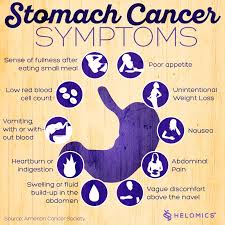
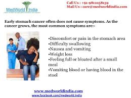
Early on, stomach cancer may cause:
- Indigestion
- Feeling bloated after you eat a meal
- Heartburn
- Slight nausea
- Loss of appetite As stomach tumors grow, you may have more serious symptoms, such as:
- Stomach pain
- Blood in your stool
- Vomiting
- Weight loss for no reason
- Trouble swallowing
- Yellowish eyes or skin
- Swelling in your stomach
- Constipation or diarrhea
- Weakness or feeling tired
- Heartburn
Just having indigestion or heartburn after a meal doesn’t mean you have cancer. But if you feel these symptoms a lot, talk to your doctor. He can see if you have other risk factors and test you to look for any problems.
Stomach cancers are usually found when a person goes to the doctor because of signs or symptoms they are having. The doctor will take a history and examine the patient. If stomach cancer is suspected, tests will be needed to confirm the diagnosis.
Medical history and physical exam
When taking your medical history, the doctor will ask you questions about your symptoms (eating problems, pain, bloating, etc.) and possible risk factors to see if they might suggest stomach cancer or another cause. The physical exam gives your doctor information about your general health, possible signs of stomach cancer, and other health problems. In particular, the doctor will feel your abdomen for any abnormal changes.
If your doctor thinks you might have stomach cancer or another type of stomach problem, he or she will refer you to a gastroenterologist, a doctor who specializes in diseases of the digestive tract, who will examine you and do further testing.
Upper endoscopy
Upper endoscopy (also called esophagogastroduodenoscopy or EGD) is the main test used to find stomach cancer. It may be used when someone has certain risk factors or when signs and symptoms suggest this disease may be present.
During this test, the doctor passes an endoscope, which is a thin, flexible, lighted tube with a small video camera on the end, down your throat. This lets the doctor see the lining of your esophagus, stomach, and first part of the small intestine. If abnormal areas are seen, biopsies (tissue samples) can be taken using instruments passed through the endoscope. The tissue samples are sent to a lab, where they are looked at under a microscope to see if cancer is present.
When seen through an endoscope, stomach cancer can look like an ulcer, a mushroom-shaped or protruding mass, or diffuse, flat, thickened areas of mucosa known as linitis plastica. Unfortunately, the stomach cancers in hereditary diffuse gastric cancer syndrome often cannot be seen during endoscopy.
Endoscopy can also be used as part of a special imaging test known as endoscopic ultrasound, which is described below.
This test is usually done after you are given medication to make you sleepy (sedation). If sedation is used, you will need someone to take you home (not just a cab).
Endoscopic ultrasound
Ultrasound uses sound waves to produce images of organs such as the stomach. During a standard ultrasound, a wand-shaped probe called a transducer is placed on the skin. It gives off sound waves and detects the echoes as they bounce off internal organs. The pattern of echoes is processed by a computer to produce a black and white image on a screen.
In endoscopic ultrasound (EUS), a small transducer is placed on the tip of an endoscope. While you are sedated, the endoscope is passed down the throat and into the stomach. This lets the transducer rest directly on the wall of the stomach where the cancer is. It lets the doctor look at the layers of the stomach wall, as well as the nearby lymph nodes and other structures just outside the stomach. The picture quality is better than a standard ultrasound because of the shorter distance the sound waves have to travel.
EUS is most useful in seeing how far a cancer may have spread into the wall of the stomach, to nearby tissues, and to nearby lymph nodes. It can also be used to help guide a needle into a suspicious area to get a tissue sample (EUS-guided needle biopsy).
Biopsy
Your doctor may suspect cancer if an abnormal-looking area is seen on endoscopy or an imaging test, but the only way to tell for sure if it is really cancer is by doing a biopsy. During a biopsy, the doctor removes a sample of the abnormal area.
Biopsies to check for stomach cancer are most often obtained during upper endoscopy. If the doctor sees any abnormal areas in the stomach lining during the endoscopy, instruments can be passed down the endoscope to biopsy them.
Some stomach cancers are deep within the stomach wall, which can make them hard to biopsy with standard endoscopy. If the doctor suspects cancer might be deeper in the stomach wall, endoscopic ultrasound can be used to guide a thin, hollow needle into the wall of the stomach to get a biopsy sample.
Biopsies may also be taken from areas of possible cancer spread, such as nearby lymph nodes or suspicious areas in other parts of the body.
Testing biopsy samples
Biopsy samples are sent to a lab to be looked at under a microscope. The samples are checked to see if they contain cancer, and if they do, what kind it is (for example, adenocarcinoma, carcinoid, gastrointestinal stromal tumor, or lymphoma).
If a sample contains adenocarcinoma cells, it may be tested to see if it has too much of a growth-promoting protein called HER2/neu (often just shortened to HER2). The HER2/neu gene instructs the cells to make this protein. Tumors with increased levels of HER2/neu are called HER2-positive.
Stomach cancers that are HER2-positive can be treated with drugs that target the HER2/neu protein, such as trastuzumab (Herceptin®).
The biopsy sample may be tested in 2 different ways:
- Immunohistochemistry (IHC): In this test, special antibodies that stick to the HER2/neu protein are applied to the sample, which cause cells to change color if many copies are present. This color change can be seen under a microscope. The test results are reported as 0, 1+, 2+, or 3+.
- Fluorescent in situ hybridization (FISH): This test uses fluorescent pieces of DNA that specifically stick to copies of the HER2/neu gene in cells, which can then be counted under a special microscope.
Often the IHC test is used first.
- If the results are 0 or 1+, the cancer is HER2-negative. People with HER2-negative tumors are not treated with drugs (like trastuzumab) that target HER2.
- If the test comes back 3+, the cancer is HER2-positive. Patients with HER2-positive tumors may be treated with drugs like trastuzumab.
- When the result is 2+, the HER2 status of the tumor is not clear. This often leads to testing the tumor with FISH.
Imaging tests
Imaging tests use x-rays, magnetic fields, sound waves, or radioactive substances to create pictures of the inside of your body. Imaging tests may be done for a number of reasons, including:
- To help find out whether a suspicious area might be cancerous
- To learn how far cancer may have spread
- To help determine if treatment has been effective
Upper gastrointestinal (GI) series
This is an x-ray test to look at the inner lining of the esophagus, stomach, and first part of the small intestine. This test is used less often than endoscopy to look for stomach cancer or other stomach problems, as it may miss some abnormal areas and does not allow the doctor to take biopsy samples. But it is less invasive than endoscopy, and it might be useful in some situations.
For this test, the patient drinks a white chalky solution containing a substance called barium. The barium coats the lining of the esophagus, stomach, and small intestine. Several x-ray pictures are then taken. Because x-rays can’t pass through the coating of barium, this will outline any abnormalities of the lining of these organs.
A double-contrast technique may be used to look for early stomach cancer. With this technique, after the barium solution is swallowed, a thin tube is passed into the stomach and air is pumped in. This makes the barium coating very thin, so even small abnormalities will show up.
Computed tomography (CT or CAT) scan
The CT scan is an x-ray test that produces detailed cross-sectional images of your body. Instead of taking one picture, like a standard x-ray, a CT scanner takes many pictures as it rotates around you. A computer then combines these pictures into images of slices of the part of your body being studied.
Before the test, you may be asked to drink 1 or 2 pints of a contrast solution and/or receive an intravenous (IV) line through which a contrast dye is injected. This helps better outline structures in your body.
The IV contrast can cause some flushing (redness and warm feeling). Some people are allergic and get hives, or rarely have more serious reactions like trouble breathing and low blood pressure. Be sure to tell the doctor if you have any allergies or have ever had a reaction to any contrast material used for x-rays.
A CT scanner has been described as a large donut, with a narrow table that slides in and out of the middle opening. You will need to lie still on the table while the scan is being done. CT scans take longer than regular x-rays, and you might feel a bit confined by the ring while the pictures are being taken.
CT scans show the stomach fairly clearly and often can confirm the location of the cancer. CT scans can also show the organs near the stomach, such as the liver, as well as lymph nodes and distant organs where cancer might have spread. The CT scan can help determine the extent (stage) of the cancer and whether surgery may be a good treatment option.
CT-guided needle biopsy: CT scans can also be used to guide a biopsy needle into a suspected area of cancer spread. The patient remains on the CT scanning table while a doctor moves a biopsy needle through the skin toward the mass. CT scans are repeated until the needle is within the mass. A fine-needle biopsy sample (tiny fragment of tissue) or a core-needle biopsy sample (a thin cylinder of tissue) is then removed and looked at under a microscope.
Magnetic resonance imaging (MRI) scan
MRI scans use radio waves and strong magnets instead of x-rays. The energy from the radio waves is absorbed by the body and then released in a pattern formed by the type of body tissue and by certain diseases. A computer translates the pattern into a very detailed image of parts of the body. A contrast material might be injected just as with CT scans, but this is used less often.
Most doctors prefer to use CT scans to look at the stomach. But an MRI may sometimes provide more information. MRIs are often used to look at the brain and spinal cord.
MRI scans take longer than CT scans, often up to an hour. You may have to lie inside a narrow tube, which is confining and can upset people with a fear of enclosed spaces. Special, open MRI machines can help with this if needed, although the images may not be as sharp in some cases. The MRI machine makes loud buzzing noises that you may find disturbing. Some places provide headphones to block this noise out.
Positron emission tomography (PET) scan
In this test, radioactive substance (usually a type of sugar related to glucose, known as FDG) is injected into a vein. (The amount of radioactivity used is very low and will pass out of the body over the next day or so.) Because cancer cells are growing faster than normal cells, they use sugar much faster, so they take up the radioactive material. After about an hour, you are moved onto a table in the PET scanner. You lie on the table for about 30 minutes while a special camera creates a picture of areas of radioactivity in the body.
PET is sometimes useful if your doctor thinks the cancer might have spread but doesn’t know where. The picture is not finely detailed like a CT or MRI scan, but it provides helpful information about the whole body. Although PET scans can be useful for finding areas of cancer spread, they aren’t always helpful in certain kinds of stomach cancer because these types don’t take up glucose very much.
Some machines can do both a PET and CT scan at the same time (PET/CT scan). This lets the doctor compare areas of higher radioactivity on the PET with the more detailed appearance of that area on the CT. PET/CT may be more helpful than PET alone for stomach cancer. This can help show if the cancer has spread beyond the stomach to other parts of the body, in which case surgery might not be a good treatment.
Chest x-ray
This test can help find out if the cancer has spread to the lungs. It might also determine if there are any serious lung or heart diseases present. This test is not needed if a CT scan of the chest has been done.
Other tests
Laparoscopy
If this procedure is done, it is usually only after stomach cancer has already been found. Although CT or MRI scans can create detailed pictures of the inside of the body, they can miss some tumors, especially if they are very small. Doctors might do a laparoscopy before any other surgery to help confirm a stomach cancer is still only in the stomach and can be removed completely with surgery. It may also be done before chemotherapy and/or radiation if these are planned before surgery.
This procedure is done in an operating room with the patient under general anesthesia (in a deep sleep). A laparoscope (a thin, flexible tube) is inserted through a small surgical opening in the patient’s side. The laparoscope has a small video camera on its end, which sends pictures of the inside of the abdomen to a TV screen. Doctors can look closely at the surfaces of the organs and nearby lymph nodes, or even take small samples of tissue. If it doesn’t look like the cancer has spread, sometimes the doctor will “wash” the abdomen with saline (salt water). The fluid (called peritoneal washings) is then removed and checked to see if it contains cancer cells. If it does, the cancer has spread, even if the spread couldn’t be seen.
Sometimes laparoscopy is combined with ultrasound to give a better picture of the cancer.
Lab tests
When looking for signs of stomach cancer, a doctor may order a blood test called a complete blood count (CBC) to look for anemia (which could be caused by the cancer bleeding into the stomach). A fecal occult blood test may be done to look for blood in stool (feces) that isn’t visible to the naked eye.
The doctor might recommend other tests if cancer is found, especially if you are going to have surgery. For instance, blood tests will be done to make sure your liver and kidney functions are normal and that your blood clots normally. If surgery is planned or you are going to get medicines that can affect the heart, you may also have an electrocardiogram (EKG) and echocardiogram (an ultrasound of the heart) to make sure your heart is functioning well.
Part III Part Treatment to Stomach Cancer:
Many treatments can fight stomach cancer. The one you and your doctor choose will depend on how long you’ve had the disease or how much it has spread in your body, called the stage of your cancer.
Surgery. Your doctor might remove part of your stomach or other tissues nearby that have cancer cells. Surgery gets rid of the tumor and stops cancer from spreading to other parts of your body. If your disease is in a more advanced stage, your doctor might need to remove all of your stomach=Gastrectomy or in some other cases the Surgeon may only have to remove part of the stomach=Partial Gastrectomy.
Some tumors can keep food from moving in and out of your stomach. In that case, you might have surgery to put in a stent, a device that keeps the pathways open.
Chemotherapy. Drugs kill your cancer cells or keep them from growing. You can take them as pills or through an IV at a clinic. Chemo usually takes several weeks. The drugs can cause side effects, but your doctor can help you find ways to feel better during treatment.
Radiation. High-energy waves or particles can kill cancer cells and shrink tumors. Your doctor may use an X-ray or other machine to beam radiation at the spot where your tumor is.
Chemoradiation. Your doctor might use this mix of chemotherapy and radiation to shrink your tumor before surgery.
Targeted drugs. These newer drugs are different because they fight only cancer cells. Other treatments, like chemo and radiation, can kill healthy cells along with diseased ones. As a result, targeted therapies have fewer side effects than these other treatments.
How Can I Prevent Stomach Cancer?
Treat stomach infections. If you have ulcers from an H. pylori infection, get treatment. Antibiotics can kill the bacteria, and other drugs will heal the sores in the lining of your stomach to cut your risk of cancer.
Eat healthy. Get more fresh fruits and vegetables on your plate every day. They’re high in fiber and in some vitamins that can lower your cancer risk. Avoid very salty, pickled, cured, or smoked foods like hot dogs, processed lunch meats, or smoked cheeses. Keep your weight at a healthy level, too. Being overweight or obese can also raise your risk of the disease.
QUOTE FOR THE DAY:
“After a cancer diagnosis, staging provides important information about the extent of cancer in the body and anticipated response to treatment.”
American Cancer Society
Go to striveforgoodhealth.com and learn more on the types of treatments given to patients with stom

