Archive | July 2023

The Immune System


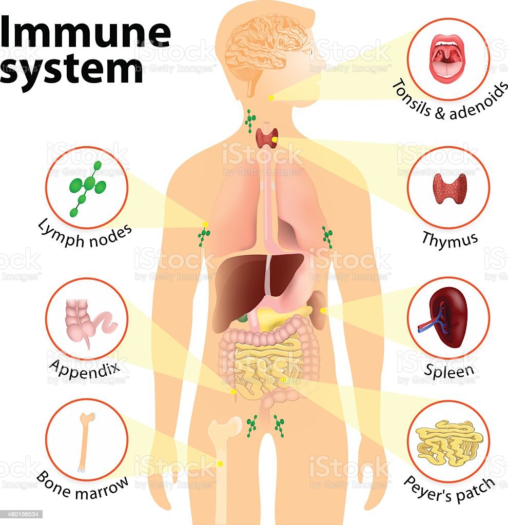
Immune system. Human anatomy. Human silhouette with internal organs.

We hear those 2 words “Immune System “a lot; what is the immune system?  The main parts of the immune system are:

  • white blood cells.
  • antibodies.
  • complement system.
  • lymphatic system.
  • spleen.
  • bone marrow.
  • thymus.

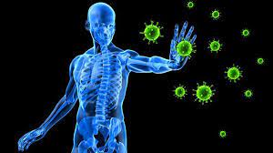
Your immune system is a large network of organs, white blood cells, proteins (antibodies) and chemicals. This system works together to protect you from foreign invaders (bacteria, viruses, parasites, and fungi) that cause infection, illness and disease.

What does the immune system do and how does it work?

Your immune system works hard to keep you healthy. Its job is to keep germs out of your body, destroy them or limit the extent of their harm if they get in.

When your immune system is working properly: When your immune system is working properly, it can tell which cells are yours and which substances are foreign to your body. It activates, mobilizes, attacks and kills foreign invader germs that can cause you harm. Your immune system learns about germs after you’ve been exposed to them too. Your body develops antibodies to protect you from those specific germs. An example of this concept occurs when you get a vaccine. Your immune system builds up antibodies to foreign cells in the vaccine and will quickly remember these foreign cells and destroy them if you are exposed to them in the future. Sometimes doctors can prescribe antibiotics to help your immune system if you get sick. But antibiotics only kill certain bacteria. They don’t kill viruses.

When your immune system is not working properly: When your immune system can’t mount a winning attack against an invader, a problem, such as an infection, develops. Also, sometimes your immune system mounts an attack when there is no invader or doesn’t stop an attack after the invader has been killed. These activities result in such problems as autoimmune diseases and allergic reactions.

Your immune system is made of up a complex collection of cells and organs. They all work together to protect you from germs and help you get better when you’re sick. The main parts of the immune system are:

  • White blood cells: Serving as an army against harmful bacteria and viruses, white blood cells search for, attack and destroy germs to keep you healthy. White blood cells are a key part of your immune system. There are many white blood cell types in your immune system. Each cell type either circulates in your bloodstream and throughout your body or resides in a particular tissue, waiting to be called into action. Each cell type has a specific mission in your body’s defense system. Each has a different way of recognizing a problem, communicating with other cells on the defense team and performing their function.
  • Lymph nodes: These small glands filter and destroy germs so they can’t spread to other parts of your body and make you sick. They also are part of your body’s lymphatic system. Lymph nodes contain immune cells that analyze the foreign invaders brought into your body. They then activate, replicate and send the specific lymphocytes (white blood cells) to fight off that particular invader. You have hundreds of lymph nodes all over your body, including in your neck, armpits, and groin. Swollen, tender lymph nodes are a clue that your body is fighting an infection.
  • Spleen: Your spleen stores white blood cells that defend your body from foreign invaders. It also filters your blood, destroying old and damaged red blood cells.
  • Tonsils and adenoids: Because they are located in your throat and nasal passage, tonsils and adenoids can trap foreign invaders (for example, bacteria or viruses) as soon as they enter your body. They have immune cells that produce antibodies to protect you from foreign invaders that cause throat and lung infections.
  • Thymus: This small organ in your upper chest beneath your breast bone helps mature a certain type of white blood cell. The specific task of this cell is to learn to recognize and remember an invader so that an attack can be quickly mounted the next time this invader is encountered.
  • Bone marrow: Stem cells in the spongy center of your bones develop into red blood cells, plasma cells and a variety of white blood cells and other types of immune cells. Your bone marrow makes billions of new blood cells every day and releases them into your bloodstream.
  • Skin, mucous membranes and other first-line defenses: Your skin is the first line of defense in preventing and destroying germs before they enter your body. Skin produces oils and secretes other protective immune system cells. Mucous membranes line the respiratory, digestive, urinary and reproductive tracts. These membranes secrete mucus, which lubricates and moistens surfaces. Germs stick to mucus in the respiratory tract and then are moved out of the airways by hair-like structures called cilia. Tiny hairs in your nose catch germs. Enzymes found in sweat, tears, saliva and mucus membranes as well as secretions in the vagina all defend and destroy germs.
  • Stomach and bowel: Stomach acid kills many bacteria soon after they enter your body. You also have beneficial (good) bacteria in your intestines that kill harmful bacteria.

QUOTE FOR THE WEEKEND:

“Menopause is a point in time when you’ve gone 12 consecutive months without a menstrual cycle. The time leading up to menopause is called perimenopause. This is when a lot of women or people assigned female at birth (AFAB) start to transition to menopause. They may notice changes in their menstrual cycles or have symptoms like hot flashes.  Natural menopause is the permanent ending of menstruation that doesn’t happen because of any type of medical treatment. The process is gradual and happens in 3 stages.  The average age of menopause in the United States is approximately 51 years old. However, the transition to menopause usually begins in your mid-40s.  “.

Cleveland Clinic (https://my.clevelandclinic.org/health/diseases/21841-menopause)

Menopause and it’s effects on women’s health.

What is Menopause exactly?

Menopause is when the ovaries naturally stop producing 2 hormones called estrogen and progesterone. Your ovaries are similar to what a car does in that over years it wears down, well so does the mechanism that regulates your hormones which is the ovaries. You go 12 consecutive months without having a period with no reasons to be explained for its occurrence, both biological or physical with it never returning. If both ovaries are removed surgically the menopause kicks in immediately. Menopause has signs and symptoms (s/s) that kick in which have varying intensities (it depends on the individual). You may experience mild to severe s/s.

Menopause is the time that marks the end of your menstrual cycles. It’s diagnosed after you’ve gone 12 months without a menstrual period. Menopause can happen in your 40s or 50s, but the average age is 51 in the United States. Menopause is a natural biological process.

The signs and symptoms of Menopause :

1- Hot Flashes 2-Irregular Periods 3- Breast Pains 4- Night Sweats 5- Mood Swings 6- Loss of Labido 7- Vaginal Dryness 8-Brittle Nails 9-Bloating 10-Irritability 11-Depression 12- Weight Gain 13-Sleep Problems 14- Osteoporosis-one of the worst symptoms of menopause. 15-Loss of breast fullness in some. 16- Thinning of the hair and dry skin.

Did you know over 60% of adult Americans are considered obese or overweight?
Weight gain happens when a person increases their body mass, whether it is a result of fat deposits, additional muscle tissue, or excess fluid. However, weight gain associated with menopause typically involves increased amounts of fat around the abdomen. One of the most accurate ways to see if you are obese is to measure your body mass index which is free online, check out the internet. I do every so often.
Go to Free BMI Calculator – AICR.org‎
www.aicr.org/bmi calculator‎.

On average, a women gains about 12 to 15 pounds between the ages of 45 and 55, this is usually when menopause typically occurs. This extra weight generally does not evenly distribute itself throughout a woman’s body. The weight tends to accumulate around the abdomen instead and women often notice the shape of their bodies slowly losing their hour-glass figure.

What can resolve this issue?

We can’t stop menopause but their are things we can do in dealing with it.

As years progress the metabolism slows down; setting the physiological stage for weight gain. As a woman’s hormones fluctuate prior to menopause and preparing for a permanently reduced hormonal level, it is likely to experience weight gain.

So things that can be done:

Eat well and nutritious foods.

Exercise but balance with rest.

Keep up with regular visits with your doctor for preventive health care and any medical concerns. Continue getting these appointments during and after menopause.

Preventive health care as you age may include recommended health screening tests, such as colonoscopy, mammography and triglyceride screening. Your doctor might recommend other tests and exams, too, including thyroid testing if suggested by your history, and breast and pelvic exams.

QUOTE FOR FRIDAY:

“The aorta carries blood from the heart to the body. Aortic aneurysms can occur anywhere in the aorta. They may be tube shaped or round.

Aortic aneurysms include:

  • Abdominal aortic aneurysm. An abdominal aortic aneurysm occurs along the part of the aorta that passes through the belly area.
  • Thoracic aortic aneurysm. A thoracic aortic aneurysm occurs along the part of the aorta that passes through the chest cavity.”

MAYO CLINIC (https://www.mayoclinic.org/diseases-conditions/aortic-aneurysm/symptoms-causes/syc-20369472)

Some people may have both types of aortic aneurysms.

Having an aortic aneurysm increases the risk of developing a tear in the inner layer of the wall of the aorta. This tear is called an aortic dissection.

Part II Aortic Aneurysms –What’s inside it, What are the risk factors in getting this & the most common causes!

What is inside an aortic aneurysm?

The inside walls of aneurysms are often lined with a blood clot that forms because there is stagnant blood. The wall of an aneurysm is layered, like a piece of plywood.

Who is most likely to have an abdominal aortic aneurysm?

Abdominal aortic aneurysms tend to occur in white males over the age of 60. In the United States, these aneurysms occur in up to 3.0% of the population. Aneurysms start to form at about age 50 and peak at age 80. Women are less likely to have aneurysms than men and African Americans are less likely to have aneurysms than Caucasians.

There is a genetic component that predisposes one to developing an aneurysm; the prevalence in someone who has a first-degree relative with the condition can be as high as 25%.

Collagen vascular diseases that can weaken the tissues of the aortic walls are also associated with aortic aneurysms. These diseases include Marfan and Ehlers-Danlos syndrome.

What are risk factors for aortic aneurysms?

The risk factors for aortic aneurysm are the same as those for atherosclerotic heart disease, stoke, and peripheral artery disease and include:

  • Cigarette smoking: This not only increases the risk of developing an abdominal aortic aneurysm, but also increases the risk of aneurysm rupture. Aortic rupture is a life-threatening event where blood escapes the aorta and the patient can quickly bleed to death.
  • High blood pressure
  • Elevated blood cholesterol levels
  • Diabetes mellitus The most common cause of aortic aneurysms is “hardening of the arteries” called arteriosclerosis. At a majority of aortic aneurysms are from arteriosclerosis. The arteriosclerosis can weaken the aortic wall and the increased pressure of the blood being pumped through the aorta causes weakness of the inner layer of the aortic wall.
  • The aortic wall has three layers, the tunica adventitia, tunica media, and tunica intima. The layers add strength to the aorta as well as elasticity to tolerate changes in blood pressure. Chronically increased blood pressure causes the media layer to break down and leads to the continuous, slow dilation of the aorta.
  • What is the most common cause of aortic aneurysms?

Smoking is a major cause of aortic aneurysm. Studies have shown that the rate of aortic aneurysm has fallen at the same rate as population smoking rates.

Know a aneurysm can be located in different arteries, like the brain with the same common causes, risk factors and make up inside the aneurysm.

aortic aneurysm 2 

QUOTE FOR THURSDAY:

“An aortic aneurysm is a balloon-like bulge in the aorta, the large artery that carries blood from the heart through the chest and torso.  Facts about aortic aneurysms:

  • Aortic aneurysms or aortic dissections were the cause of 9,904 deaths in 2019.1
  • In 2019, about 59% of deaths due to aortic aneurysm or aortic dissection happen among men.1
  • A history of smoking accounts for about 75% of all abdominal aortic aneurysms.2
  • The U.S. Preventive Services Task Force recommends that men 65 to 75 years old who have ever smoked should get an ultrasound screening for abdominal aortic aneurysms, even if they have no symptoms.3“.

Centers for Disease Control and Prevention – CDC (https://www.cdc.gov/heartdisease/aortic_aneurysm.htm)

Part I What is an aortic aneurysm?

aortic aneurysm 3aortic aneurysm 4aortic aeurysm 5

The aorta is the large artery that exits in the heart and delivers blood to the body. It begins at the aortic valve that separates the left ventricle of the heart from the aorta and prevents blood from leaking back into the left ventricle after a contraction, which is actually when the heart pumps blood. The various sections of the aorta are named based upon “arch-like” initial design and the location of the aorta in the body. Thus, the beginning of the aorta is referred to as the ascending aorta (basically meaning the blood going against resistance due to the vessel being a hill for the blood to go up), followed by the arch of the aorta, then the descending aorta (which is the blood going downward via gravity with the help of the heart pumping the blood of course). The portion of the aorta that is located in the chest (called thorax) is referred to as the thoracic aorta, while the abdominal aorta (the part of the aorta below the thorax region) is located in the abdomen. The abdominal aorta extends from the diaphragm (at the bottom of the lungs like a floor to divide the lungs from the organs in the abdomen) to the mid-abdomen where it splits into the iliac arteries and when it reaches the legs the femoral arteries now start which supplies to the legs oxygenated blood. This is why commonly a cardiac catheterization to visualize the aorta and sometimes the left side of the heart is done starting in the femoral artery since in time it diverts into starting the abdominal aorta.

An aneurysm is an area of a localized widening (dilation) of a blood vessel. The word “aneurysm” is borrowed from the Greek “aneurysma” meaning “a widening”.

An aortic aneurysm involves the aorta, the major artery that leaves the heart to supply blood to the body. An aortic aneurysm is a dilation or bulging of the aorta..

Most aortic aneurysms are fusiform. They are shaped like a spindle (“fusus” means spindle in Latin) with widening all around the circumference of the aorta. (Saccular aneurysms just involve a portion of the aortic wall with a localized out pocketing).

What is inside an aortic aneurysm?

The inside walls of aneurysms are often lined with a blood clot that forms because there is stagnant blood. The wall of an aneurysm is layered, like a piece of plywood.

Who is most likely to have an abdominal aortic aneurysm?

Abdominal aortic aneurysms tend to occur in white males over the age of 60. In the United States, these aneurysms occur in up to 3.0% of the population. Aneurysms start to form at about age 50 and peak at age 80. Women are less likely to have aneurysms than men and African Americans are less likely to have aneurysms than Caucasians.

There is a genetic component that predisposes one to developing an aneurysm; the prevalence in someone who has a first-degree relative with the condition can be as high as 25%.

Collagen vascular diseases that can weaken the tissues of the aortic walls are also associated with aortic aneurysms. These diseases include Marfan syndrome and Ehlers-Danlos syndrome

Aortic aneurysms can develop anywhere along the length of the aorta but the majority are located in the abdominal aorta. Most of these abdominal aneurysms are located below the level of the renal arteries, the vessels that provide blood to the kidneys. Abdominal aortic aneurysms can extend into the iliac arteries.

What shape are most aortic aneurysms?

Most aortic aneurysms are fusiform. They are shaped like a spindle (“fusus” means spindle in Latin) with widening all around the circumference of the aorta. (Saccular aneurysms just involve a portion of the aortic wall with a localized out pocketing).

Stayed tune for Part II on Aortic Aneurysms tomorrow!


QUOTE FOR WEDNESDAY:

“Gaseous chemical element, symbol: O, atomic number: 8 and atomic weight 15,9994. It’s of great interest because it’s the essential element in the respiratory processes of most of the living cells and in combustion processes. It’s the most abundant element in The Earth’s crust. Nearly one fifth (in volume) of the air is oxygen. Non-combined gaseous oxygen normally exists in form of diatomic molecules, O2, but it also exists in triatomic form, O3, named ozone.  In normal conditions oxygen is a colourless, odourless and insipid gas; it condensates in a light blue liquid. Oxygen is part of a small group of gasses literally paramagnetic, and it’s the most paramagnetic of all. Liquid oxygen is also slightly paramagnetic.

Oxygen is essential for all forms of life since it is a constituent of DNA and almost all other biologically important compounds. Is it even more drammatically essential, in that animals must have minute by minute supply of the gas in order to survive. Oxygen in the lungs is picked up by the iron atom at the center of hemoglobin in the blood and thereby transported to where it is needed.

Every human being needs oxygen to breathe, but as in so many cases too much is not good. If one is exposed to large amounts of oxygen for a long time, lung damage can occur. Breathing 50-100% oxygen at normal pressure over a prolonged period causes lung damage. Those people who work with frequent or potentially high exposures to pure oxygen, should take lung function tests before beginning employment and after that. Oxygen is usually stored under very low temperatures and therefore one should wear special clothes to prevent the freezing of body tissues.”

LennTech  (https://www.lenntech.com/periodic/elements/o.htm#ixzz88ZXf1arK)


Raising awareness of both pros and cons of oxygenation medically and in our environment!

oxygenationparti3 part-i-oxygenation

oxygenationpart14 oxygenation1a oxygenation1

Now don’t get me wrong oxygen is an element that is a must for most creatures that live in the world both now and since it began but there is dangers to any element especially if mixed with some other element causing a negative result in the end. So you wonder how oxygenation can have pros and cons and why oxygen would ever have dangers to it, well let’s take a deeper look.

Oxygenation may refer to:

Oxygen saturation (medicine), the process by which concentrations of oxygen increase within a tissue

Oxygenation (environmental), a measurement of dissolved oxygen concentration in soil or water

Great Oxygenation Event, an ancient event that led to the rise of oxygen within our atmosphere

Water oxygenation, the process of increasing the oxygen saturation of the water

Dioxygen complex, the chemical details of how metals bind oxygen

Of course, oxygen has its good points. Besides being necessary for respiration and the reliable combustion engine, it can be liquefied and used as rocket fuel. Oxygen is also widely used in the world of medicine as a means to imbue the body with a greater amount of the needed gas. But recent studies indicate that administering oxygen might be doing less good than hoped—and in fact be causing harm. No one is immune to the dangers of oxygen, but the people who might most suffer the ill effects are infants newly introduced to breathing, and those who are clinically deceased.

Oxygen regarding the medical view:

There are a variety of injuries and ailments for which modern medicine dictates oxygen therapy. Look at the medical aspect, the common wisdom is that by filling the lungs with pure O2, one is pushing more of the vital gas into the blood, and thus to organs that are weakened and in need of support. It has also long been known that even at partial pressures, pure oxygen can be toxic—a fact with which scuba divers and astronauts are intimately familiar. Recent studies have indicated that the human body responds to pure oxygen, even at normal pressures, in a negative way.

When pure O2 is introduced to the lungs, autonomic reflex increases respiration. The increased rate of breathing means that a much larger load of carbon dioxide is released from the body, which causes the blood vessels to constrict. Despite the increased amount of available oxygen in the lungs, the circulatory system is hampered, and cannot deliver precious O2 as well as it could when breathing normal atmosphere.

Ronald Harper, a neurobiology professor at UCLA, conducted observations on a group of healthy teenagers breathing various gas mixes using functional magnetic resonance imaging (fMRI). His findings showed that in some subjects the pure O2 caused the brain to go clinically bonkers. Brain structures such as the hippocampus, the insula, and the cingulate cortex all displayed an adverse reaction; they in turn spurred the hypothalamus, the body’s main regulatory gland, into a fervor. The hypothalamus regulates a myriad of things, including heart rate, body temperature, and is the master of a variety of other glands. The introduction of pure oxygen prompts the hypothalamus to flood the body with a cocktail of hormones and neurotransmitters which serve to hamper heart rate, and further reduce the circulatory system’s effectiveness. But Harper also found that by adding a mere 5% CO2, all the detrimental effects found in pure oxygen are negated.

There are circumstances, however, where even the proper mix of gases would prove inadequate. Modern medicine has long taught that after respiration stops, the brain can only survive for six to seven minutes without oxygen before its cells begin to die in droves. In order to combat this, standard procedure has been to aggressively attempted to restore breathing and heartbeat immediately upon cessation, CPR. The base premise on which this protocol is designed may be in error but only if continuing longer than the AHA guides us to do CPR. For there is more than just to lack of oxygen in patients who die having CPR done to them for death (Ex Exacerbation of a disease, multi – organ failure, years of CHF, etc… Even thought lack of 02 is part of the reason for the death in the end. There was a cause for it happening and leading to lack of 02 is the prime entity to death of all diseases leading up to this in a human.).

Upon examining heart cells and neurons deprived of oxygen under a microscope, Dr Lance Becker of the University of Pennsylvania found there was no indication that the cells were dying after five or six minutes. In fact, they seemed to endure the state for up to an hour without adverse affect. Given this unexpected observation, the researchers were forced to investigate why human resuscitation becomes impossible after only a few minutes of clinical death. The answer they uncovered was that the body’s cells were not dying of oxygen starvation; they were expiring due to Reperfusion—the sudden reintroduction of oxygen to a dormant cell = Programmed cell death! The cells reintroducing oxygen back into the cell from outside the cell in the bloodstream caused the destruction of the red blood cells, the RBCs carry oxygen to all our tissues sites. You would think that would save the cells in sending more oxygen out to the tissues but like we’re told from childhood too much of almost anything can hurt or kill you (Ex. Food/work/stress…)

Take a patient with severe emphysema they do get oxygen in their body but the problem is that oxygen gets air spaced elsewhere rather than all the 02 breathed in going in the red blood cells at the lungs exchange for 02 at the bottom of the lungs with CO2 (carbon dioxide) sent from the cells to the lungs to leave the body. Than the cells go off throughout the bloodstream having our tissues utilize from the red blood cells the oxygen it needs (a transfer of 02 to our tissues).   Upon return of the red blood cells that took the CO2 from the tissues to keep the tissues more oxygenated, so they can do their function as an organ. Oxygen deprivation to a severe state is Oxygen Starvation to our bodies leading to death, if not reversed. Also with the severe COPD emphysema pt their body adjusts to having high C02 levels compared to a person without emphysema. A normal person’s brain functions to sending messages out to cause us to breath when our 02 level is low but to a severe emphysema pt the low C02 levels causes their brain to send out messages to breath, so if you give an emphysema pt over 2L of 02 for several hours if will turn the brain off and the pt deceases (except when a emphysema pt is in respiratory distress since it is needed and temporary support of higher oxygen levels than when stable and out of respiratory distress their at 2L of 02 again).

Inside the cells, the culprit seems to be in the mitochondria, which is the cell’s power plant where sugar and oxygen are converted to usable energy. Mitochondria are also responsible for apoptosis—the organized, controlled self-destruction of a cell. Normally apoptosis occurs in situations such as the cell being damaged beyond repair, infected by a virus, an attempt to prevent cancer, or aiding in initial tissue development. The process effectively kills and dismantles the cell allowing the body’s usual waste management functions to carry the cell’s remains away. For reasons not entirely clear, reperfusion triggers apoptosis—the oxygen intended to save the cell actually causes cellular suicide.

Armed with this new information about how cells react to oxygen, it is clear that current emergency care is not altogether ideal, and new protocols are under investigation. Dr Becker proposes that induced hypothermia may slow cell degradation, and if a means can be found to safely reintroduce oxygen to tissues, a clinically dead person—who still has trillions of living cells—could be resuscitated after being an hour dead.

This glorious future is still on the horizon, but to imagine the practical application leads one to ponder the multitude of accidents and injuries that are currently fatal, but will one day be treatable. Emergency Medical Personnel could arrive on the scene, and inject the patient with a slurry of ice and salt that lowers the body temperature to about 92° F. In a hypothermic state, the patient is hauled to the hospital, where instead of frantically trying to restart the heart, doctors patch up the problem, prevent apoptosis , and then restart the heart. Though it won’t save everyone, these findings may lead to a future where a person made up of perfectly good human cells is not written off as dead merely because their heart has stopped beating. The miracle of modern medicine, it seems, is on the cusp of determining the true distinction between dead and mostly dead.


QUOTE FOR TUESDAY:

“Nothing compares to the joy of coming home to a loyal companion. The unconditional love of a pet can do more than keep you company. Pets may also decrease stress, improve heart health, and even help children with their emotional and social skills.  An estimated 68% of U.S. households have a pet.  Interacting with animals has been shown to decrease levels of cortisol (a stress-related hormone) and lower blood pressure. Other studies have found that animals can reduce loneliness, increase feelings of social support, and boost your mood.”

National Institute of Health NIH News in Health (https://newsinhealth.nih.gov/2018/02/power-pets)