Archive | June 2023

QUOTE FOR FRIDAY:

“Hemochromatosis can be difficult to diagnose. Early symptoms such as stiff joints and fatigue may be due to conditions other than hemochromatosis.

Many people with the disease don’t have any symptoms other than high levels of iron in their blood. Hemochromatosis may be identified because of irregular blood test results after testing is done for other reasons.  There are two key tests to detect iron overload.

The goal is to remove some of your blood so that your iron levels return to normal.”.

MAYO Clinic


Part II Hemochromatosis – How its Diagnosed & Rx.

Diagnosis

It can be tricky for your doctor to diagnose hemochromatosis, because other conditions have the same symptoms. He might want you to get tested if:

  • You’re having symptoms.
  • You have one of the problems listed above.
  • A family member has the disorder.

There are some other ways your doctor can figure out if you have it:

Checking your history. He’ll ask about your family and if anyone has hemochromatosis or signs of it. He might also ask about things like arthritis and liver disease, which might mean you or someone in your family has hemochromatosis but doesn’t know it.

Physical exam. Your doctor will examine your body. This involves using a stethoscope to listen to what’s going on inside. He might also tap on different parts of your body.

Blood tests. Two tests can give your doctor a clue about hemochromatosis:

  • Transferrin saturation. This shows how much iron is stuck to transferrin, a protein that carries iron in your blood.
  • Serum ferritin. This test measures the amount of ferritin, a protein that stores iron, in your blood. – If either of these show you have more iron than you should, your doctor might order a third test to see if you have a gene that causes hemochromatosis.
  • Liver biopsy. Your doctor will take a small piece of your liver. He’ll look at it under a microscope to see if there’s any liver damage.
  • MRI. This is a scan that uses magnets and radio waves to take a picture of your organs.

    Treatment

    If you have primary hemochromatosis, doctors treat it by removing blood from your body on a regular basis. It’s alot like donating blood. Your doctor will insert a needle into a vein in your arm or leg. The blood flows through the needle and into a tube that’s attached to a bag.

    • Initial treatment schedule. In the beginning, you may have a pint (about 470 milliliters) of blood taken once or twice a week — usually in a hospital or your provider’s office. While you lean back in a chair, a needle is inserted into a vein in your arm. The blood flows from the needle into a tube that’s attached to a blood bag. The process of removing blood is referred to as therapeutic blood removal.  In the Initial treatment. You’ll visit your doctor’s office or a hospital once or twice a week to have your blood drawn. You may have up to a pint taken at a time.
    • Maintenance treatment schedule. Once your iron levels go down, blood can be removed less often, typically every 2 to 3 months. Some people may maintain typical iron levels without having any blood taken. Some may need to have blood removed monthly. The schedule depends on how quickly iron builds up in your body.  So Maintenance treatment starts when once your blood iron levels have gone back to normal, you’ll still have to have to have blood taken, but not as often. It’ll be based on how fast iron builds back up in your body.

    The goal is to remove some of your blood so that your iron levels return to normal. This could take up to a year or more. Blood removal is divided into two parts: initial treatment and maintenance treatment.


Part II Stomach Cancer – Signs/Symptoms and how it’s diagnosed.

Symptoms

Early on, stomach cancer may cause:

  • Indigestion
  • Feeling bloated after you eat a meal
  • Heartburn
  • Slight nausea
  • Loss of appetite                                                                                                As stomach tumors grow, you may have more serious symptoms, such as:
    • Stomach pain
    • Blood in your stool
    • Vomiting
    • Weight loss for no reason
    • Trouble swallowing
    • Yellowish eyes or skin
    • Swelling in your stomach
    • Constipation or diarrhea
    • Weakness or feeling tired
    • Heartburn

Just having indigestion or heartburn after a meal doesn’t mean you have cancer. But if you feel these symptoms a lot, talk to your doctor. He can see if you have other risk factors and test you to look for any problems.

Stomach cancers are usually found when a person goes to the doctor because of signs or symptoms they are having. The doctor will take a history and examine the patient. If stomach cancer is suspected, tests will be needed to confirm the diagnosis.

Medical history and physical exam

When taking your medical history, the doctor will ask you questions about your symptoms (eating problems, pain, bloating, etc.) and possible risk factors to see if they might suggest stomach cancer or another cause. The physical exam gives your doctor information about your general health, possible signs of stomach cancer, and other health problems. In particular, the doctor will feel your abdomen for any abnormal changes.

If your doctor thinks you might have stomach cancer or another type of stomach problem, he or she will refer you to a gastroenterologist, a doctor who specializes in diseases of the digestive tract, who will examine you and do further testing.

Upper endoscopy

Upper endoscopy (also called esophagogastroduodenoscopy or EGD) is the main test used to find stomach cancer. It may be used when someone has certain risk factors or when signs and symptoms suggest this disease may be present.

During this test, the doctor passes an endoscope, which is a thin, flexible, lighted tube with a small video camera on the end, down your throat. This lets the doctor see the lining of your esophagus, stomach, and first part of the small intestine. If abnormal areas are seen, biopsies (tissue samples) can be taken using instruments passed through the endoscope. The tissue samples are sent to a lab, where they are looked at under a microscope to see if cancer is present.

When seen through an endoscope, stomach cancer can look like an ulcer, a mushroom-shaped or protruding mass, or diffuse, flat, thickened areas of mucosa known as linitis plastica. Unfortunately, the stomach cancers in hereditary diffuse gastric cancer syndrome often cannot be seen during endoscopy.

Endoscopy can also be used as part of a special imaging test known as endoscopic ultrasound, which is described below.

This test is usually done after you are given medication to make you sleepy (sedation). If sedation is used, you will need someone to take you home (not just a cab).

Endoscopic ultrasound

Ultrasound uses sound waves to produce images of organs such as the stomach. During a standard ultrasound, a wand-shaped probe called a transducer is placed on the skin. It gives off sound waves and detects the echoes as they bounce off internal organs. The pattern of echoes is processed by a computer to produce a black and white image on a screen.

In endoscopic ultrasound (EUS), a small transducer is placed on the tip of an endoscope. While you are sedated, the endoscope is passed down the throat and into the stomach. This lets the transducer rest directly on the wall of the stomach where the cancer is. It lets the doctor look at the layers of the stomach wall, as well as the nearby lymph nodes and other structures just outside the stomach. The picture quality is better than a standard ultrasound because of the shorter distance the sound waves have to travel.

EUS is most useful in seeing how far a cancer may have spread into the wall of the stomach, to nearby tissues, and to nearby lymph nodes. It can also be used to help guide a needle into a suspicious area to get a tissue sample (EUS-guided needle biopsy).

Biopsy

Your doctor may suspect cancer if an abnormal-looking area is seen on endoscopy or an imaging test, but the only way to tell for sure if it is really cancer is by doing a biopsy. During a biopsy, the doctor removes a sample of the abnormal area.

Biopsies to check for stomach cancer are most often obtained during upper endoscopy. If the doctor sees any abnormal areas in the stomach lining during the endoscopy, instruments can be passed down the endoscope to biopsy them.

Some stomach cancers are deep within the stomach wall, which can make them hard to biopsy with standard endoscopy. If the doctor suspects cancer might be deeper in the stomach wall, endoscopic ultrasound can be used to guide a thin, hollow needle into the wall of the stomach to get a biopsy sample.

Biopsies may also be taken from areas of possible cancer spread, such as nearby lymph nodes or suspicious areas in other parts of the body.

Testing biopsy samples

Biopsy samples are sent to a lab to be looked at under a microscope. The samples are checked to see if they contain cancer, and if they do, what kind it is (for example, adenocarcinoma, carcinoid, gastrointestinal stromal tumor, or lymphoma).

If a sample contains adenocarcinoma cells, it may be tested to see if it has too much of a growth-promoting protein called HER2/neu (often just shortened to HER2). The HER2/neu gene instructs the cells to make this protein. Tumors with increased levels of HER2/neu are called HER2-positive.

Stomach cancers that are HER2-positive can be treated with drugs that target the HER2/neu protein, such as trastuzumab (Herceptin®).

The biopsy sample may be tested in 2 different ways:

  • Immunohistochemistry (IHC): In this test, special antibodies that stick to the HER2/neu protein are applied to the sample, which cause cells to change color if many copies are present. This color change can be seen under a microscope. The test results are reported as 0, 1+, 2+, or 3+.
  • Fluorescent in situ hybridization (FISH): This test uses fluorescent pieces of DNA that specifically stick to copies of the HER2/neu gene in cells, which can then be counted under a special microscope.

Often the IHC test is used first.

  • If the results are 0 or 1+, the cancer is HER2-negative. People with HER2-negative tumors are not treated with drugs (like trastuzumab) that target HER2.
  • If the test comes back 3+, the cancer is HER2-positive. Patients with HER2-positive tumors may be treated with drugs like trastuzumab.
  • When the result is 2+, the HER2 status of the tumor is not clear. This often leads to testing the tumor with FISH.

Imaging tests

Imaging tests use x-rays, magnetic fields, sound waves, or radioactive substances to create pictures of the inside of your body. Imaging tests may be done for a number of reasons, including:

  • To help find out whether a suspicious area might be cancerous
  • To learn how far cancer may have spread
  • To help determine if treatment has been effective

Upper gastrointestinal (GI) series

This is an x-ray test to look at the inner lining of the esophagus, stomach, and first part of the small intestine. This test is used less often than endoscopy to look for stomach cancer or other stomach problems, as it may miss some abnormal areas and does not allow the doctor to take biopsy samples. But it is less invasive than endoscopy, and it might be useful in some situations.

For this test, the patient drinks a white chalky solution containing a substance called barium. The barium coats the lining of the esophagus, stomach, and small intestine. Several x-ray pictures are then taken. Because x-rays can’t pass through the coating of barium, this will outline any abnormalities of the lining of these organs.

A double-contrast technique may be used to look for early stomach cancer. With this technique, after the barium solution is swallowed, a thin tube is passed into the stomach and air is pumped in. This makes the barium coating very thin, so even small abnormalities will show up.

Computed tomography (CT or CAT) scan

The CT scan is an x-ray test that produces detailed cross-sectional images of your body. Instead of taking one picture, like a standard x-ray, a CT scanner takes many pictures as it rotates around you. A computer then combines these pictures into images of slices of the part of your body being studied.

Before the test, you may be asked to drink 1 or 2 pints of a contrast solution and/or receive an intravenous (IV) line through which a contrast dye is injected. This helps better outline structures in your body.

The IV contrast can cause some flushing (redness and warm feeling). Some people are allergic and get hives, or rarely have more serious reactions like trouble breathing and low blood pressure. Be sure to tell the doctor if you have any allergies or have ever had a reaction to any contrast material used for x-rays.

A CT scanner has been described as a large donut, with a narrow table that slides in and out of the middle opening. You will need to lie still on the table while the scan is being done. CT scans take longer than regular x-rays, and you might feel a bit confined by the ring while the pictures are being taken.

CT scans show the stomach fairly clearly and often can confirm the location of the cancer. CT scans can also show the organs near the stomach, such as the liver, as well as lymph nodes and distant organs where cancer might have spread. The CT scan can help determine the extent (stage) of the cancer and whether surgery may be a good treatment option.

CT-guided needle biopsy: CT scans can also be used to guide a biopsy needle into a suspected area of cancer spread. The patient remains on the CT scanning table while a doctor moves a biopsy needle through the skin toward the mass. CT scans are repeated until the needle is within the mass. A fine-needle biopsy sample (tiny fragment of tissue) or a core-needle biopsy sample (a thin cylinder of tissue) is then removed and looked at under a microscope.

Magnetic resonance imaging (MRI) scan

MRI scans use radio waves and strong magnets instead of x-rays. The energy from the radio waves is absorbed by the body and then released in a pattern formed by the type of body tissue and by certain diseases. A computer translates the pattern into a very detailed image of parts of the body. A contrast material might be injected just as with CT scans, but this is used less often.

Most doctors prefer to use CT scans to look at the stomach. But an MRI may sometimes provide more information. MRIs are often used to look at the brain and spinal cord.

MRI scans take longer than CT scans, often up to an hour. You may have to lie inside a narrow tube, which is confining and can upset people with a fear of enclosed spaces. Special, open MRI machines can help with this if needed, although the images may not be as sharp in some cases. The MRI machine makes loud buzzing noises that you may find disturbing. Some places provide headphones to block this noise out.

Positron emission tomography (PET) scan

In this test, radioactive substance (usually a type of sugar related to glucose, known as FDG) is injected into a vein. (The amount of radioactivity used is very low and will pass out of the body over the next day or so.) Because cancer cells are growing faster than normal cells, they use sugar much faster, so they take up the radioactive material. After about an hour, you are moved onto a table in the PET scanner. You lie on the table for about 30 minutes while a special camera creates a picture of areas of radioactivity in the body.

PET is sometimes useful if your doctor thinks the cancer might have spread but doesn’t know where. The picture is not finely detailed like a CT or MRI scan, but it provides helpful information about the whole body. Although PET scans can be useful for finding areas of cancer spread, they aren’t always helpful in certain kinds of stomach cancer because these types don’t take up glucose very much.

Some machines can do both a PET and CT scan at the same time (PET/CT scan). This lets the doctor compare areas of higher radioactivity on the PET with the more detailed appearance of that area on the CT. PET/CT may be more helpful than PET alone for stomach cancer. This can help show if the cancer has spread beyond the stomach to other parts of the body, in which case surgery might not be a good treatment.

Chest x-ray

This test can help find out if the cancer has spread to the lungs. It might also determine if there are any serious lung or heart diseases present. This test is not needed if a CT scan of the chest has been done.

Other tests

Laparoscopy

If this procedure is done, it is usually only after stomach cancer has already been found. Although CT or MRI scans can create detailed pictures of the inside of the body, they can miss some tumors, especially if they are very small. Doctors might do a laparoscopy before any other surgery to help confirm a stomach cancer is still only in the stomach and can be removed completely with surgery. It may also be done before chemotherapy and/or radiation if these are planned before surgery.

This procedure is done in an operating room with the patient under general anesthesia (in a deep sleep). A laparoscope (a thin, flexible tube) is inserted through a small surgical opening in the patient’s side. The laparoscope has a small video camera on its end, which sends pictures of the inside of the abdomen to a TV screen. Doctors can look closely at the surfaces of the organs and nearby lymph nodes, or even take small samples of tissue. If it doesn’t look like the cancer has spread, sometimes the doctor will “wash” the abdomen with saline (salt water). The fluid (called peritoneal washings) is then removed and checked to see if it contains cancer cells. If it does, the cancer has spread, even if the spread couldn’t be seen.

Sometimes laparoscopy is combined with ultrasound to give a better picture of the cancer.

Lab tests

When looking for signs of stomach cancer, a doctor may order a blood test called a complete blood count (CBC) to look for anemia (which could be caused by the cancer bleeding into the stomach). A fecal occult blood test may be done to look for blood in stool (feces) that isn’t visible to the naked eye.

The doctor might recommend other tests if cancer is found, especially if you are going to have surgery. For instance, blood tests will be done to make sure your liver and kidney functions are normal and that your blood clots normally. If surgery is planned or you are going to get medicines that can affect the heart, you may also have an electrocardiogram (EKG) and echocardiogram (an ultrasound of the heart) to make sure your heart is functioning well.


Part III Part Treatment to Stomach Cancer:

Many treatments can fight stomach cancer. The one you and your doctor choose will depend on how long you’ve had the disease or how much it has spread in your body, called the stage of your cancer.

Surgery. Your doctor might remove part of your stomach or other tissues nearby that have cancer cells. Surgery gets rid of the tumor and stops cancer from spreading to other parts of your body. If your disease is in a more advanced stage, your doctor might need to remove all of your stomach=Gastrectomy or in some other cases the Surgeon may only have to remove part of the stomach=Partial Gastrectomy.

Some tumors can keep food from moving in and out of your stomach. In that case, you might have surgery to put in a stent, a device that keeps the pathways open.

Chemotherapy. Drugs kill your cancer cells or keep them from growing. You can take them as pills or through an IV at a clinic. Chemo usually takes several weeks. The drugs can cause side effects, but your doctor can help you find ways to feel better during treatment.

Radiation. High-energy waves or particles can kill cancer cells and shrink tumors. Your doctor may use an X-ray or other machine to beam radiation at the spot where your tumor is.

Chemoradiation. Your doctor might use this mix of chemotherapy and radiation to shrink your tumor before surgery.

Targeted drugs. These newer drugs are different because they fight only cancer cells. Other treatments, like chemo and radiation, can kill healthy cells along with diseased ones. As a result, targeted therapies have fewer side effects than these other treatments.

How Can I Prevent Stomach Cancer?

Treat stomach infections. If you have ulcers from an H. pylori infection, get treatment. Antibiotics can kill the bacteria, and other drugs will heal the sores in the lining of your stomach to cut your risk of cancer.

Eat healthy. Get more fresh fruits and vegetables on your plate every day. They’re high in fiber and in some vitamins that can lower your cancer risk. Avoid very salty, pickled, cured, or smoked foods like hot dogs, processed lunch meats, or smoked cheeses. Keep your weight at a healthy level, too. Being overweight or obese can also raise your risk of the disease.

QUOTE FOR THE DAY:


“After a cancer diagnosis, staging provides important information about the extent of cancer in the body and anticipated response to treatment.”


American Cancer Society


Go to striveforgoodhealth.com and learn more on the types of treatments given to patients with stom

QUOTE FOR THURSDAY:

“Hemochromatosis is a disorder in which the body can build up too much iron in the skin, heart, liver, pancreas, pituitary gland, and joints. Hemochromatosis is a disorder in which the body can build up too much iron in the skin, heart, liver, pancreas, pituitary gland, and joints. Too much iron is toxic to the body and over time the high levels of iron can damage tissues and organs and lead to serious problems with these organs mentioned above.”

Centers for Disease Control and Prevention CDC (https://www.cdc.gov/genomics/disease/hemochromatosis.htm)


What is Hemochromatosis?


This is a problem with the Liver and in a nutshell it is IRON OVERLOAD!

Hemochromatosis (Hemo meaning blood, Chroma meaning change in color and Osis meaning swelling)  is a condition where there is abnormal accumulation of iron in the organs resulting in organ toxicity. It is the most common autosomal recessive genetic condition along with being the most common cause of severe iron overload. 75 percent of patients who have hereditary hemochromatosis are asymptomatic. The diagnosis can be made based on the clinical symptoms of the disease. Since most patients are asymptomatic, they are diagnosed when their serum iron levels are noted to be elevated during a routine chemistry screening panel. Some of the tests performed on an individual suspected with hemochromatosis include genetic testing to test for HFE mutations, transferrin saturation levels, hepatic iron concentration, and serum ferritin studies. Imaging studies such as a chest radiography and echocardiography might be beneficial to help evaluate cardiac disease in patients with hemochromatosis.

Early diagnosis is important in hemochromatosis, like in most every other diagnosis.

The aim of therapy is to remove iron before it causes irreversible parenchymal damage. Once the diagnosis is confirmed, it is treated by phlebotomy to maintain normal iron stores and rid the body of excess iron. Chelation therapy uses agents such as deferoxamine, deferiprone, or deferasirox. Surgery may be required if there is end stage liver disease or severe arthropathy. In the United States, hereditary hemochromatosis is found in 1 of 200 to 500 individuals. Most of these cases are seen in individuals of northern European origin. It has the same prevalence in Australia, Europe, and other western countries. The highest prevalence is among individuals of Celtic origin and less among those of African descent.

Symptoms would be:

1.) Fatigue and 2 types of fatigue, that would be:

Physical fatigue occurs when there is transient inability to achieve peak physical performance.

Mental fatigue is the temporary inability to maintain optimal cognitive performance. It is a common and non-specific symptom that can be seen in stress, overwork, depression, boredom, anemia, inadequate sleep, malnutrition, pregnancy, infections, and more. It can manifest as somnolence in patients.

2.) Impotence in patients with hemochromatosis is due to a result of hypogonadism. Hypogonadism can occur in hemochromatosis patients due to pituitary or testicular iron deposition.

3.) Arthropathy that’s a disease of a joint. In arthropathy, there can be joint pain, stiffness, swelling, and decreased range of motion. Patients can see this occuring because of the accumulation of iron in the joint tissues. The most commonly affected joints are the knees, feet, wrists, neck, back, metacarpophalangeal joints, and proximal interphalangeal joints.

4.) It has been observed that 62 percent of patients with hemochromatosis experience partial loss of body hair. The most commonly affected area is the pubic area. Approximately 12 percent of patients experience total body hair loss. In some patients, the thinning and loss of hair may be reversed through therapy.

5.) Koilonychia refers to spoon nails where the nails are abnormally thin and have lost their convexity resulting in flat or concave nails. Causes of koilonychia include iron deficiency anemia and it can be seen in 25 to 50 percent of these patients.

6.) Skin Hyperpigmentation or Bronzing of the skin refers to darkening of the skin. It is due to the combination of melanin and iron deposition which is characteristic. It is one of the classic triad symptoms with the other two being cirrhosis and diabetes in the late stages of the disease. This usually occurs only when the total iron content in the body is more than 5 times the normal levels.

7.) Amenorrhea refers to the absence of menstruation in females who are in their reproductive age.

8.) In the heart it can cause the following: Atrial flutter is a problem with the way your heart beats. Such problems, whether in the rhythm or speed of the heartbeat, are known as arrhythmias. Heart Failure is the other.

In addition to these symptoms it can also cause problems with the spleen, adrenal glands, pituitary gland, gallbladder or thyroid.

For helping in possible PREVENTION of this diagnosis and most others stay as healthy as possible and get yearly check ups to pick up this or any other diagnosis EARLY!! In so many cases of diseases the early picked up or diagnosed the better the results turn out in most cases.  When a disease just start in your body is being treated compared to late in the disease higher the odds will resolve or you will last longer with a better quality of life, just like with so many other diseases!

QUOTE FOR WEDNESDAY:

“Scoliosis, a common, sometimes hereditary spinal condition, is a curvature of the spine that affects 2% to 3% of the population in the United States, or an estimated 6 million to 9 million people.  “If your child has scoliosis, don’t be afraid of it,” Vitale says. “The key is being aware of it and getting the diagnosis early so you can begin treatment. When treated early, there’s a lot of hope for stopping curve progression and allowing kids to live a perfectly active, normal life.”.

(https://www.cuimc.columbia.edu/news/8-facts-know-about-scoliosis)

Columbia University Irving Medical Center Dr. Michael G. Vitale                  (https://www.cuimc.columbia.edu/news/8-facts-know-about-scoliosis)

What is Scoliosis?

u15784442k14738439

Scoliosis is a problem with the spine where the spine is curved instead of straight, with the upper back being rounded and the lower back having a “swayback,” or inner curved problem, reports WebMD.

According to the Scoliosis Research Society, 85 percent of all scoliosis causes are idiopathic, meaning the cause is unknown. The remaining causes of scoliosis include birth defects, such as vertebrae that form abnormally before birth, and certain disorders such as cerebral palsy, Marfan’s syndrome, muscular dystrophy and Down syndrome. Infections and spinal fractures can also cause scoliosis.curvature of the spine during surgical correction of this condition. Screws and rods are placed in order to stabilize and straighten the spine.

What You Should Know About Adult Scoliosis

Scoliosis is defined as a curve of the spine of 10 degrees. Adult scoliosis is broadly defined as a curve in your spine of 10 degrees or greater in a person 18 years of age or older. Adult scoliosis is separated into 2 common categories:

  • Adult Idiopathic Scoliosis patients have had scoliosis since childhood or as a teenager and have grown into adulthood.  We do not yet know the cause of idiopathic scoliosis, but there is a lot of genetic work going on in an attempt to answer this question.
  • Adult “De Novo” or Degenerative Scoliosis develops in adulthood. Degenerative scoliosis develops as a result of disc degeneration. As the disc degenerates, it loses height. If one side of the disc degenerates more rapidly than the other, the disc begins to tilt. As it tilts, more pressure is placed on one side of your spine and gravity tends to cause the spine to bend and curve. The more discs that degenerate, the more the spine begins to curve.

Scoliosis is more common in girls than in boys, and the diagnosis is usually made after a child reaches 10 years of age. A doctor performs a physical examination and may take X-rays to definitively diagnose the disease. An X-ray tells if there is any growth left in the growth plates of the femur or humerus, and scoliosis can become worse if the patient has more growing to do, states MedicineNet. Serial X-rays are performed to track the changes of the spinal curve, which helps determine the best course of treatment.

Types of idiopathic scoliosis are categorized by both age at which the curve is detected and by the type and location of the curve.

When grouped by age, scoliosis usually is categorized into three age groups:

  • Infantile scoliosis: from birth to 3 years old
  • Juvenile scoliosis: from 3 to 9 years old
  • Adolescent scoliosis: from 10 to 18 years old

This last category of scoliosis, adolescent scoliosis, occurs in children age 10 to 18 years old, and comprises approximately 80% of all cases of idiopathic scoliosis. This age range is when rapid growth typically occurs, which is why the detection of a curve at this stage should be monitored closely for progression as the child’s skeleton develops.

Terms Used to Describe Spinal Curvature

Scoliosis curves are often described based on the direction and location of the curve. Physicians have several detailed systems to classify specific curves, but here are some common terms used to describe scoliosis:

Terms that describe the direction of the curve:

  • Dextroscoliosis describes a spinal curve to the right (“dextro” = right). Usually occurring in the thoracic spine, this is the most common type of curve. It can occur on its own (forming a “C” shape) or with another curve bending the opposite way in the lower spine (forming an “S”).

Severe scoliosis can lead to heart and lung problems if not treated, as the ribs press against the chest, making breathing more difficult, states Mayo Clinic. Adults who had scoliosis as a child may experience more back pain throughout their lives as compared to people without scoliosis.

Symptoms of scoliosis include an uneven waist, uneven shoulders, disjointed hip and a protruding shoulder blade, according to Mayo Clinic. The spine also curves or twists in acute cases, and the disease can cause one side of the ribs to protrude more than the other. Severe cases also induce labored breathing and back pain.

Diagnosis

Scoliosis can be recognized and diagnosed with a clinical exam, but xrays are necessary to fully evaluate the magnitude and type of scoliosis present. For a proper scoliosis evaluation, full length, whole spine xrays need to be performed. An MRI may also be recommended if there are symptoms of leg pain that may be associated with stenosis or if there is concern about possible spinal cord compression or abnormalities.

Treatment

The treatment of adult scoliosis is very individualized and based on the specific symptoms and age of the patient. Many patients have scoliosis and have very minor symptoms and live with it without treatment. Patients with predominant symptoms of back pain would typically be treated with physical therapy. Patients with back pain and leg pain may receive some benefit from injection treatment to help relieve the leg pain.  If lumbar stenosis (narrowing of the spinal canal) is present and is unresponsive to non-surgical treatment, then a decompression( removal of bone and ligaments pressing on the nerves) may be recommended. If the scoliosis is greater than 30 degrees, a fusion procedure will most likely be recommended along with the decompression. The fusion is recommended to prevent the curve from progressing when the spine is destabilized by the bone removal that is necessary to  decompress the nerves. Fusions are usually accompanied with metal rod and screw placement into the spine to help correct and stabilize the scoliosis and help the bone heal or fuse together. The length of the fusion, or the number of spine levels included, depends on the type of scoliosis and the area of the spine involved. The goal of adult scoliosis surgery is to first remove pressure on the nerves, and second to keep the scoliosis from progressing further.

QUOTE FOR TUESDAY:

“On an airplane, an oxygen mask descends in front of you. What do you do? As we all know, the first rule is to put on your own oxygen mask before you assist anyone else. Only when we first help ourselves can we effectively help others. Caring for yourself is one of the most important—and one of the most often forgotten—things you can do as a caregiver. When your needs are taken care of, the person you care for will benefit, too.”.

Family Caregiver Alliance

Health and Safety for Caregivers

Safety Tips for Health Givers in Hospitals and Long Term Care Facilities through All American Homecare:

1 – Medical facilities such as hospitals and long-term care facilities will have people that maintain the grounds and buildings. However, as an at-home caregiver, you’ll be visiting patients’ homes that are in a variety of conditions. Be sure to watch your step while approaching the home, making sure that you don’t trip on uneven concrete and keep an eye out for rotting wood.

Once inside, do not remove your shoes, or change into a pair of shoes that you only wear indoors (it is important to keep your safety in mind while also respecting the wishes or culture of your patient). Your shoes will protect you from slipping or stubbing a toe.

Also be mindful of slippery hard surfaces, open cupboards, and other things that can cause injury when you are engrossed in helping your patient.

2 –When visiting patients, keep your guard up and be aware of your surroundings. Unlike the controlled environments of hospitals and other facilities, the homes of patients can be unpredictable.

Trust your instincts and never go into a situation that makes you feel unsafe. Be sure to call your supervisor to figure out how to proceed in these situations. Also learn to recognize warning signs and if you feel threatened, immediately leave and inform your supervisor of the situation.

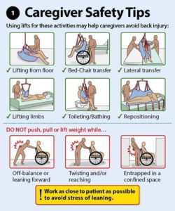
3 – It’s vital that you use safe lifting practices each time you transfer a patient. This will help reduce incidents of back injury and muscle strains. Here’s a refresher on proper lifting techniques:

  • Keep your head and neck in proper alignment with your spine
  • Maintain the natural curve of your spine; bend with your hips and knees, not your spine
  • Avoid twisting your body when carrying the person
  • Always keep the patient being moved close to your body
  • Keep your feet shoulder-width apart to maintain your balance
  • Use the muscles in your legs to lift and/or pull

4 – There are plenty of tools for home healthcare workers to aid them in their daily tasks. These can include lifting and transfer aides. If you are helping a patient change a light bulb, don’t stand on a chair to do it; instead, use a step stool.

5 – Your uniform (often scrubs or a pair of slacks and a polo) ensures that you are properly dressed for the job. If you don’t have a company uniform, ensure that your clothing allows you to move freely. Also, wear close-toed shoes with good traction and support.

6 – Household spills and incontinence accidents increase the risk of falls for both the caregiver and the patient. Clutter can also lead to tripping and falling. Clean up these and other safety hazards immediately.

7 – Many of your patients will have pets. Not only can animals cause you to become distracted or interfere with your work, even the friendliest pet can turn on you. Your best bet is to avoid the animals in the home and focus on your patient.

8 – One of the biggest risks to home healthcare workers is going past their limits. Make sure that you know your limits. If a patient is too heavy for you to transfer on your own, ask for help. Also, pace yourself and build flexibility into your schedule. Injury-inducing shortcuts just aren’t worth it!

Taking time to stretch throughout the day can help reduce muscle fatigue and strains. When it’s time to take a break, take it. Keep yourself hydrated throughout the day and eat a meal and snacks. Being dehydrated and not eating can lead to feelings of tiredness and making dangerous mistakes.

9 – Here are a few other basic safety protocols you need to follow:

  • Confirm with your clients by phone before you visit.
  • Have detailed directions to your client’s home or use a GPS device.
  • Keep your vehicle in good working condition and fill up your gas tank.
  • Don’t text or talk on the phone while driving.
  • Keep your car doors locked and your windows closed.
  • Lock your bag and other possessions in the trunk of your car.
  • Most importantly, make sure that someone knows where you are at all times.

By following these safety guidelines, you’ll be able to provide better care for your patients and have a satisfying career that you enjoy each day!

QUOTE FOR MONDAY:

“Sickle cell disease (SCD) is a group of conditions that affect the red blood cells. Normal red blood cells are round and flexible. A person with sickle cell disease has red blood cells that are hard, sticky, and crescent-shaped like crescent moons or sickles. People with sickle cell disease often experience intense pain and anemia because of their abnormal red blood cells.

SCD is one of the most common hereditary disorders in the United States, affecting 1 in 1,900 infants. Of the 66,000 infants born in Minnesota annually, about 25 infants are born with SCD and more than 1,000 infants are born with a sickle cell trait (SCT).

Currently, the only known cure for SCD is a blood or marrow transplant (BMT). A blood or marrow transplant is a procedure that takes healthy stem cells from a donor and puts them into someone else”

Department of Health in Minnesota (https://www.health.state.mn.us/diseases/sicklecell/about.html)