QUOTE FOR TUESDAY:

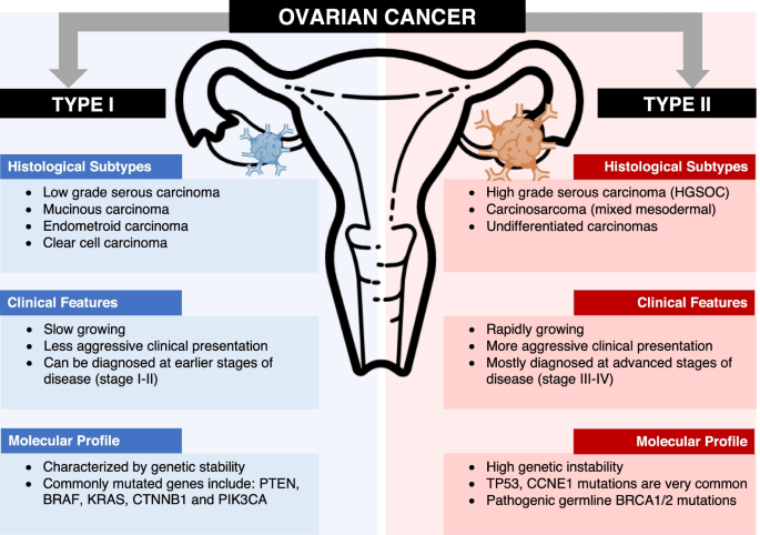
“Ovarian cancer affects 1 in 70 women across their lifetime and is the second most common type of gynecologic cancer in the United States. The most common risk factor is age. About two-thirds of all ovarian cancers are diagnosed in women between ages 50-75. Only 5 percent of ovarian cancers diagnosed occur in women under the age of 30.

About 1 in 10 women who are diagnosed will have either a strong family history of ovarian cancer or a mutation in a gene that increases risk of the disease.

Ovarian cancer rarely has noticeable symptoms when it is in its earliest stages. As the cancer progresses, subtle signs begin to appear, but you might not notice them right away, or they may be blamed on other common conditions.”

Memorial Sloan Kettering Cancer Center (Ovarian Cancer | Memorial Sloan Kettering Cancer Center)

Part I Ovarian Cancer – Learn about the silent sister to Breast Cancer with symptoms and risk factors!

Most people are aware that October is Breast Cancer Awareness Month, but how many of you are also aware that September is Ovarian Cancer Awareness Month?

This cancer, Ovarian Cancer, is the more silent sister to breast cancer-which takes over the month of October with a worldwide pink party and numerous product promotions, some tasteful and some less so. Maybe people and product promoters are just drawn to pink versus the more reserved teal blue color for ovarian cancer. More likely it’s because breasts are visual and ovaries are invisible to the eye.

Remember ovarian cancer is very visible to those diagnosed with it and to their loved ones.  We need to make more noise about ovarian cancer awareness.  First you have to listen… to your body. Ovarian cancer can be sneaky.

Symptoms such as indigestion, bloating, painful intercourse, menstrual irregularities and back pain, can point to other less invasive conditions. While breast cancer has screening protocols like mammograms and breast self-examination, there is no reliable screening for ovarian cancer. Unfortunately for many women the disease is often detected at an advanced stage. Both breast and ovarian cancer are diagnosed in women of all ages and ethnic backgrounds.

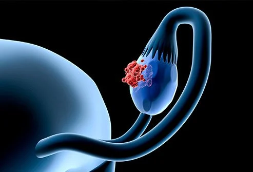
Ovarian cancer is a type of cancer that begins in the ovaries. Women have two ovaries, one on each side of the uterus. The ovaries — each about the size of an almond — produce eggs (ova) as well as the hormones estrogen and progesterone.

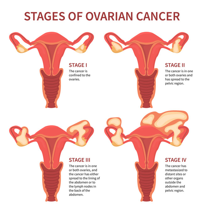
Ovarian cancer often goes undetected until it has spread within the pelvis and abdomen. At this late stage, ovarian cancer is more difficult to treat, just like most other cancers in late stages as well, and is unfortunately frequently fatal. Early-stage ovarian cancer, in which the disease is confined to the ovary, is more likely to be treated successfully.

Early-stage ovarian cancer rarely causes any symptoms. Advanced-stage ovarian cancer may cause few and nonspecific symptoms that are often mistaken for more common benign conditions, such as constipation or irritable bowel.

Signs and symptoms of ovarian cancer may include and don’t ever ignore them:

  • Abdominal bloating or swelling
  • Quickly feeling full when eating
  • Weight loss
  • Discomfort in the pelvis area
  • Changes in bowel habits, such as constipation or diarrhea
  • A frequent need to urinate (urgency including difficulty to void)
  • Increased Abdominal Size
  • Painful Sex
  • Heavy menstrual bleedingWhen to see a doctorIf you have a family history of ovarian cancer or breast cancer, talk to your doctor about your risk of ovarian cancer. Your doctor may refer you to a genetic counselor to discuss testing for certain gene mutations that increase your risk of breast and ovarian cancers. Only a small number of women are found to have genetic mutations that can lead to ovarian cancer.
  • Certain factors may increase your risk of ovarian cancer:
  • Make an appointment with your doctor if you have any signs or symptoms that worry you. 

Risk Factors:

  • Age. Ovarian cancer can occur at any age but is most common in women ages 50 to 60 years.
  • Inherited gene mutation. A small percentage of ovarian cancers are caused by an inherited gene mutation. The genes known to increase the risk of ovarian cancer are called breast cancer gene 1 (BRCA1) and breast cancer gene 2 (BRCA2). These genes were originally identified in families with multiple cases of breast cancer, which is how they got their names, but women with these mutations also have a significantly increased risk of ovarian cancer.
  • The gene mutations that cause Lynch syndrome, which is associated with colon cancer, also increase a woman’s risk of ovarian cancer.
  • Estrogen hormone replacement therapy, especially with long-term use and in large doses.
  • Age when menstruation started and ended. If you began menstruating before age 12 or underwent menopause after age 52, or both, your risk of ovarian cancer may be higher.
  • Never being pregnant.
  • Fertility treatment.
  • Smoking.
  • Use of an intrauterine device.
  • Polycystic ovary syndrome.  In years past ovarian cancer used to be call  the silent killer but it’s really not completely silent, at least in some patients.  You shouldn’t ignore your symptoms!


QUOTE FOR MONDAY:

“Key Messages from the CDC:

  1. International Overdose Awareness Day 2026 marks 25 years of global remembrance, awareness, and action. This year’s theme, “25 Years On. Still Needed.”, reminds us that overdose continues to affect people, families, and communities everywhere.
  2. Behind every statistic is a person. IOAD is a time to honor people who have died from overdose, recognize the grief of families and communities, and support people affected by substance use and in recovery.
  3. Overdose prevention is still needed. Open conversations, compassion, person-centered language, and evidence-based action can help connect people to support.”

Centers for Disease Control and Prevention – CDC

(International Overdose Awareness Day (IOAD) Toolkit | Overdose Prevention | CDC)

Opioid Misuse Prevention Day is August 31st – How to prevent misuse of opoids and know the symptoms!

Opioid addiction remains one of the primary public health crises in the nation. In order to fight it, everyone needs to do their part. The medical community is taking steps to address the frequency with which they prescribe opioid-based pain medications. Law enforcement has stepped up their efforts to reduce the influx of synthetic opioids such as fentanyl and carfentanyl. Unison Health has increased its capacity to treat substance abuse disorders through its Sub-Acute Detox facility and Recovery Housing.

PREVENT MISUSE IN THE FIRST PLACE:

One of the best ways to prevent you or the people around you from misusing opioids is to not have any around in the first place! If you’ve been prescribed opioid-based prescription pain medications in the past, check your medicine cabinet to make sure you don’t have any leftovers. Over half of all people with a history of opioid misuse report having gotten or stolen them from a friend or family member.

The best way to dispose of extra medications is through a program in your community. In the meantime, there are places throughout the greater Toledo area where you can deposit unwanted prescription medications safely and easily or check out what places are nearer to you in your county online.

Recognizing the Signs of Opioid Misuse:

According to the AMA, 45% of people who use heroin started with an addiction to prescription opioids. Because people usually receive their initial prescriptions from honest doctors trying to alleviate legitimate pain, some individuals labor under the misconception that these medications are safer than illegal drugs. The fact of the matter is that the potential for abuse and even addiction is very real, and addiction has been known to take hold after just one week of regular use.

For people who are concerned that their child, loved one or friend is misusing opioids, look for the signs. Mood swings or constant irritability may describe the majority of teenagers, but in their extreme form it could indicate a more serious problem. Losing a job or failing in school can also be a red flag, as can disappearing for long periods of time, stealing money or frequent flu-like symptoms. Of course, be on the lookout for more obvious signs such as puncture marks on the arms or excessively lethargic behavior.

Stage 3: Getting Help for People Fighting Opioid Addiction

The AMA also offers pointers on what to do when you’re dealing with someone contending with serious opioid misuse, including knowing the signs of an overdose. These may include:

  • Slowed or no breathing
  • Unconsciousness
  • Confusion
  • Nervousness
  • Pinpoint pupils
  • Clammy skin
  • Fatigue
  • Seizures

If someone you know is misusing opioids, it may be a good idea to keep Naloxone – Narcon (a narcotic antidote) on hand. Naloxone reverses the effects of opioids and can help preventing death from an overdose until healthcare professionals can arrive. Naloxone is available at a number of pharmacies throughout the U.S. without a prescription, and is recommend you confer with your doctor to see if Naloxone is right for your loved one.


QUOTE FOR THE WEEKEND:

“7 Disorders are part of or closely related to Autism. Each disorder has symptoms commonly seen with autism, as well as its own specific symptoms.  These disorders are:

  • Williams Syndrome
  • Fragile X Syndrome
  • Landau-Kleffner Syndrome
  • Prader-Willi Syndrome
  • Angelman Syndrome
  • Rett Syndrome
  • Tardive Dyskinesia”

Autism Research Institute (7 Disorders Closely Related to Autism – Autism Research Institute)


WILLIAMS SYNDROME

Williams Syndrome1 williams-syndrome-2

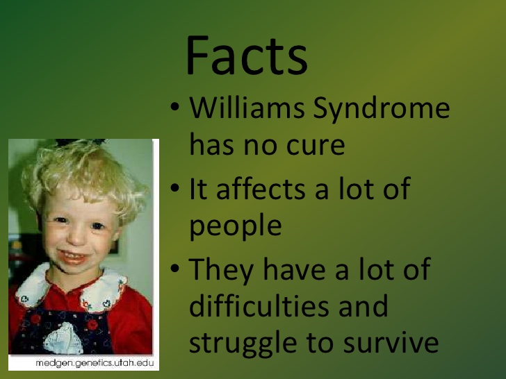
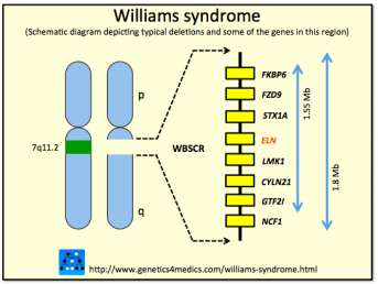
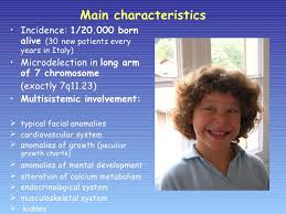
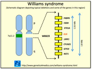
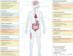
Williams syndrome (WS) is a genetic condition that is present at birth and can affect anyone.  It is characterized by medical problems, including cardiovascular disease, developmental delays, and learning disabilities.  The most significant medical problem associated with WS is the cardiovascular disease caused by the narrowed arteries. WS is also associated with elevated blood calcium levels in infancy. A random genetic mutation (deletion of a small piece of chromosome 7), rather than inheritance, most often causes the disorder. Williams syndrome is considered an autosomal dominant condition because one copy of the altered chromosome 7 in each cell is sufficient to cause the disorder. In a small percentage of cases, people with Williams syndrome inherit the chromosomal deletion from a parent with the condition.

Most cases of Williams syndrome are not inherited but occur as random events during the formation of reproductive cells (eggs or sperm) in a parent of an affected individual. These cases occur in people with no history of the disorder in their family.

However, individuals who have WS have a 50 percent chance of passing it on if they decide to have children. These often occur side by side with striking verbal abilities, highly social personalities and an affinity for music.

WS affects 1 in 7,500 – 10,000 people worldwide – an estimated 20,000 to 30,000 people in the United States. It is known to occur equally in both males and females and in every culture.

Unlike disorders that can make connecting with your child difficult, children with Williams syndrome tend to be social, friendly and endearing.  Parents often say the joy and perspective a child with WS brings into their lives had been unimaginable.

But there are major struggles as well.  Many babies have life-threatening cardiovascular problems.  Children with WS need costly and ongoing medical care and early interventions (such as speech or occupational therapy) that may not be covered by insurance or state funding.  As they grow, they struggle with things like spatial relations, numbers, and abstract reasoning, which can make daily tasks a challenge. As adults, most people with Williams syndrome will need supportive housing to live to their fullest potential.  Many adults with WS contribute to their communities as volunteers or paid employees; often working at assisted living homes for senior citizens, hospitals and libraries, or as store greeters or veterinary aides.

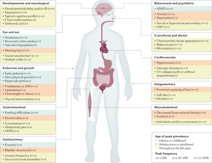
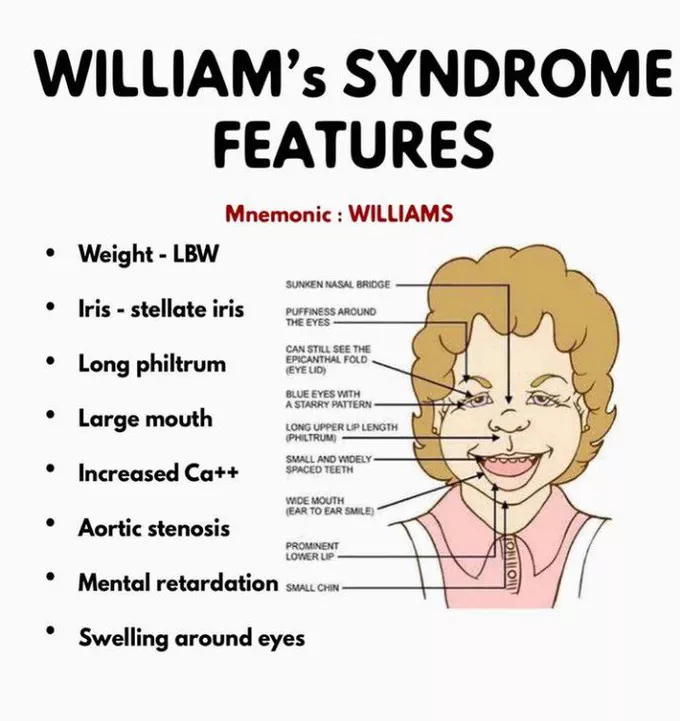
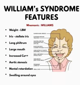
However, individuals who have WS have a 50 percent chance of passing it on if they decide to have children. The characteristic facial features of WS include puffiness around the eyes, a short nose with a broad nasal tip, wide mouth, full cheeks, full lips, and a small chin. People with WS are also likely to have a long neck, sloping shoulders, short stature, limited mobility in their joints, and curvature of the spine. Some individuals with WS have a star-like pattern in the iris of their eyes. Infants with WS are often irritable and colicky, with feeding problems that keep them from gaining weight. Chronic abdominal pain is common in adolescents and adults. By age 30, the majority of individuals with WS have diabetes or pre-diabetes and mild to moderate sensorineural hearing loss (a form of deafness due to disturbed function of the auditory nerve). For some people, hearing loss may begin as early as late childhood. WS also is associated with a characteristic “cognitive profile” of mental strengths and weaknesses composed of strengths in verbal short-term memory and language, combined with severe weakness in visuospatial construction (the skills used to copy patterns, draw, or write). Most older children and adults with WS speak fluently and use good grammar. More than 50% of children with WS have attention deficit disorders (ADD or ADHD), and about 50% have specific phobias, such as a fear of loud noises. The majority of individuals with WS worry excessively.

Unfortunately there is no cure for Williams syndrome, nor is there a standard course of treatment.

The prognosis for individuals with WS varies. Some degree of impaired intellect is found in most people with the disorder. Some adults are able to function independently, complete academic or vocational school, and live in supervised homes or on their own; most live with a caregiver.
   

Where you can find additional information about Williams syndrome:

You may find the following resources about Williams syndrome helpful. These materials are written for the general public.


QUOTE FOR FRIDAY:

“Homocysteine is a sulfur-containing amino acid produced during the metabolism of methionine, an essential amino acid found in dietary protein. Normally, homocysteine is converted into other substances with the help of B vitamins—folate (B9), B6, and B12.  Epidemiological studies have consistently shown that higher homocysteine levels correlate with increased risk of coronary artery disease and stroke.”

Cleveland Clinic (Homocysteine: Function, Levels & Health Effects)

The relationship between High Homocysteine Levels and Heart DIsease!

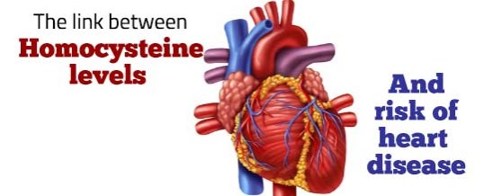
Homocysteine is a common amino acid in your blood. You get it mostly from eating meat. High levels of it are linked to early development of heart disease.

In fact, a high level of homocysteine is a risk factor for heart disease. It’s associated with low levels of vitamins B6, B12, and folate, as well as renal disease. Research has shown, however, that getting your homocysteine levels down with vitamins doesn’t reduce your chance of having heart disease.

How Does Homocysteine Increase Heart Disease Risk?

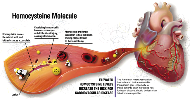
There does appear to be a relationship between high levels of homocysteine and artery damage. That can lead to atherosclerosis (hardening of the arteries) and blood clots.  Arteries filling up with blockages

Studies have shown that high levels of homocysteine are caused by a lack of nutrients in the diet, particularly the B group of vitamins. Without these essential vitamins your body is unable to produce the enzymes necessary to remove homocysteine efficiently from your blood. Homocysteine will cause damage to your arteries when present in very high concentrations.

Other causes may include aging, drug and alcohol use, impaired kidney function, problems with B12 absorption, smoking and obesity.

Do I Need to Have My Homocysteine Level Checked?

There’s no universal recommendation for checking homocysteine levels. The test is still relatively expensive, it isn’t widely available, and insurance rarely covers it but check with your insurance.The normal level of homocysteine in your blood should be up to 15 micro mol/L. This is level in the average healthy person.The optimal level of homocysteine in your blood would be under 7 micro mol/L.Make sure you get a homocysteine test as part of your next visit to the doctor, or on your own at a licensed medical facility.

Can High Homocysteine Levels Be Prevented?

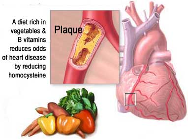
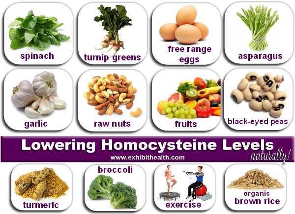
A lack of B Vitamins leads to elevated homocysteine levels which is why vegetarians are particularly at risk due to the lack of meat in their diets. Fortunately the situation is easily treatable. In the late 60’s Dr. Kilmer McCully determined through extensive research that taking adequate amounts of folic acid (vitamin B9), along with vitamins B6 and B12 will help homocysteine levels normalize.

If you have high homocysteine levels, talk to your doctor about how to change your diet.

Don’t Forget:


QUOTE FOR THURSDAY:

“Fibrodysplasia ossificans progressiva (FOP)  can be caused by a genetic condition where people are born with bunions and their body’s muscle tissue and connective tissues, like tendons and ligaments, turn into bone on the outside of their skeleton. This condition restricts movement and can cause a loss of mobility over time in people diagnosed with the condition.

Fibrodysplasia ossificans progressiva can also affect anyone because it’s most often the result of a new genetic mutation that wasn’t inherited from a parent. Genetic mutations are unpredictable. They occur during fertilization due to an error when cells divide and replicate but their can be outside factors as causes also.”

Cleveland Clinic (Fibrodysplasia Ossificans Progressiva: Causes, Symptoms, Treatment, Outlook)

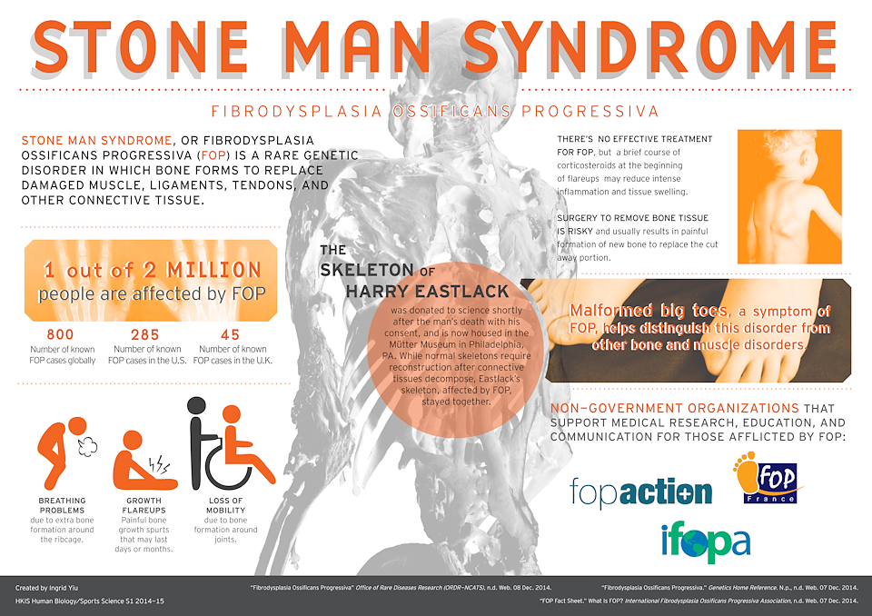
Fibrodysplasia Ossificans Progressiva/Stone Man Syndrome – What it is, the symptoms, how its diagnosed and treated!

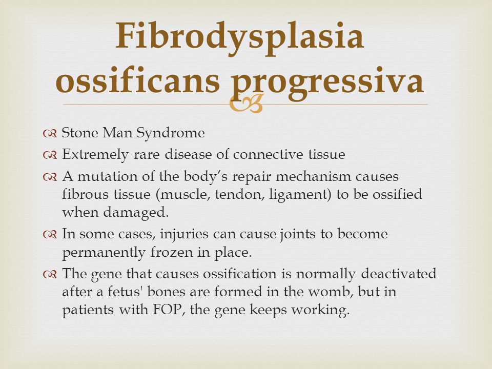
Fibrodysplasia ossificans progressiva (FOP) 

It is a disorder in which skeletal muscle and connective tissue, such as tendons and ligaments, are gradually replaced by bone (ossified). This condition leads to bone formation outside the skeleton (extra-skeletal or heterotopic bone) that restricts movement.

This process generally becomes noticeable in early childhood, starting with the neck and shoulders and moving down the body and into the limbs. People with FOP are born with abnormal big toes (hallux valgus) which can be helpful in making the diagnosis. Trauma, such as a fall or invasive medical procedure, or a viral illness may trigger episodes of muscle swelling and inflammation (myositis). These flareups lasts for several days to months and often result in permanent bone growth in the injured area.

FOP is almost always caused by a mutation at the same place in the ACVR1 gene (The ACVR1 gene provides instructions for making the activin receptor type-1 (ACVR1) protein, which is a member of a protein family called bone morphogenetic protein (BMP) type I receptors.)  and is inherited in an autosomal dominant manner. This condition occurs in about 1 in 1,600,000 newborns and about 800 people worldwide are known to have FOP.

SIGNS AND SYMPTOMS

-Fibrodysplasia ossificans progressiva (FOP) is characterized by the gradual replacement of muscle tissue and connective tissue (such as tendons and ligaments) by bone, restricting movement. This process generally becomes noticeable in early childhood, starting with the neck and shoulders and proceeding down the body and into the limbs.

-The formation of extra-skeletal bone causes progressive loss of mobility as the joints become affected. Speaking and eating may also become difficult as the mouth becomes affected. Over time, people with FOP may become malnourished because of the inability to eat. They may also develop breathing difficulties as a result of extra bone formation around the rib cage that restricts expansion of the lungs.

-Any trauma to the muscles of an individual with FOP (a fall or an invasive medical procedure) may trigger episodes of muscle swelling and inflammation followed by more rapid ossification in the injured area. Flare-ups may also be caused by viral illnesses such as the flu.

-Affected individuals may also have short thumbs and other skeletal abnormalities.

Inheritance

-Fibrodysplasia ossificans progressiva is inherited in an autosomal dominant pattern, which means one copy of the altered gene in each cell is sufficient to cause the disorder.

-Most cases of fibrodysplasia ossificans progressiva result from new mutations in the gene. These cases occur in people with no history of the disorder in their family. In only a small number of cases, an affected person has inherited the mutation from one affected parent.

How this disease is diagnosed:

-Making a diagnosis for a genetic or rare disease can often be challenging. Healthcare professionals typically look at a person’s medical history, symptoms, physical exam, and laboratory test results in order to make a diagnosis. The following resources provide information relating to diagnosis and testing for this condition. If you have questions about getting a diagnosis, you should contact a healthcare professional.

-The Genetic Testing Registry (GTR) provides information about the genetic tests for this condition. The intended audience for the GTR is health care providers and researchers. Patients and consumers with specific questions about a genetic test should contact a health care provider or a genetics professional.

TREATMENT:

There is currently no definitive treatment.  However, a brief course of high-dose corticosteroids, such as Prednisone, started within the first 24 hours of a flare-up, may help reduce the intense inflammation and tissue swelling seen in the early stages of fibrodysplasia ossificans progressiva.  Other medications, such as muscle relaxants, mast cell inhibitors, and aminobisphosphonates, if appropriate, should be closely monitored by a physician.  Surgery to remove heterotopic and extra-skeletal bone is risky and can potentially cause painful new bone growth.

References:


  • Fibrodysplasia ossificans progressiva. Genetics Home Reference (GHR). August 2007; http://ghr.nlm.nih.gov/condition/fibrodysplasia-ossificans-progressiva.
  • FOP Fact Sheet. International Fibrodysplasia Ossificans Progressiva Association. http://www.ifopa.org/what-is-fop/overview.html. Accessed 6/5/2014.
  • Pignolo R, Kaplan F. Pediatric Fibrodysplasia Ossificans Progressiva. E-medicine. July 30, 2009; http://emedicine.medscape.com/article/1007104-overview. Accessed 3/17/2011