Part II National Brain Injury Awareness Month!

Overview

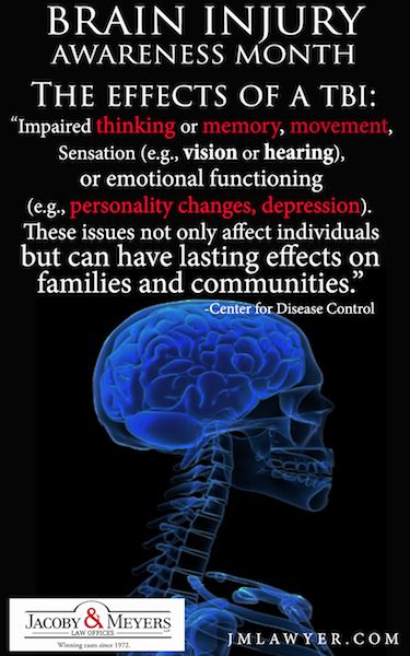
Traumatic brain injury (TBI) is sudden damage to the brain caused by a blow or jolt to the head. Common causes include car or motorcycle crashes, falls, sports injuries, and assaults. Injuries can range from mild concussions to severe permanent brain damage. While treatment for mild TBI may include rest and medication, severe TBI may require intensive care and life-saving surgery. Those who survive a brain injury can face lasting effects in their physical and mental abilities as well as emotions and personality. Most people who suffer moderate to severe TBI will need rehabilitation to recover and relearn skills.

What is a traumatic brain injury?

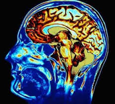
TBI is an injury to the brain caused by a blow or jolt to the head from blunt or penetrating trauma. The injury that occurs at the moment of impact is known as the primary injury. Primary injuries can involve a specific lobe of the brain or can involve the entire brain. Sometimes the skull may be fractured, but not always. During the impact of an accident, the brain crashes back and forth inside the skull causing bruising, bleeding, and tearing of nerve fibers (Fig. 1). Immediately after the accident the person may be confused, not remember what happened, have blurry vision and dizziness, or lose consciousness. At first the person may appear fine, but their condition can decline rapidly. After the initial impact occurs, the brain undergoes a delayed trauma – it swells – pushing itself against the skull and reducing the flow of oxygen-rich blood. This is called secondary injury, which is often more damaging than the primary injury.

TBI

Figure 1. During impact to the head, the soft brain crashes back and forth against the inside of the hard skull causing bruising, bleeding, and shearing of the brain.

Traumatic brain injuries are classified according to the severity and mechanism of injury:

  • Mild: person is awake; eyes open. Symptoms can include confusion, disorientation, memory loss, headache, and brief loss of consciousness.
  • Moderate: person is lethargic; eyes open to stimulation. Loss of consciousness lasting 20 minutes to 6 hours. Some brain swelling or bleeding causing sleepiness, but still arousable.
  • Severe: person is unconscious; eyes do not open, even with stimulation. Loss of consciousness lasting more than 6 hours.

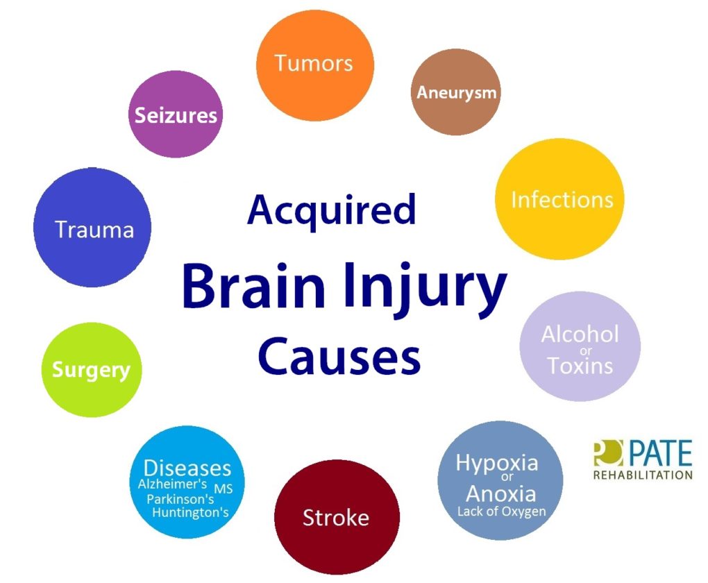
Types of traumatic brain injuries

  • Concussion is a mild head injury that can cause a brief loss of consciousness and usually does not cause permanent brain injury.
  • Contusion is a bruise to a specific area of the brain caused by an impact to the head; also called coup or contrecoup injuries. In coup injuries, the brain is injured directly under the area of impact, while in contrecoup injuries it is injured on the side opposite the impact.
  • Diffuse axonal injury (DAI) is a shearing and stretching of the nerve cells at the cellular level. It occurs when the brain quickly moves back and forth inside the skull, tearing and damaging the nerve axons. Axons connect one nerve cell to another throughout the brain, like telephone wires. Widespread axonal injury disrupts the brain’s normal transmission of information and can result in substantial changes in a person’s wakefulness.
  • Traumatic Subarachnoid Hemorrhage (tSAH) is bleeding into the space that surrounds the brain. This space is normally filled with cerebrospinal fluid (CSF), which acts as a floating cushion to protect the brain. Traumatic SAH occurs when small arteries tear during the initial injury. The blood spreads over the surface of the brain causing widespread effects.
  • Hematoma is a blood clot that forms when a blood vessel ruptures. Blood that escapes the normal bloodstream starts to thicken and clot. Clotting is the body’s natural way to stop the bleeding. A hematoma may be small or it may grow large and compress the brain. Symptoms vary depending on the location of the clot. A clot that forms between the skull and the dura lining of the brain is called an epidural hematoma. A clot that forms between the brain and the dura is called a subdural hematoma. A clot that forms deep within the brain tissue itself is called an intracerebral hematoma. Over time the body reabsorbs the clot. Sometimes surgery is performed to remove large clots.

Although described as individual injuries, a person who has suffered a TBI is more likely to have a combination of injuries, each of which may have a different level of severity. This makes answering questions like “what part of the brain is hurt?” difficult, as more than one area is usually involved.

Secondary brain injury occurs as a result of the body’s inflammatory response to the primary injury. Extra fluid and nutrients accumulate in an attempt to heal the injury. In other areas of the body, this is a good and expected result that helps the body heal. However, brain inflammation can be dangerous because the rigid skull limits the space available for the extra fluid and nutrients. Brain swelling increases pressure within the head, which causes injury to parts of the brain that were not initially injured. The swelling happens gradually and can occur up to 5 days after the injury.

What are the symptoms?

Depending on the type and location of the injury, the person’s symptoms may include:

  • Loss of consciousness
  • Confusion and disorientation
  • Memory loss / amnesia
  • Fatigue
  • Headaches
  • Visual problems
  • Poor attention / concentration
  • Sleep disturbances
  • Dizziness / loss of balance
  • Irritability / emotional disturbances
  • Feelings of depression
  • Seizures
  • Vomiting

Diffuse injuries (such as a concussion or diffuse axonal injury) will typically cause an overall decreased level of consciousness. Whereas, focal injuries (such as an ICH or a contusion) will have symptoms based on the brain area affected (Fig. 2).

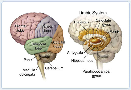
Lobes of the brain that can get affected by TBI:

brain anatomy

Figure 2. The brain is composed of three parts: the brainstem, cerebellum, and cerebrum, which is divided into lobes. The table lists the lobes of the brain and their normal functions as well as problems that may occur when injured. While an injury may occur in a specific area, it is important to understand that the brain functions as a whole by interrelating its component parts.

Every patient is unique and some injuries can involve more than one area or a partial section, making it difficult to predict which specific symptoms the patient will experience.

QUOTE FOR WEDNESDAY:

“Common Causes of Traumatic Brain Injury:

▪Falls
▪Motor vehicle crashes
▪Sports related injuries
▪Explosive blast/military
combat injuries

Common Causes of Non-Traumatic Brain Injury:

▪Stroke
▪Near-drowning
▪Aneurysm
▪Tumor
▪ Infectious disease that affects
the brain
▪Lack of oxygen supply to the brain”

Brain Injury Association of America (Brain Injury Awareness Month – Brain Injury Association of America)


Part I National Brain Injury Awareness Month

  BRAIN INJURY IMAGE  brain Lobe Regions


An acquired brain injury (ABI) is an injury to the brain that is not hereditary, congenital, degenerative, or induced by birth trauma. Essentially, this type of brain injury is one that has occurred after birth. The injury results in a change to the brain’s neuronal activity, which affects the physical integrity, metabolic activity, or functional ability of nerve cells in the brain.

There are two types of acquired brain injury: traumatic and non-traumatic.

A traumatic brain injury (TBI) is defined as an alteration in brain function, or other evidence of brain pathology, caused by an external force. Traumatic impact injuries can be defined as closed (or non-penetrating) or open (penetrating).

Often referred to as an acquired brain injury, a non-traumatic brain injury causes damage to the brain by internal factors, such as a lack of oxygen, exposure to toxins, pressure from a tumor, etc.

A Brain Injury is damage to the brain that results in a loss of function such as mobility or feeling.

Traumatic Brain Injuries can result from a closed head injury or a penetrating head injury.

Closed Injury:  A closed injury occurs when the head suddenly and violently hits an object but the object does not break through the skull.

Penetrating Injury: A penetrating injury occurs when an object pierces the skull and enters brain tissue. As the first line of defense, the skull is particularly vulnerable to injury. Skull fractures occur when the bone of the skull cracks or breaks. A depressed skull fracture occurs when pieces of the broken skull press into the tissue of the brain. A penetrating skull fracture occurs when something pierces the skull, such as a bullet, leaving a distinct and localized injury to brain tissue. Skull fractures can cause cerebral contusion.

Brain trauma occurs when a person has an injury to the brain, and can be mild or severe. When a person sustains trauma to the brain, he or she may lose motor functions along with cognitive and physical abilities. Physicians use the Glasgow Coma Scale to determine the extent of brain trauma. This is a neurological scale that measures the level of a person’s consciousness. The Rancho Los Amigos Scale is used to monitor the recovery of the brain.

There are several different types of brain injuries. A mild injury may cause temporary symptoms while a severe injury could require years of rehabilitation. The two most common types of brain trauma are:

1. Traumatic Brain Injuries 

2. Acquired Brain Injuries.

1-Traumatic brain injury occurs from external force on the head or neck. These injuries can occur from blows to the head or aggressive twisting of the neck. Some ways this could happen include falls, motor vehicle accidents, sports, and vigorous shaking. In infants, Shaken Baby Syndrome is a type of traumatic brain injury.

2-An acquired brain injury means simply you got this injury after you were born and it was caused by a condition or illness after birth. This type of injury can result from several different causes like strokes, toxic poisoning or brain tumors. Degenerative diseases and lack of oxygen may also cause this type of brain trauma. Here are some examples of acquired brain injuries:

-Bleeding in the brain which can lead to brain injury.   Blood Vessels in the brain can rupture resulting in an intra-cerebral hemorrhagic (one of the causes of a stroke, its what we called Hemorrhagic Stroke). Symptoms may include headaches, loss of vision, weakness to one side of the body and eye pain to even garbled speech.

Other Causes of Brian Injury:

-Anoxia is another insult to the brain that can cause injury. Anoxia is a condition in which there is an absence of oxygen supply to an organ’s tissues, even if there is adequate blood flow to the tissue.  Common causes of anoxia are near drowning, choking, suffocation, strangulation, heart attacks, lung damage, or very low blood pressure.  They all decrease oxygen intake to the red blood cells that feed the organs nutrients (being oxygen) to our body.  Anoxia is starvation to our body tissues.  

-Hypoxia:

Hypoxia refers to a decrease in oxygen supply rather than a complete absence of oxygen, and ischemia is inadequate blood supply, as is seen in cases in which the brain swells that causes compressing on the brain vessels near where the swelling is. In any of these cases, without adequate oxygen, a biochemical cascade called the ischemic cascade is unleashed, and the cells of the brain can die within several minutes. This type of injury is often seen in near-drowning victims, in heart attack patients, or in people who suffer significant blood loss from other injuries that decrease blood flow to the brain.

-Toxemia:

This occurs due to poisoning from chemical or biological factors that can damage the brain. Toxemia can be caused by drugs, chemicals, gases or even toxic foods.

-Viruses and bacteria. An infection of the brain can be very damaging like:

*Meningitis is a inflammation of the lining around the brain or spinal cord, usually due to infection; Neck stiffness, headache, fever, and confusion are common symptoms.

*Encephalitis (en-sef-uh-LIE-tis) is inflammation of the brain. Viral infections are the most common cause of the condition. Encephalitis can cause flu-like symptoms, such as a fever or severe headache. It can also cause confused thinking, seizures, or problems with senses or movement.

*HIV can lead to brain injury. HIV, can affect the brain in different ways. HIV-meningoencephalitis is infection of the brain and the lining of the brain by the HIV virus. It occurs shortly after the person is first infected with HIV and may cause headache, neck stiffness, drowsiness, confusion and/or seizures. HIV-encephalopathy (HIV-associated dementia) is the result of damage to the brain by longstanding HIV infection.  It is a form of dementia and occurs in advanced HIV infection. Mild Neurocognitive Disorder is problems with thinking and memory in HIV, however is not as severe as HIV-encephalopathy. Unlike HIV-encephalopathy it can occur early in HIV infection and is not a feature of Aquired Immune Deficiency Syndrome – AIDS.

*Lastly, Herpes. There are two types of herpes simplex virus (HSV). Either type can cause encephalitis. HSV type 1 (HSV-1) is usually responsible for cold sores or fever blisters around your mouth, and HSV type 2 (HSV-2) commonly causes genital herpes. Encephalitis caused by HSV-1 is rare, but it has the potential to cause significant brain damage or death.

*Other herpes viruses. Other herpes viruses that may cause encephalitis include the Epstein-Barr virus, which commonly causes infectious mononucleosis, and the varicella-zoster virus, which commonly causes chickenpox and shingles.*Viral infections due to blood sucking insects like mosquitoes and ticks to animals with rabies a rapid progression to encephalitis once symptoms begin. Rabies is a rare cause of encephalitis in the U.S.

When a person is diagnosed with a brain trauma, doctors will decide if rehabilitation is needed.

Rehabilitation programs may vary depending on the type of brain injury and estimated recovery time. Treatment usually consists of physical therapy and daily activities. In extreme cases, patients may need to learn how to read and write again.

Therapy for brain trauma typically takes place on an outpatient basis or through an assisted living facility. Therapy may last several weeks, months or even years, and sometimes the patient is not able to make a full recovery.

It may not always be obvious when a person has sustained a brain injury. The patient may have hit his or her head and not have symptoms until a few hours later. Some signs of a possible brain injury are headaches, confusion and loss of memory. If brain trauma is not treated, it could cause permanent damage or death.

Brain injuries can affect the patient and the patient’s family, with emotional and financial hardship. When problems arise with treatment or financial issues, a brain injury lawyer or specialist may need to intervene.

REVISED 2/25/2021 By Elizabeth Lynch RN BSN Cardiac/Stroke Certified (RN 34.5 Years)


QUOTE FOR THE WEEKEND:

“Colon cancer is a growth of cells that begins in a part of the large intestine called the colon. The colon is the first and longest part of the large intestine. The large intestine is the last part of the digestive system. The digestive system breaks down food for the body to use.

Colon cancer typically affects older adults, though it can happen at any age. It usually begins as small clumps of cells called polyps that form inside the colon. Polyps generally aren’t cancerous, but some can turn into colon cancers over time.

Polyps often don’t cause symptoms. For this reason, doctors recommend regular screening tests to look for polyps in the colon. Finding and removing polyps helps prevent colon cancer.”

MAYO  CLINIC (Colon cancer – Symptoms and causes – Mayo Clinic)

QUOTE FOR FRIDAY:

“Coronary artery disease (CAD)

  • Coronary heart disease is the most common type of heart disease. It killed 371,506 people in 2022.1
  • About 1 in 20 adults age 20 and older have CAD (about 5%).3
  • In 2022, about 1 out of every 5 deaths from cardiovascular diseases (CVDs) was among adults younger than 65 years old.1

Heart attack

  • In the United States, someone has a heart attack every 40 seconds.3
  • Every year, about 805,000 people in the United States have a heart attack.3 Of these, 605,000 are a first heart attack, and 200,000 happen to be people who have already had a heart attack.3
  • About 1 in 5 heart attacks are silent—the damage is done, but the person is not aware of it.”

Centers for Disease Prevention – CDC (Heart Disease Facts | Heart Disease | CDC)

QUOTE FOR THURSDAY:

“Knowledge is power. If you understand the risks for heart attack, you can take steps to improve your health.

Risk factors are traits and lifestyle habits that can increase your chance of having a heart attack. So, it’s important to know them. You can change some risk factors, some you can’t.

It’s important to know your risk factors for a heart attack.

  • You can control some risk factors, such as tobacco use, physical activity, blood pressure, blood cholesterol and blood glucose.
  • Work with your health care team to manage your risk of heart disease.”

American Heart Association (Understand Your Risks to Prevent a Heart Attack | American Heart Association)

What are the risk factors for myocardial infarction/heart attack?


Continuation of February being the month of the heart I decided to cover the risks of a myocardial infarction (heart attack).

People who are at risk for the development of coronary artery disease and myocardial infarction include those who fall into any of the categories listed below:

-People with a history of heart disease.

-Males.

-Smokers.

-People with high cholesterol.

-People with high blood pressure.

-Obese people.

-People with diabetes.

-People who suffer stress.

-People who live a sedentary life style.

-Heredity is a powerful factor that contributes to early heart disease. Being male is a risk factor, but the incidence of heart disease in women increases dramatically after menopause.

-The risk factors to concentrate upon are those that can be modified. These include cigarette smoking, high blood pressure, cholesterol, obesity, sedentary life style and stress. Cigarette smoking causes many deaths from myocardial infarction and other heart diseases. Smoking contributes to almost half of the heart attacks of women under age 55.

-Stopping smoking can greatly reduce your chances of having a heart attack. Controlling blood pressure can reduce your risk of heart attack. Lowering cholesterol to safe levels through diet and medications can reduce your risk and may even lead to some regression of the plaques already present. Lean body weight and a regular exercise program are helpful.

-If you are diabetic, precise control of your diabetes will help reduce your risk of blood vessel damage due to diabetes. Stress is a risk factor that is common, difficult to quantify and difficult to control effectively over time. Methods of stress reduction include meditation, regular exercise, time management, and a supportive environment.

How is a heart attack diagnosed?

Chest pain is the most common symptom of a heart attack. The chest pain is usually a burning or pressure sensation beneath the mid or upper breast bone. The pain may radiate into the upper mid back, neck, jaw or arms. The pain may be severe but often is only moderate in severity.

There may be associated shortness of breath or sweating. If patients have had angina previously, the heart attack pain will feel the same as their usual angina only stronger and more prolonged. If you have a pain like this that lasts longer than 15 minutes, it is best to be evaluated immediately.

Calling your medic unit is the fastest and safest way to ask for help. If you have symptoms like this that wax and wane, this is often a warning sign that a heart attack is about to occur and prompt medical attention is needed.

Once you are in an emergency room or a doctor’s office an electrocardiogram (ECG or EKG) will be obtained. This is often helpful in diagnosing a heart attack. Sometimes, however, the test is normal even when the heart injury is present but usually a great diagnostic tool.

When heart cells die, certain enzymes present in heart cells are released into the bloodstream that serve as a marker of heart injury (troponin I and CPK or CK-MB). These enzymes can be measured by blood tests. The amount of enzyme released into the blood stream also helps assess how much heart damage has occurred.

TREATMENT:

The best way to limit the size of a heart attack is to restore the flow of blood to the heat muscle as fast as possible. There are two basic methods to do this.

Because most heart attacks are caused by clots forming within the coronary artery, dissolving the clot quickly will restore blood flow. Drugs called thrombolytics are quite effective.

The sooner these drugs are given, the quicker the blood flow will be restored. An alternative method involves the use of balloon angioplasty.

This involves taking the heart attack victim promptly to the cardiac cath lab in the hospital.

An angiogram is performed to show the blocked blood vessel leading to the heart attack. Then a balloon catheter is placed across the blockage and flow is restored.

Sometimes a stent (a device that assists in holding the blood vessel open) is placed to create a large channel.

Smaller heart attacks, often those not producing significant abnormalities on the ECG are often treated with bedrest and blood thinners such as heparin as well as drugs to reduce the work the heart does.

These heart attacks are called non-transmural myocardial infarctions. Before discharge, x-ray studies of the heart arteries are often carried out to see if angioplasty or surgery will be necessary.

Following thrombolytic (clot reducing) therapy, angiogram are often performed to outline the coronary anatomy to help determine if additional therapy such as angioplasty or bypass surgery is indicated. This may be done during the initial hospitalization or later as an outpatient procedure based on the severity of results in the diagnostic tooling the MD orders (tests).

QUOTE FOR WEDNESDAY:

“SARS (severe acute respiratory syndrome, SARS-CoV-1) was a respiratory illness that spread in many countries around the world in 2002 and 2003. For many people, SARS caused flu-like symptoms (like fever and headache), but it progressed to severe illness in about 10% to 20% of people during the outbreak.

More than 8,000 people in 29 countries had SARS during the 2002-2003 outbreak. There was a small outbreak related to occupational exposure in 2004. Preventive measures stopped its spread, and only nine people were infected. There haven’t been any reported cases since then.

A virus called SARS-associated coronavirus (SARS-CoV-1) causes SARS.”

Cleveland Clinic (Severe Acute Respiratory Syndrome (SARS): Cause & Treatment)

What is SARS that first occurred February 2003 in China?

  4 days ago

World Health Organization on this disease SARS coronavirus:

Severe acute respiratory syndrome (SARS) is a viral respiratory disease caused by a SARS-associated coronavirus. It was first identified at the end of February 2003 during an outbreak that emerged in China and spread to 4 other countries.

The virus identified in 2003. SARS-CoV is thought to be an animal virus from an as-yet-uncertain animal reservoir, perhaps bats, that spread to other animals (civet cats) and first infected humans in the Guangdong province of southern China in 2002.

Transmission

An epidemic of SARS affected 26 countries and resulted in more than 8000 cases in 2003. Since then, a small number of cases have occurred as a result of laboratory accidents or, possibly, through animal-to-human transmission (Guangdong, China).

Transmission of SARS-CoV is primarily from person to person. It appears to have occurred mainly during the second week of illness, which corresponds to the peak of virus excretion in respiratory secretions and stool, and when cases with severe disease start to deteriorate clinically. Most cases of human-to-human transmission occurred in the health care setting, in the absence of adequate infection control precautions. Implementation of appropriate infection control practices brought the global outbreak to an end.

Nature of the disease

Symptoms are influenza-like and include fever, malaise, myalgia, headache, diarrhoea, and shivering (rigors). No individual symptom or cluster of symptoms has proved to be specific for a diagnosis of SARS. Although fever is the most frequently reported symptom, it is sometimes absent on initial measurement, especially in elderly and immunosuppressed patients.

Cough (initially dry), shortness of breath, and diarrhoea are present in the first and/or second week of illness. Severe cases often evolve rapidly, progressing to respiratory distress and requiring intensive care.

Geographical distribution

The distribution is based on the 2002–2003 epidemic. The disease appeared in November 2002 in the Guangdong province of southern China. This area is considered as a potential zone of re-emergence of SARS-CoV.

Other countries/areas in which chains of human-to-human transmission occurred after early importation of cases were Toronto in Canada, Hong Kong Special Administrative Region of China, Chinese Taipei, Singapore, and Hanoi in Viet Nam.

Risk for travellers

Currently, no areas of the world are reporting transmission of SARS. Since the end of the global epidemic in July 2003, SARS has reappeared four times – three times from laboratory accidents (Singapore and Chinese Taipei), and once in southern China where the source of infection remains undetermined although there is circumstantial evidence of animal-to-human transmission.

Should SARS re-emerge in epidemic form, WHO will provide guidance on the risk of travel to affected areas. Travellers should stay informed about current travel recommendations. However, even during the height of the 2003 epidemic, the overall risk of SARS-CoV transmission to travellers was low.

Prophylaxis

None. Experimental vaccines are under development.

The National Institute of Health (NIH) states:

The virus that causes coronavirus disease 2019 (COVID-19) is stable for several hours to days in aerosols and on surfaces, according to a new study from National Institutes of Health, CDC, UCLA and Princeton University scientists in The New England Journal of Medicine. The scientists found that severe acute respiratory syndrome coronavirus 2 (SARS-CoV-2) was detectable in aerosols for up to three hours, up to four hours on copper, up to 24 hours on cardboard and up to two to three days on plastic and stainless steel. The results provide key information about the stability of SARS-CoV-2, which causes COVID-19 disease, and suggests that people may acquire the virus through the air and after touching contaminated objects. The study information was widely shared during the past two weeks after the researchers placed the contents on a preprint server to quickly share their data with colleagues.

The NIH scientists, from the National Institute of Allergy and Infectious Diseases’ Montana facility at Rocky Mountain Laboratories, compared how the environment affects SARS-CoV-2 and SARS-CoV-1, which causes SARS. SARS-CoV-1, like its successor now circulating across the globe, emerged from China and infected more than 8,000 people in 2002 and 2003. SARS-CoV-1 was eradicated by intensive contact tracing and case isolation measures and no cases have been detected since 2004. SARS-CoV-1 is the human coronavirus most closely related to SARS-CoV-2. In the stability study the two viruses behaved similarly, which unfortunately fails to explain why COVID-19 has become a much larger outbreak.

The NIH study attempted to mimic virus being deposited from an infected person onto everyday surfaces in a household or hospital setting, such as through coughing or touching objects. The scientists then investigated how long the virus remained infectious on these surfaces.

The scientists highlighted additional observations from their study:

  • If the viability of the two coronaviruses is similar, why is SARS-CoV-2 resulting in more cases? Emerging evidence suggests that people infected with SARS-CoV-2 might be spreading virus without recognizing, or prior to recognizing, symptoms. This would make disease control measures that were effective against SARS-CoV-1 less effective against its successor.
  • In contrast to SARS-CoV-1, most secondary cases of virus transmission of SARS-CoV-2 appear to be occurring in community settings rather than healthcare settings.  However, healthcare settings are also vulnerable to the introduction and spread of SARS-CoV-2, and the stability of SARS-CoV-2 in aerosols and on surfaces likely contributes to transmission of the virus in healthcare settings.

The findings affirm the guidance from public health professionals to use precautions similar to those for influenza and other respiratory viruses to prevent the spread of SARS-CoV-2:

  • Avoid close contact with people who are sick.
  • Avoid touching your eyes, nose, and mouth.
  • Stay home when you are sick.
  • Cover your cough or sneeze with a tissue, then throw the tissue in the trash.
  • Clean and disinfect frequently touched objects and surfaces using a regular household cleaning spray or wipe.

QUOTE FOR TUESDAY:

The CDC states:

“-Seasonal influenza (flu) vaccines are designed to protect against the three influenza viruses that are expected to circulate most widely during the upcoming season.

-CDC is working with other federal agencies and the private sector, to improve flu vaccine technologies.

-Three different flu vaccine production technologies are FDA approved for use in the United States: egg-based flu vaccine, cell-culture based flu vaccine, and recombinant flu vaccine.

Influenza (flu) vaccines cause antibodies to develop in the body about two weeks after vaccination. These antibodies provide protection against infection with the influenza viruses that are used to make vaccine. The seasonal flu vaccine protects against the influenza viruses that research suggests will be most common during the upcoming season. These include three different main groups of influenza Type A and B viruses.”

Center for Disease Control and Prevention – CDC (How Influenza (Flu) Vaccines Are Made | Influenza (Flu) | CDC&Different Types of Flu Vaccines | Influenza (Flu) | CDC)