“I would not have traded two minutes of the joy and the grief with that man for two decades of anything with another.”
– Lou Gehrig’s wife, Eleanor
“I would not have traded two minutes of the joy and the grief with that man for two decades of anything with another.”
– Lou Gehrig’s wife, Eleanor
ALS, or amyotrophic lateral sclerosis, is a progressive neurodegenerative disease that affects nerve cells in the brain and the spinal cord. A-myo-trophic comes from the Greek language. “A” means no. “Myo” refers to muscle, and “Trophic” means nourishment – “No muscle nourishment.” When a muscle has no nourishment, it “atrophies” or wastes away. “Lateral” identifies the areas in a person’s spinal cord where portions of the nerve cells that signal and control the muscles are located. As this area degenerates it leads to scarring or hardening (“sclerosis”) in the region.
Motor neurons reach from the brain to the spinal cord and from the spinal cord to the muscles throughout the body. The progressive degeneration of the motor neurons in ALS eventually leads to their demise. When the motor neurons die, the ability of the brain to initiate and control muscle movement is lost. With voluntary muscle action progressively affected, people may lose the ability to speak, eat, move and breathe. The motor nerves that are affected when you have ALS are the motor neurons that provide voluntary movements and muscle control. Examples of voluntary movements are making the effort to reach for a smart phone or step off a curb. These actions are controlled by the muscles in the arms and legs. Motor neurons are nerve cells located in the brain, brain stem, and spinal cord that serve as controlling units and vital communication links between the nervous system and the voluntary muscles of the body. Messages from motor neurons in the brain called upper motor neurons are transmitted to motor neurons in the spinal cord which are lower motor neurons and from them to particular muscles. The problem with ACL, both the upper motor niurons and the ower motor niurons degenerate or die which causes stoppage of sending messages to muscles. Unable to function, the muscles gradually weaken, waste away (atrophy), and have very fine twitches (called fasciculations). Eventually, the ability of the brain to start voluntary movement with messages is unable to work anymore.
Symptoms:
The onset of ALS may be so subtle that the symptoms are overlooked. The earliest symptoms may include fasciculations, cramps, tight and stiff muscles (spasticity), muscle weakness affecting an arm or a leg, slurred and nasal speech, or difficulty chewing or swallowing. These general complaints then develop into more obvious weakness or atrophy that may cause a physician to suspect ALS. Regardless of the part of the body first affected by the disease, muscle weakness and atrophy spread to other parts of the body as the disease progresses.
The parts of the body showing early symptoms of ALS depend on which muscles in the body are affected. Many individuals first see the effects of the disease in a hand or arm as they experience difficulty with simple tasks requiring manual dexterity such as buttoning a shirt, writing, or turning a key in a lock. In other cases, symptoms initially affect one of the legs, and people experience awkwardness when walking or running or they notice that they are tripping or stumbling more often.
There are two different types of ALS, sporadic and familial. Sporadic which is the most common form of the disease in the U.S., is 90 – 95 percent of all cases. It may affect anyone, anywhere. Familial ALS (FALS) accounts for 5 to 10 percent of all cases in the U.S. Familial ALS means the disease is inherited. In those families, there is a 50% chance each offspring will inherit the gene mutation and may develop the disease. French neurologist Jean-Martin Charcot discovered the disease in 1869.
The cause of ALS is not known, and scientists do not yet know why ALS strikes some people and not others. An important step toward answering this question was made in 1993 when scientists supported by the National Institute of Neurological Disorders and Stroke (NINDS) discovered that mutations in the gene that produces the SOD1 enzyme were associated with some cases of familial ALS.
Recent years have brought a wealth of new scientific understanding regarding the physiology of this disease. There is currently one FDA approved drug, riluzole, that modestly slows the progression of ALS in some people. Although there is not yet a cure or treatment that halts or reverses ALS, scientists have made significant progress in learning more about this disease. In addition, people with ALS may experience a better quality of life in living with the disease by participating in support groups and attending an ALS Association Certified Treatment Center of Excellence or a Recognized Treatment Center. Such Centers provide a national standard of best-practice multidisciplinary care to help manage the symptoms of the disease and assist people living with ALS to maintain as much independence as possible for as long as possible. According to the American Academy of Neurology’s Practice Paramater Update, studies have shown that participation in a multidisciplinary ALS clinic may prolong survival and improve quality of life. To find a Center near you, visit http://www.alsa.org/community/certified-centers/.
ALS usually strikes people between the ages of 40 and 70, and approximately 20,000 Americans can have the disease at any given time (although this number fluctuates). For unknown reasons, military veterans are approximately twice as likely to be diagnosed with the disease than the general public. Notable individuals who have been diagnosed with ALS include baseball great Lou Gehrig, Hall of Fame pitcher Jim “Catfish” Hunter, Toto bassist Mike Porcaro, Senator Jacob Javits, actor David Niven, “Sesame Street” creator Jon Stone, boxing champion Ezzard Charles, NBA Hall of Fame basketball player George Yardley, golf caddie Bruce Edwards, , musician Lead Belly (Huddie Ledbetter), photographer Eddie Adams, entertainer Dennis Day, jazz musician Charles Mingus, former vice president of the United States Henry A. Wallace, U.S. Army General Maxwell Taylor, and NFL football players Steve Gleason, O.J. Brigance and Tim Shaw.
More than 12,000 people in the U.S. have a definite diagnosis of ALS, for a prevalence of 3.9 cases per 100,000 persons in the U.S. general population, according to a report on data from the National ALS Registry. ALS is one of the most common neuromuscular diseases worldwide, and people of all races and ethnic backgrounds are affected. ALS is more common among white males, non-Hispanics, and persons aged 60–69 years, but younger and older people also can develop the disease. Men are affected more often than women.
In 90 to 95 percent of all ALS cases, the disease occurs apparently at random with no clearly associated risk factors. Individuals with this sporadic form of the disease do not have a family history of ALS, and their family members are not considered to be at increased risk for developing it.
About 5 to 10 percent of all ALS cases are inherited. The familial form of ALS usually results from a pattern of inheritance that requires only one parent to carry the gene responsible for the disease. Mutations in more than a dozen genes have been found to cause familial ALS.
About one-third of all familial cases (and a small percentage of sporadic cases) result from a defect in a gene known as “chromosome 9 open reading frame 72,” or C9orf72. The function of this gene is still unknown. Another 20 percent of familial cases result from mutations in the gene that encodes the enzyme copper-zinc superoxide dismutase 1 (SOD1).
Treatment:
No cure has yet been found for ALS. However, the Food and Drug Administration (FDA) approved the first drug treatment for the disease—riluzole (Rilutek)—in 1995. Riluzole is believed to reduce damage to motor neurons by decreasing the release of glutamate. Clinical trials with ALS patients showed that riluzole prolongs survival by several months, mainly in those with difficulty swallowing. The drug also extends the time before an individual needs ventilation support. Riluzole does not reverse the damage already done to motor neurons, and persons taking the drug must be monitored for liver damage and other possible side effects. However, this first disease-specific therapy offers hope that the progression of ALS may one day be slowed by new medications or combinations of drugs.
A Japanese legend says that if you can’t sleep at night its because you are awake in someone else’s dream. (I must be in a lot of people’s dreams).
Insomnia is a sleeping disorder that is characterized by difficulty falling and/or staying asleep. People with Insomnia have one or more of the following symptoms:
ChronicInsomnia caused by:
*Symptoms of insomnia can include:
“A Baker’s cyst, also called a popliteal (pop-luh-TEE-ul) cyst, is usually the result of a problem with your knee joint, such as arthritis or a cartilage tear.”
MAYO CLINIC
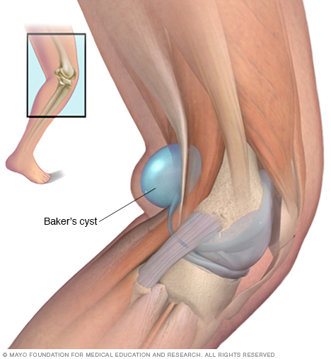
A Baker cyst is swelling caused by fluid from the knee joint protruding to the back of the knee. The back of the knee is also referred to as the popliteal area of the knee. A Baker cyst is sometimes referred to as a popliteal cyst. When an excess of knee joint fluid is compressed by the body weight between the bones of the knee joint, it can become trapped and separate from the joint to form the fluid-filled sac of a Baker cyst. The name of the cyst is in memory of the physician who originally described the condition, the British surgeon William Morrant Baker (1839-1896).
When cartilage tears or other internal knee problems are associated, physical therapy or surgery can be the best treatment option. During a surgical operation, the surgeon can remove the swollen tissue (synovium) that leads to the cyst formation. This is most commonly done with arthroscopic surgery.
There is no disease more conducive to clinical humility than aneurysm of the aorta.
William Osler (July 12, 1849 – December 29, 1919 — was a Canadian physician and one of the four founding professors of Johns Hopkins Hospital.)
The aorta is the large artery that exits in the heart and delivers blood to the body. It begins at the aortic valve that separates the left ventricle of the heart from the aorta and prevents blood from leaking back into the left ventricle after a contraction, which is actually when the heart pumps blood. The various sections of the aorta are named based upon “arch-like” initial design and the location of the aorta in the body. Thus, the beginning of the aorta is referred to as the ascending aorta (basically meaning the blood going against resistance due to the vessel being a hill for the blood to go up), followed by the arch of the aorta, then the descending aorta (which is the blood going downward via gravity with the help of the heart pumping the blood of course). The portion of the aorta that is located in the chest (called thorax) is referred to as the thoracic aorta, while the abdominal aorta (the part of the aorta below the thorax region) is located in the abdomen. The abdominal aorta extends from the diaphragm (at the bottom of the lungs like a floor to divide the lungs from the organs in the abdomen) to the mid-abdomen where it splits into the iliac arteries and when it reaches the legs the femoral arteries now start which supplies to the legs oxygenated blood. This is why commonly a cardiac catheterization to visualize the aorta and sometimes the left side of the heart is done starting in the femoral artery since in time it diverts into starting the abdominal aorta.
An aneurysm is an area of a localized widening (dilation) of a blood vessel. The word “aneurysm” is borrowed from the Greek “aneurysma” meaning “a widening”.
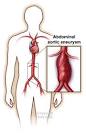
An aortic aneurysm involves the aorta, the major artery that leaves the heart to supply blood to the body. An aortic aneurysm is a dilation or bulging of the aorta..
Most aortic aneurysms are fusiform. They are shaped like a spindle (“fusus” means spindle in Latin) with widening all around the circumference of the aorta. (Saccular aneurysms just involve a portion of the aortic wall with a localized out pocketing).
What is inside an aortic aneurysm?
The inside walls of aneurysms are often lined with a blood clot that forms because there is stagnant blood. The wall of an aneurysm is layered, like a piece of plywood.
Who is most likely to have an abdominal aortic aneurysm?
Abdominal aortic aneurysms tend to occur in white males over the age of 60. In the United States, these aneurysms occur in up to 3.0% of the population. Aneurysms start to form at about age 50 and peak at age 80. Women are less likely to have aneurysms than men and African Americans are less likely to have aneurysms than Caucasians.
There is a genetic component that predisposes one to developing an aneurysm; the prevalence in someone who has a first-degree relative with the condition can be as high as 25%.
Collagen vascular diseases that can weaken the tissues of the aortic walls are also associated with aortic aneurysms. These diseases include Marfan syndrome and Ehlers-Danlos syndrome
Aortic aneurysms can develop anywhere along the length of the aorta but the majority are located in the abdominal aorta. Most of these abdominal aneurysms are located below the level of the renal arteries, the vessels that provide blood to the kidneys. Abdominal aortic aneurysms can extend into the iliac arteries.
What shape are most aortic aneurysms?
Most aortic aneurysms are fusiform. They are shaped like a spindle (“fusus” means spindle in Latin) with widening all around the circumference of the aorta. (Saccular aneurysms just involve a portion of the aortic wall with a localized out pocketing).
What is inside an aortic aneurysm?
The inside walls of aneurysms are often lined with a blood clot that forms because there is stagnant blood. The wall of an aneurysm is layered, like a piece of plywood.
Who is most likely to have an abdominal aortic aneurysm?
Abdominal aortic aneurysms tend to occur in white males over the age of 60. In the United States, these aneurysms occur in up to 3.0% of the population. Aneurysms start to form at about age 50 and peak at age 80. Women are less likely to have aneurysms than men and African Americans are less likely to have aneurysms than Caucasians.
There is a genetic component that predisposes one to developing an aneurysm; the prevalence in someone who has a first-degree relative with the condition can be as high as 25%.
Collagen vascular diseases that can weaken the tissues of the aortic walls are also associated with aortic aneurysms. These diseases include Marfan syndrome and Ehlers-Danlos syndrome.
What are risk factors for aortic aneurysms?
The risk factors for aortic aneurysm are the same as those for atherosclerotic heart disease, stroke, and peripheral artery disease and include:
“It is easier to find men who will volunteer to die, than to find those who are willing to endure pain with patience.”
Julius Caesar
‘This is potentially a very important discovery which may go a long way to explain the marked differences in pain sensitivity and chronicity between women and men.”
says James McRoberts, a pain researcher at the University of California-Los Angeles