Gerard Way (born April 9, 1977) is an American musician and comic book writer who was the lead vocalist and co-founder of the American alternative-rock band My Chemical Romance from its formation in September 2001 until its split in March 2013. He is also executive producer of a band.
Part 2 Depression vs. Anger
September is SUICIDE prevention month.
Depression is a real thing and know there is treatment for it; know what considerations with red flags to look for and what the key factor is for the person who has depression must have to take the first step in controlling it if not resolving depression completely.
Treatments
Treatment for anger and depression is based on the risk factors present, and is individualized for your specific needs. Treatment may include group therapy to discuss and rationalize anger, stress management exercises or even medications. Learning what causes anger and what can be done to avoid becoming angry are among the main focuses of treatment. Also important is learning what to do when becoming angry, and positive ways to focus feelings instead of becoming aggressive in response.
Considerations
It’s important to consider all options when deciding to deal with feelings of depression and anger. A physician can make recommendations as to which type of treatment may be best, or he can refer individuals to someone else. People shouldn’t be embarrassed or feel inadequate because they need help but should focus on the strength shown to solicit the help.
Warnings
Constant feelings of anger and depression can have a negative effect on overall health. People who are depressed and angry more than most are much more likely to suffer from heart-related problems. Anyone who experiences thoughts of harming themselves or suicide should contact a physician immediately for the appropriate treatment. If you know someone who maybe with this REACH OUT and SPEAK UP to someone significant to that individual and bring that person to an expert to help this individual. Look what happen to Robin Williams but alcoholism was the key factor in this unique talented man’s cause of suicide, by media. Now lets look at the average American; the majority of us are not millionaires to billionaires who have everything financially going for us and more. Now unless the millionaire or billionaire puts themselves in a situation that they gamble their money away and lose everything remember they could have put away a safe chest of money if that happened but if not done that was their choice. Unfortunately they have to deal with that poor judgment call caused a crash financially but life does not have to be over. It is the choice of everyone at an adult age (21 y/o and up) in where they let their money go but to those not in that situation going through depression its up to you to make your priorities regarding what is most important to you and making them reachable. That step to make priorities in what most important to you is all up to you starting it. Those things bringing you down, reach out to yourself and list what is bringing you down and address them. If its work search for something new but don’t walk out until you have s0mething new (but money doesn’t make you happy alone). Again look at Robin Williams, who at one point had everything going for him. If its your marriage well it takes not 1 but 2 to work on it. One thing you both could do that would only help you understand each other better is both address their hurts, needs, desires with each other and if none to express to each other with both parties not wanting to work on it or ending line no resolution than part to allow each one live life to its fullest with letting misery start to heal. If your in the scenario staying together just for the children if you think fighting and being unhappy in front of them all the time; you may want to think twice (their not blind). If parting to stay on a friends note is better for everyone maybe that is the choice. Of course without question if its stay together because of quilt or embarrassed in front of others if you split; just FYI these people are not blind in seeing you happy or unhappy with each other and over a long time of seeing the couple fight or unhappy they might even be thinking to themselves they wish you guys were so each individual could be possible happier. The key factor in starting to figure out what’s best for yourself is to use self discipline and give yourself a chance to figure out the best way to go to resolve it or control it is get HELP. Where the MD, or group, or psychologist, etc… can assist you whether the depression be alone or with a significant other you might be having depression over (Ex. Marriage). Ending line if you could have resolved it on your own you wouldn’t still be in depression. If this is the situation your in at this time of your life, set up what your top 3 to 5 things most important in your life is that would make life better for you and DO SOMETHING ABOUT IT don’t stay in the dark hole! Your just drowning yourself in misery. It may sound easier than it actually is but it initially takes YOU to make the first move (if you can’t help yourself than who can you help-Noone). So make yourself a happier individual to some extent to be a good impact to others or those who would want to be around you. First to reach there its advised to go to a professional (from MD to counselor to group meetings but not simply yourself (its not recommended to try to resolve this on your own if this depression has been going on for a longtime or over 6mths after a devastating situation in your life.) It’s better than doing nothing to allow yourself and others around you to be happier. Good luck.
The current data available from the Centers for Disease Control and Prevention. In 2013, there were 41,149 deaths by suicide in the United States. Suicide is the 10th leading cause of death; homicide ranks 16th. It is the second leading cause of death for 15 – 24 year olds. *  The chart above which shows the Ten Leading Causes of Death in the U.S. in 2013. For more data, you may visit www.cdc.gov or www.who.int. For more comprehensive data, visit the CDC website’s Web-based Injury Statistics Query and Reporting System (WISQARS)
General Statistics
- Suicide is the 10th leading cause of death in the US for all ages. (CDC)
- The suicide rates decreased from 1990-2000 from 12.5 suicides per 100,000 to 10.4 per 100,000. Over the past decade, however, the rate has again increased to 12.1 per 100,000. Every day, approximately 105 Americans die by suicide. (CDC)
- There is one death by suicide in the US every 13 minutes. (CDC)
- Depression affects 20-25% of Americans ages 18+ in a given year. (CDC)
- Suicide takes the lives of over 38,000 Americans every year. (CDC)
- Only half of all Americans experiencing an episode of major depression receive treatment. (NAMI)
- 80% -90% of people that seek treatment for depression are treated successfully using therapy and/or medication. (TAPS study)
- An estimated quarter million people each year become suicide survivors (AAS).
- There is one suicide for every estimated 25 suicide attempts. (CDC)
- There is one suicide for every estimated 4 suicide attempts in the elderly. (CDC)
Gender Disparaties
- Suicide among males is 4x’s higher than among females. Male deaths represent 79% of all US suicides. (CDC)
- Firearms are the most commonly used method of suicide among males (51%). (CDC)
- Access to firearms is associated with a significantly increased risk of suicide. (NAMI)
- Females are more likely than males to have had suicidal thoughts. (CDC)
- Females experience depression at roughly 2x’s the rate of men.(SMH)
- Females attempt suicide 3x’s as often as males. (CDC)
- Poisoning is the most common method of suicide for females. (CDC)
Age Disparaties
- 1 in 100,000 children ages 10 to 14 die by suicide each year. (NIMH)
- 7 in 100,000 youth ages 15 to 19 die by suicide each year. (NIMH)
- 12.7 in 100,000 young adults ages 20-24 die by suicide each year. (NIMH)
- The prevalence of suicidal thoughts, suicidal planning and suicide attempts is significantly higher among adults aged 18-29 than among adults aged 30+. (CDC)
- Suicide is the 2nd leading cause of death for 15 to 24 year old Americans. (CDC)
- Suicide is the 4th leading cause of death for adults ages 18-65. (CDC)
- The highest increase in suicide is in males 50+ (30 per 100,000). (CDC)
- Suicide rates for females are highest among those aged 45-54 (9 per 100,000). (CDC)
- Suicide rates for males are highest among those aged 75+ (36 per 100,000). (CDC)
- Suicide rates among the elderly are highest for those who are divorced or widowed. (SMH)
Racial and Ethnic Disparaties
- The highest suicide rates in the US are among Whites, American Indians and Alaska Natives.
Worldwide
- Over 800,000 people die by suicide every year. (WHO)
- There is one death by suicide in the world every 40 seconds. (WHO)
- Suicide is the 3rd leading cause of death in the world for those aged 15-44 years. (WHO)
- Depression is the leading cause of disability worldwide. (WHO)
(Sources: CDC – Center for Disease Control, WHO – World Health Organization, AAS – American Association of Suicidology, NAMI- National Alliance on Mental Illness, NIMH – National Institute of Mental Health, SMH – Screening for Mental Health).
QUOTE FOR FRIDAY:
Robin Williams said before he traveled for work again he would return home to San Francisco to spend time with family members, who encouraged him to get help and who keep him going by telling him they love him.
“That’s the bottom line,” he said. “That helps the most, and then the rest is up to me, and God. And then you got to keep working it every day.”
Robin William
QUOTE FOR THURSDAY:
Acne is the most common skin condition in the United States. Although it’s common, accurate information about acne can be scarce.
American Academy of Dermatology
Acne
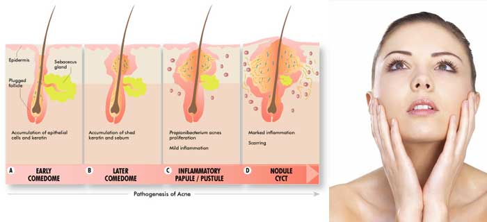
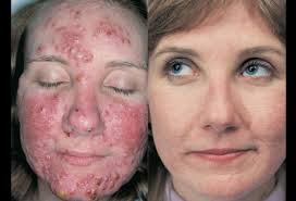
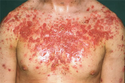
Acne is a skin condition that occurs when your hair follicles become plugged with oil and dead skin cells. Acne usually appears on your face, neck, chest, back and shoulders. Effective treatments are available, but acne can be persistent. The pimples and bumps heal slowly, and when one begins to go away, others seem to crop up.
Acne is most common among teenagers, with a reported prevalence of 70 to 87 percent. Increasingly, younger children are getting acne as well.
Depending on its severity, acne can cause emotional distress and scar the skin. The earlier you start treatment, the lower your risk of lasting physical and emotional damage.
Acne signs and symptoms vary depending on the severity of your condition:
- Whiteheads (closed plugged pores)
- Blackheads (open plugged pores — the oil turns brown when it is exposed to air)
- Small red, tender bumps (papules)
- Pimples (pustules), which are papules with pus at their tips
- Large, solid, painful lumps beneath the surface of the skin (nodules)
- Painful, pus-filled lumps beneath the surface of the skin (cystic lesions) If home care remedies don’t work to clear up your acne, see your primary care doctor. He or she can prescribe stronger medications. If acne persists or is severe, you may want to seek medical treatment from a doctor who specializes in the skin (dermatologist).Seek emergency medical help if after using a nonprescription skin product you experience:
- The Food and Drug Administration warns that some popular nonprescription acne lotions, cleansers and other skin products can cause a serious reaction. This type of reaction is quite rare, so don’t confuse it with the redness, irritation or itchiness where you’ve applied medications or products.
- When to see a doctor
- Faintness
- Difficulty breathing
- Swelling of the eyes, face, lips or tongue
- Tightness of the throat
- Four main factors cause acne:
- Oil production
- Dead skin cells
- Clogged pores
- BacteriaHair follicles are connected to oil glands. These glands secrete an oily substance (sebum) to lubricate your hair and skin. Sebum normally travels along the hair shafts and through the openings of the hair follicles onto the surface of your skin.
- Acne typically appears on your face, neck, chest, back and shoulders. These areas of skin have the most oil (sebaceous) glands. Acne occurs when hair follicles become plugged with oil and dead skin cells.
When your body produces an excess amount of sebum and dead skin cells, the two can build up in the hair follicles. They form a soft plug, creating an environment where bacteria can thrive. If the clogged pore becomes infected with bacteria, inflammation results.
The plugged pore may cause the follicle wall to bulge and produce a whitehead. Or the plug may be open to the surface and may darken, causing a blackhead. A blackhead may look like dirt stuck in pores. But actually the pore is congested with bacteria and oil, which turns brown when it’s exposed to the air.
Pimples are raised red spots with a white center that develop when blocked hair follicles become inflamed or infected. Blockages and inflammation that develop deep inside hair follicles produce cyst-like lumps beneath the surface of your skin. Other pores in your skin, which are the openings of the sweat glands, aren’t usually involved in acne.
Factors that may worsen acne
These factors can trigger or aggravate an existing case of acne:
- Hormones. Androgens are hormones that increase in boys and girls during puberty and cause the sebaceous glands to enlarge and make more sebum. Hormonal changes related to pregnancy and the use of oral contraceptives also can affect sebum production. And low amounts of androgens circulate in the blood of women and can worsen acne.
- Certain medications. Drugs containing corticosteroids, androgens or lithium can worsen acne.
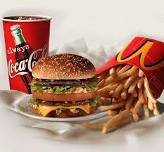
- Diet. Studies indicate that certain dietary factors, including dairy products and carbohydrate-rich foods — such as bread, bagels and chips — may trigger acne. Chocolate has long been suspected of making acne worse. A recent study of 14 men with acne showed that eating chocolate was related to an increase in acne. Further study is needed to examine why this happens or whether acne patients need to follow specific dietary restrictions.
- Stress. Stress can make acne worse.
Acne myths
These factors have little effect on acne:
- Greasy foods. Eating greasy food has little to no effect on acne. Though working in a greasy area, such as a kitchen with fry vats, does because the oil can stick to the skin and block the hair follicles. This further irritates the skin or promotes acne.
- Dirty skin. Acne isn’t caused by dirt. In fact, scrubbing the skin too hard or cleansing with harsh soaps or chemicals irritates the skin and can make acne worse. Though it does help to gently remove oil, dead skin and other substances.
- Cosmetics. Cosmetics don’t necessarily worsen acne, especially if you use oil-free makeup that doesn’t clog pores (noncomedogenics) and remove makeup regularly. Nonoily cosmetics don’t interfere with the effectiveness of acne drugs.
- Risk factors for acne include:
- Hormonal changes. Such changes are common in teenagers, women and girls, and people using certain medications, including those containing corticosteroids, androgens or lithium.
- Family history. Genetics plays a role in acne. If both parents had acne, you’re likely to develop it, too.
- Greasy or oily substances. You may develop acne where your skin comes into contact with oily lotions and creams or with grease in a work area, such as a kitchen with fry vats.
- Friction or pressure on your skin. This can be caused by items such as telephones, cellphones, helmets, tight collars and backpacks.
Stress. This doesn’t cause acne, but if you have acne already, stress may make it worse.
REFERENCE: MAYO CLINIC
QUOTE FOR WEDNESDAY:
Chronic traumatic encephalopathy (CTE) is the term coined for the neurodegenerative disease often suspected in athletes with histories of repeated concussion and progressive dementia.
Frontiers in Human Science (Published online 2013 May 24. doi: 10.3389/fnhum.2013.00222).
QUOTE FOR TUESDAY:
“I want every person who leaves this to be as healthy as possible when they leave. We all give up stiffness or pain in knees, backs, joints. You don’t want to give up your brain.”
Indianapolis Colts Center-Jeff Saturday.
QUOTE FOR MONDAY
“All natural sweeteners, including sugar, honey and high fructose corn syrup, contain carbohydrates that feed bacteria in the mouth and can contribute to tooth decay if consumed in excess.”
The Facts about High Fructose Corn Syrup
Risks of high levels of fructose.


You might think that the increase of the use of “high-fructose corn syrup during the past 30 years, would be safe. High Fructose Corn Syrup (HFCS) is an “artificial” sweetener made from a complex process with corn; a process of brewing, separating, breaking down, injecting enzymes, filtering, mixing and blending. Sounds safe enough, doesn’t it?
High fructose corn syrup is extremely soluble and mixes well in many foods. It is cheap to produce, sweet and easy to store. It’s used in everything from bread to pasta sauces to bacon to beer as well as in “health products” like protein bars and “natural” sodas.
HFCS is less expensive, lasts longer, and is more easily transported and handled than natural sugar; thus food producers prefer it for their manufacturing processes.
Research has shown that “high-fructose corn syrup” goes directly to the liver, releasing enzymes that instruct the body to then store fat! This may elevate triglyceride (fat in blood) levels and elevate cholesterol levels. Because it is metabolized by the liver, fructose does not cause the pancreas to release insulin the way it normally does. Fructose converts to fat more than any other sugar. Fructose reduces the affinity of insulin for its receptor, which is the hallmark of type-2 diabetes.
Some research claims that HFCS does not metabolize in the body like regular “natural” sugars; and that it might cause obesity-related glitches within the liver and other organs which normally deal with metabolizing, storing and using sugars in the body.
HFCS is Often Contaminated with Mercury. Recent studies of samples of HFCS and food products containing it in the United States conducted via two studies found that between 31% and 45% of the samples contained mercury. Mercury is toxic in even small quantities. For years, there have been suspicions that mercury used in vaccines may be related to the rise in autism in the United States. But this mercury contamination issue is much bigger and affects common foods widespread throughout the nation’s food supply. Products tested from big-name manufacturers such as Minute Maid, Coca-Cola, Hershey’s, Quaker, Hunt’s, Manwich, Smucker’s, Kraft, Nutri-Grain, and Yoplait had detectable levels of mercury.
Today, Commercial fruit juices and any products containing high fructose corn syrup are more dangerous than sugar and should be removed from the diet.
Read Labels! You’ll quickly see that this ingredient has been added to half the supermarket. So read under Ingredients carefully and look for High Fructose Corn Syrup or even just Corn Syrup.
Americans are being poisoned by a common additive present in a wide array of processed foods like soft drinks and salad dressings, commercially made cakes and cookies, and breakfast cereals and brand-name breads.
This commonplace additive silently increases our risk of obesity, diabetes, hypertension, and atherosclerosis.
The name of this toxic additive is high-fructose corn syrup. It is so ubiquitous in processed foods and so over-consumed by the average American that many experts believe our nation faces the prospect of an epidemic of metabolic disease in the future, related in significant degree to excess consumption of high-fructose corn syrup.
The food industry has long known that “a spoonful of sugar helps the medicine go down in the most delightful way.” And cane sugar had been America’s most delightful sweetener of choice, that is, until the 1970s, when the much less expensive corn-derived sweeteners like maltodextrin and high-fructose corn syrup were developed. While regular table sugar (sucrose) is 50% fructose and 50% glucose, high-fructose corn syrup can contain up to 80% fructose and 20% glucose, almost twice the fructose of common table sugar. Both table sugar and high-fructose sweetener contain four calories per gram, so calories alone are not the key problem with high-fructose corn syrup. Rather, metabolism of excess amounts of fructose is the major concern.
The alarming rise in diseases1,2 related to poor lifestyle habits has been mirrored by an equally dramatic increase in fructose consumption, particularly in the form of the corn-derived sweetener, high-fructose corn syrup.3-12 In this article, we’ll examine the evidence for these associations, and we’ll attempt to determine if high-fructose corn syrup is a benign food additive, as the sweetener industry has lobbied us (and the FDA) to believe, or a dangerously overlooked threat to public health.
Rising Concern
While cardiovascular disease remains the number one killer in America,1 scientists have noted that “we are experiencing an epidemic of [heart and kidney] disease characterized by increasing rates of obesity, hypertension, the metabolic syndrome, type 2 diabetes, and kidney disease.”2 Add to this list a disturbing rise in new cases of non-alcoholic fatty liver disease, and you have a public health crisis of enormous proportions.
With a growing sense of urgency, scientists are examining the relationship between consumption of high-fructose corn syrup (HFCS) and numerous adverse medical conditions. And they’re coming away with a sour taste in the mouth. Emerging research shows that excessive dietary fructose, largely from consumption of HFCS, represents “an important, but not well-appreciated dietary change,” which has “…rapidly become an important causative factor in the development of the metabolic syndrome,”9 a conglomeration of risk factors that greatly elevates the risk of cardiovascular disease and diabetes. Other research suggests that high dietary fructose consumption contributes to obesity and insulin resistance,5,7 encourages kidney stone formation,13 promotes gout,14-17 and is contributing to an upsurge in cases of non-alcoholic fatty liver disease.4,18,19 Furthermore, high dietary fructose consumption is associated with increased production of advanced glycation end products (AGEs), which are linked with the complications of diabetes and with the aging process itself
High dietary intake of fructose is problematic because fructose is metabolized differently from glucose. Like fructose, glucose is a simple sugar. Derived from the breakdown of carbohydrates, glucose is a primary source of ready energy. Sucrose (table sugar) comprises one molecule of glucose and one molecule of fructose. Thus, excessive sucrose intake also contributes to the rise in overall daily fructose consumption. Glucose can be metabolized and converted to ATP, which is readily “burned” for energy by the cells’ mitochondria. Alternatively, glucose can be stored in the liver as a carbohydrate for later conversion to energy. Fructose, on the other hand, is more rapidly metabolized in the liver, flooding metabolic pathways and leading to increased triglyceride synthesis and fat storage in the liver. This can cause a rise in serum triglycerides, promoting an atherogenic lipid profile and elevating cardiovascular risk. Increased fat storage in the liver may lead to an increased incidence in non-alcoholic fatty liver disease, and this is one of several links between HFCS consumption and obesity as well as the metabolic syndrome.7
Fructose may have less impact on appetite than glucose, so processed foods rich in fructose can contribute to weight gain, obesity, and its related consequences by failing to manage appetite.20 Additionally, loading of the liver with large amounts of fructose leads to increased uric acid formation, which may contribute to gout in susceptible individuals.
The high flux of fructose to the liver, the main organ capable of metabolizing this simple carbohydrate, disturbs glucose metabolism and uptake pathways and leads to metabolic disturbances that underlie the induction of insulin resistance,9 a hallmark of type 2 diabetes.
So you may want to look at labels of the food you eat more closely to prevent obesity or disease like diabetes. Just a thought.
QUOTE FOR THE WEEKEND:
“Some of these concerned people have had personal experience with ‘the silent epidemic, others have seen how osteoporosis can devastate the lives of their family and friends. They share a common belief that people should recognize whether they have risk factors for osteoporosis and that women and men should take personal responsibility for their bone health. ”
Alice Chiu, prominent philanthropist, founder and director, Sheen Hok Charitable Foundation, Hong Kong.