“By age 80, more than half of all Americans either have a cataract or have had cataract surgery.”
National Eye Institute
“By age 80, more than half of all Americans either have a cataract or have had cataract surgery.”
National Eye Institute
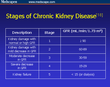
GFR = Glomerular Filtration Rate
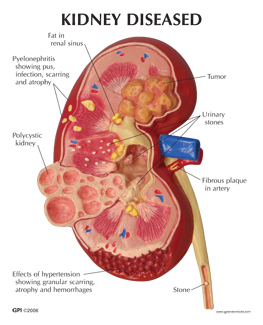
The kidneys are responsible for handling urine, so it makes sense that urine will start to change if the kidneys are failing. Some examples of urination changes include: • Urine comes out bubbly or foamy • Urine may have traces of blood • You may have the overwhelming urge to urinate during the night, waking up • Urination occurs more often and appears pale • Urination occurs less often and appears dark • You may have difficulty attempting to urinate
The kidneys are responsible for producing a specific hormone called erythropoietin (EPO). This hormone is responsible for instructing the body to produce red blood cells, which are meant to carry oxygen throughout the body. If the kidneys start to fail, they will make less EPO, which means fewer hormones are directing the body to produce the necessary amount of red blood cells. At the end of this cycle, you’re left feeling very tired and weakened throughout the day. Experiencing fatigue even when you seem to get enough sleep at night is one symptom that the kidneys are not producing enough hormones for your body.
Because of the way the kidneys interact with the body and handle the process of urination, they also are largely responsible for removing the extra fluid within your body. Kidneys that are starting to fail won’t get rid of that fluid as well as they should be. As a result, it stays inside the body — and while it’s in the body, it has to go somewhere; the fluid starts filling in pocketed areas. You may experience swelling in one or both ankles, the legs, the face, hands, as well as feet. While the swelling can be mild, it can also swell to difficult stages; for instance, it might be hard to wear a regular shoe. This is edema.
Healthy kidneys also take on the role of the body’s garbage men; that is, they’re responsible for getting rid of waste in the body. In the event of kidney problems or failure, waste won’t exit the body as efficiently as before, causing a buildup of excess waste in the bloodstream. This is known as uremia, and it can cause feelings of nausea or make you need to vomit. It should go without saying that your body doesn’t like being filled with waste, and it attempts to purge the waste by way of vomiting.
When kidneys begin to fail and cause uremia, or a buildup of waste in the body, the body may react by producing the taste of metal in your mouth or causing bad breath. Overall, you might taste a rather poor flavor in your mouth that causes you not to taste food in the same way as you did before. In particular, this may make you less interested in eating meats. In addition, you might start to notice some weight loss as a result of not eating. This could be due to the taste issue or you may simply not feel hungry enough to eat much.
Developing uremia as a result of kidney disease doesn’t stop with metallic taste or the need to vomit. The waste buildup in the bloodstream manifests further by causing patches of rashes on the skin and causing itchiness. In some cases, patches of skin can break out in what appears to be acne as well. These itchy rashes can be difficult to relieve; in more progressed instances, the itch can feel like it goes right down to the bone, making it difficult to feel relief by way of scratching.
As explained in a previous slide, the kidneys produce the hormone EPO to signal the body’s production of red blood cells. Failing this, there are fewer blood cells, which is anemia. Anemia comes with its own set of symptoms, the most prominent but overlooked being chills. If you feel cold, even inside of a warm room, you could be experiencing anemia.
One of the more characteristic symptoms of chronic kidney disease include feeling discomfort in the back or in the legs. In some cases, the feelings of discomfort could be painful. It is also possible to experience pain as far as the upper back. Problems that can cause pain include: • Kidney stones and infections, which cause severe spasms of pain • Bladder infections, which can produce a burning sensation during urination • Polcystic kidney disease, which produces painful cysts on the kidneys and liver.
If you have been experiencing shortness of breath lately, it could be connected to the kidneys in two different ways. The first possible connection is a result of the extra fluid buildup; sometimes, this extra fluid builds up in the lungs, making it more difficult to breathe. Otherwise, the shortness of breath can be a result of anemia; in this case, there are an insufficient number of red blood cells available to carry oxygen throughout the body. This leaves the brain and body starved and short of breath. If you experience shortness of breath, sit down for a moment and calmly attempt deep breaths. The experience is naturally frightening, but panicking can only lead to more difficulty breathing.
Anemia as a result of kidney disease has one more grasp on the body: It can make you dizzy and cause you to have trouble concentrating on things. When this happens, your brain is becoming starved of the oxygen it needs to be at full power. When your brain isn’t getting enough oxygen, it manifests beyond dizziness and concentration problems; you can also experience memory problems and other issues with cognitive functions. This symptom often goes hand in hand with fatigue due to the taxing effects on the brain.
” Kidneys cleanse your blood by removing waste and excess fluid, maintain the balance of salt and minerals in your blood, and help regulate blood pressure.”
Linda Bernstein MD M.D. with Web Med
“The foundation stones for a balanced success are honesty, character, integrity, faith, love and loyalty.”
Zig Ziglar (November 6, 1926 – November 28, 2012, was an American author, salesman, and motivational speaker).
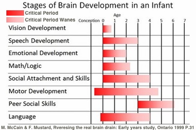
You were born preprogrammed to bond with one very significant person—your primary caregiver, probably your mother. Like all infants, you were a bundle of emotions—intensely experiencing fear, anger, sadness, and joy. The emotional attachment that grew between you and your caregiver was the first interactive relationship of your life, and it depended upon nonverbal communication. The bonding you experienced determined how you would relate to other people throughout your life, because it established the foundation for all verbal and nonverbal communication in your future relationships.
Individuals who experience confusing, frightening, or broken emotional communications during their infancy often grow into adults who have difficulty understanding their own emotions and the feelings of others. This limits their ability to build or maintain successful relationships. Attachment—the relationship between infants and their primary caregivers—is responsible for:
“Some risk factors, like a person’s age or race, can’t be changed. Others are linked to cancer-causing factors in the environment. Still others are related to personal behaviors, such as smoking, drinking, and diet.”
The American Cancer Society
QUOTE FOR THE DAY:
“Breast cancer prevention starts with healthy habits — such as limiting alcohol and staying physically active. Understand what you can do to reduce your breast cancer risk.”
MAYO CLINIC

Is there a link between birth control pills and breast cancer?
A number of older studies suggested that birth control pills slightly increased the risk of breast cancer, especially among younger women. In these studies, however, 10 years after discontinuing birth control pills women’s risk of breast cancer returned to the same level as that of women who never used oral contraceptives. Current evidence does not support an increase in breast cancer with birth control pills.
Be vigilant about breast cancer detection. If you notice any changes in your breasts, such as a new lump or skin changes, consult your doctor. Also, ask your doctor when to begin mammograms and other screenings.
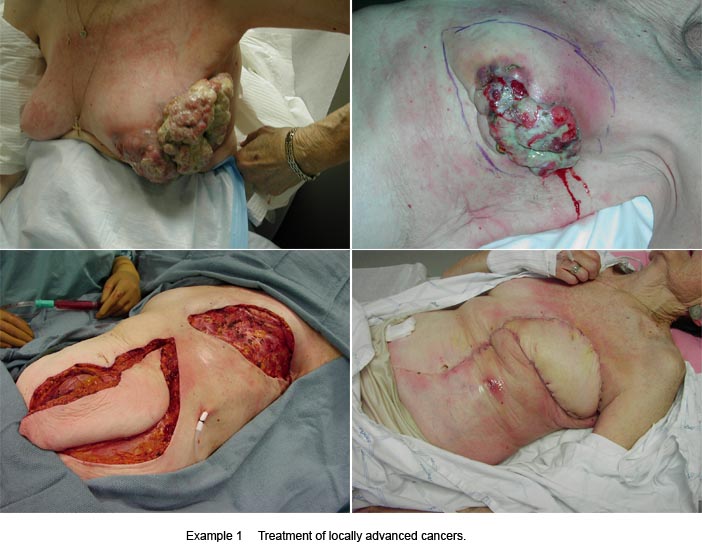
Once you’ve been diagnosed with breast cancer, your doctor works to find out the specifics of your tumor. Using a tissue sample from your breast biopsy or using your tumor if you’ve already undergone surgery, your medical team determines your breast cancer type. This information helps your doctor decide which treatment options are most appropriate for you.
Here’s what’s used to determine your breast cancer type.
Whether your cancer is invasive or noninvasive helps your doctor determine whether your cancer may have spread beyond your breast, which treatments are more appropriate for you, and your risk of developing cancer in the same breast or your other breast.
The type of tissue where your breast cancer arises determines how the cancer behaves and what treatments are most effective. Parts of the breast where cancer begins include:
“Staying at a healthy weight, being physically active, and limiting how much alcohol you drink can help reduce your risk of breast cancer.”
American Cancer Society