Archive | July 2025

Part III National Brain Injury Awareness!

What are the causes?

Common causes include falls, car or motorcycle crashes, vehicular accidents involving pedestrians, athletics, and assaults with or without a weapon.

Who is affected?

Approximately 1.5 to 2 million adults and children suffer a traumatic brain injury (TBI) each year in the United States. Most people who experience a head injury, about 1.1 million, will have a mild injury that does not require an admission to the hospital. Another 235,000 individuals will be hospitalized with a moderate to severe head injury, and approximately 50,000 will die.

How is a diagnosis made?

When a person is brought to the emergency room with a head injury, doctors will learn as much as possible about his or her symptoms and how the injury occurred. The person’s condition is assessed quickly to determine the extent of injury.

The Glasgow Coma Score (GCS) is a 15-point test used to grade a patient’s level of consciousness. Doctors assess the patient’s ability to 1) open his or her eyes, 2) ability to respond appropriately to orientation questions, (“What is your name? What is the date today?”), and 3) ability to follow commands (“Hold up two fingers, or give a thumbs up”). If unconscious or unable to follow commands, his or her response to painful stimulation is checked. A number is taken from each category and added together to get the total GCS score. The score ranges from 3 to 15 and helps doctors classify an injury as mild, moderate, or severe. Mild TBI has a score of 13-15. Moderate TBI has a score of 9-12, and severe TBI has a score of 8 and below.

Diagnostic imaging tests will be performed:

CT of TBI

Figure 3. CT scan shows a blood clot (hematoma) collecting under the bone (red arrows) and displacing brain (yellow arrow) to the other side of the skull.

  • Computed Tomography (CT) is a noninvasive X-ray that provides detailed images of anatomical structures within the brain. A CT scan of the head is taken at the time of injury to quickly identify fractures, bleeding in the brain, blood clots (hematomas) and the extent of injury (Fig. 3). CT scans are used throughout recovery to evaluate the evolution of the injury and to help guide decision-making about the patient’s care.
  • Magnetic Resonance Imaging (MRI) is a noninvasive test that uses a magnetic field and radiofrequency waves to give a detailed view of the soft tissues of the brain. A dye (contrast agent) may be injected into the patient’s bloodstream. MRI can detect subtle changes in the brain that cannot be seen on a CT scan.
  • Magnetic Resonance Spectroscopy (MRS) gives information about the metabolism of the brain. The numbers generated from this scan provide a general prognosis about the patient’s ability to recover from the injury.

What treatments are available?

Mild TBI usually requires rest and medication to relieve headache. Moderate to severe TBI require intensive care in a hospital. Bleeding and swelling in the brain can become an emergency that requires surgery. However, there are times when a patient does not require surgery and can be safely monitored by nurses and physicians in the neuroscience intensive care unit (NSICU).

The goals of treatment are to resuscitate and support the critically ill patient, minimize secondary brain injury and complications, and facilitate the patient’s transition to a recovery environment. Despite significant research, doctors only have measures to control brain swelling, but do not have a way to eliminate swelling from occurring.

Neurocritical care
Neurocritical care is the intensive care of patients who have suffered a life-threatening brain injury. Many patients with severe TBI are comatose or paralyzed; they also may have suffered injuries in other parts of the body. Their care is overseen by a neurointensivist, a specialty-trained physician who coordinates the patient’s complex neurological and medical care. Patients are monitored and awakened every hour for nursing assessments of their mental status or brain function.

Click to view larger image with labels

Figure 4. In the NSICU, the patient is connected to numerous machines, tubes, and monitors. The monitoring equipment provides information about body functions and helps guide care. Some equipment may take over certain functions, such as breathing, nutrition, and urination, until the patient’s body is able to do these things on its own.

Seeing a patient who has suffered a severe TBI can be shocking. It is possible that your loved one’s appearance will be altered because of facial injury and equipment that is used for monitoring. Numerous tubes, lines, and equipment may be used to closely monitor his or her heart rate, blood pressure, and other critical body functions. (Fig. 4)

ICP monitor

Figure 5. A brain oxygen and cerebral blood flow monitor is inserted into the brain tissue and secured to the skull with a bolt. A catheter is inserted into the ventricle of the brain to monitor intracranial pressure (ICP). If pressure is too high, the CSF fluid can be drained from the ventricles.

  • Intracranial pressure (ICP) monitor. A catheter is placed through a small hole in the skull and positioned inside the ventricle (fluid-filled area deep within the brain) to measure pressure inside the head (Fig. 5). The ICP monitor allows the NSICU team to intervene quickly if the pressure becomes too high. Typical intracranial pressure is less than 20 mmHg. However, there are times when a higher number is safe and acceptable.
  • Brain oxygen monitor (Licox). A catheter is placed through a small hole in the skull and positioned within the brain tissue. The Licox measures the oxygen level and temperature within the brain. Adjustments in the amount of oxygen given to the patient are often made to maximize the brain’s oxygen level. A cerebral blood flow monitor, called a Hemedex, is a newer monitor that is placed with the Licox and helps the NSICU team evaluate blood flow through the brain.
  • Ventilator. Some patients may require a ventilator, a machine that helps them breathe. The ventilator is connected to the patient by the endotracheal tube, or ET tube. The tube is placed into the patient’s mouth and down into the trachea, or windpipe. The tube allows the machine to push air into and out of the lungs, thereby helping the patient breathe.
  • Feeding tube. When patients are on a ventilator or have a decreased level of alertness, they may not be able to eat or get sufficient nutrition to meet their needs. A nasal-gastric feeding tube may be inserted through the patient’s nose and passed down the throat into the stomach. It delivers liquid nutrition as well as any medication that is required.
  • Seizures and EEG monitoring. A seizure is an abnormal electrical discharge from the brain. Approximately 24% of patients who suffer a TBI will have a seizure that is undetected unless they are monitored by an electroencephalogram (EEG). Seizures that are not visible to the human eye are referred to as non-convulsive seizures. Because these seizures are serious, all patients with a severe TBI are monitored with continuous EEG for 24 to 72 hours after injury.

Medication

  • Sedation and pain. After a head injury it may be necessary to keep the patient sedated with medications. These medications can be turned off quickly in order to awaken the patient and check their mental status. Because patients often have other injuries, pain medication is given to keep them comfortable.
  • Controlling intracranial pressure. Hypertonic saline is a medication used to control pressure within the brain. It works by drawing the extra water out of the brain cells into the blood vessels and allowing the kidneys to filter it out of the blood.
  • Preventing seizures. Patients who’ve had a moderate to severe traumatic brain injury are at higher risk of having seizures during the first week after their injury. Patients are given an anti-seizure medication (levetiracetam or phenytoin) to prevent seizures from occurring.
  • Preventing infection. Although every attempt is made to prevent infection, the risk is always present. Any device placed within the patient has the potential to introduce a microbe. If an infection is suspected, a test will be sent to a laboratory for analysis. If an infection is present, it will be treated with antibiotics.

Surgery

Surgery is sometimes necessary to repair skull fractures, repair bleeding vessels, or remove large blood clots (hematomas). It is also performed to relieve extremely high intracranial pressure.

  • Craniotomy involves cutting a hole in the skull to remove a bone flap so that the surgeon can access the brain. The surgeon then repairs the damage (e.g., skull fracture, bleeding vessel, remove large blood clots). The bone flap is replaced in its normal position and secured to the skull with plates and screws.

Figure 6. A large decompressive craniectomy is removed and the dura is opened to allow the brain to expand. Blood clots are removed and bleeding vessels are repaired. The bone flap is frozen and replaced about 6 weeks later.

  • Decompressive craniectomy involves removing a large section of bone so that the brain can swell and expand. This is typically performed when extremely high intracranial pressure becomes life threatening. At that time the patient is taken to the operating room where a large portion of the skull is removed to give the brain more room to swell (Fig. 6). A special biologic tissue is placed on top of the exposed brain and the skin is closed. The bone flap is stored in a freezer. One to 3 months after the swelling has resolved and the patient has stabilized from the injury, the bone flap is replaced in another surgery, called cranioplasty.

Other surgical procedures may be performed to aid in the patient’s recovery:

  • Tracheotomy involves making a small incision in the neck to insert the breathing tube directly into the windpipe. The ventilator will then be connected to this new location on the neck and the old tube is removed from the mouth.
  • Percutaneous Endoscopic Gastrostomy Tube (PEG) is a feeding tube inserted directly into the stomach through the abdominal wall. A small camera is placed down the patient’s throat into the stomach to aid with the procedure and to ensure correct placement of the PEG tube (see Surgical Procedures for Accelerated Recovery).

Clinical trials

Clinical trials are research studies in which new treatments—drugs, diagnostics, procedures, and other therapies—are tested in people to see if they are safe and effective. Research is always being conducted to improve the standard of medical care. Information about current clinical trials, including eligibility, protocol, and locations, are found on the Web. Studies can be sponsored by the National Institutes of Health (see clinicaltrials.gov) as well as private industry and pharmaceutical companies (see www.centerwatch.com).

Recovery & prevention

The recovery process varies depending on the severity of the injury, but typically progresses through stages: coma, confusion / amnesia, and recovery.

  • When a patient is in a coma, his or her eyes are closed and they show minimal reaction when spoken to or stimulated. Movements that may be seen at this time are basic reflexes or automatic responses to a stimulus. The brain wave activity in a comatose person is very different from that of a sleeping person.
  • When a patient begins to awaken, the first natural response is that of bodily protection. Patients at this stage will move away from any stimulus or tend to pull at items attached to them in an attempt to remove anything that is uncomfortable or irritating. His or her eyes may be open more often, but they may not be aware of their behavior or be able to interact in a meaningful way. It is common for a patient to respond to each stimulus (hearing, seeing, or touching) in the same way. Responses may include increased rate of breathing, moaning, moving, sweating, or a rise in blood pressure.
  • As the patient continues to wake up, their interactions may become more purposeful. They may look at a person and follow them around the room with their eyes, or follow simple commands such as “Hold up your thumb.” Patients tend to be confused and may have inappropriate or agitated behaviors.

Not all head injuries are the same. Patients recover at different rates and to varying degrees. It is difficult to determine at what point a patient will start understanding and interacting with their caregivers or family in a meaningful way. It is important to have patience; recovery from a brain injury can take weeks, months, or even years.

The Family’s Role
Many family members express feelings of helplessness when their loved one is in the NSICU. You are not alone. Please take care of yourself and use your energy wisely.

Visiting hours are limited in the NSICU. Too much stimulation can agitate the patient and raise his or her blood pressure. You can most effectively convey your concern by sitting quietly and holding your loved one’s hand. Be aware that the patient, though silent, may hear anything you say. Never speak as if the patient were not there.

As patients recover, they need help understanding what has happened to them during this “lost period of time.” Keep in mind that the recovery of consciousness is a gradual process – not just a matter of waking up. Progress is usually tracked in three areas: movement, thinking, and interacting. You can help by keeping a diary of their progress. Family photos may help with regaining memory.

Rehabilitation
Most patients are discharged from the hospital when their condition has stabilized and they no longer require intensive care. A social worker will work closely with the family as preparations are made for a return home or for transfer to a long-term care or rehabilitation center.

  • A long-term acute care (LTAC) facility is a place for patients who have stabilized from their initial injury but who still require a ventilator or frequent nursing care. Many patients are discharged to an LTAC to continue being weaned from the ventilator. Once off the ventilator, they can be moved to a rehabilitation or skilled nursing facility.
  • A rehabilitation facility is a place for patients who do not require a ventilator but who still require help with basic daily activities. Physical and occupational therapists work with patients to help them achieve their maximum potential for recovery. Rehab facilities are either Acute Inpatient Rehab that require patients to participate in 3 hours or more of rehab a day or a Skilled Nursing Facility (SNF) that provide 1-3 hours of rehab a day depending on what the patient can tolerate.

Recovering from a brain injury relies on the brain’s plasticity—the ability for undamaged areas of the brain to take over functions of the damaged areas. It also relies on regeneration and repair of nerve cells. And most importantly, on the patient’s hard work to relearn and compensate for lost abilities.

  • A physical therapist helps patients rebuild and maintain strength, balance, and coordination. They can work with the patient in any facility.
  • An occupational therapist helps patients to perform activities of daily living, such as dressing, feeding, bathing, toileting, and transferring themselves from one place to another. They also provide adaptive equipment if a patient has difficultly performing a task.
  • A speech therapist helps patients by monitoring their ability to safely swallow food and helping with communication and cognition.
  • A neuropsychologist helps patients relearn cognitive functions and develop compensation skills to cope with memory, thinking, and emotional needs.

Prevention

Tips to reduce the risk for a head injury:

  • Always wear your helmet when riding a bicycle, motorcycle, skateboard, or all-terrain vehicle.
  • Never drive under the influence of alcohol or drugs.
  • Always wear your seat belt and ensure that children are secured in the appropriate child safety seats.
  • Avoid falls in the home by keeping unsecured items off the floor, installing safety features such as non-slip mats in the bathtub, handrails on stairways, and keeping items off of stairs.
  • Avoid falls by exercising to increase strength, balance, and coordination.
  • Store firearms in a locked cabinet with bullets in a separate location.
  • Wear protective headgear while playing sports.

Reference for Part I and Part II: Mayfield Brain & Spine at 513-221-1100.

Part I – Hemochromatosis-What it is, primary/secondary, and symptoms!

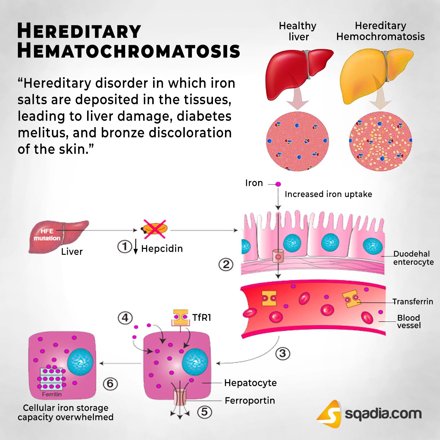
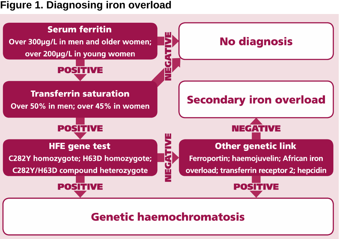
Hemochromatosis is a disorder where too much iron builds up in your body. Sometimes it’s called “iron overload.”   Hemo meaning blood and chromatosis means pigmentation specifically : deposit of pigment in a normally unpigmented area or excessive pigmentation in a normally pigmented site.

Normally, your intestines absorb just the right amount of iron from the foods you eat. But in hemochromatosis, your body absorbs too much, and it has no way to get rid of it. So, your body stores the excess iron in your joints and in organs like your liver, heart, and pancreas. This damages them. If it’s not treated, hemochromatosis can make your organs stop working.

There are two types of this condition — primary and secondary.

Primary hemochromatosis is hereditary, meaning it runs in families. If you get two of the genes that cause it, one from your mother and one from your father, you’ll have a higher risk of getting the disorder.

Secondary hemochromatosis happens because of other conditions you have. These include:

  • Certain kinds of anemia
  • Liver disease
  • Getting a lot of blood transfusions
  • White people of northern European descent are more likely to get hereditary hemochromatosis. Men are 5 times more likely to get it than women.

    Symptoms

    Up to half of people who have hemochromatosis don’t get any symptoms. In men, symptoms tend to show up between ages 30 and 50. Women often don’t show signs of this condition until they’re over 50 or past menopause. That may be because they lose iron when they get their periods and give birth.

    Symptoms of hemochromatosis include:

    • Pain in your joints, especially your knuckles
    • Feeling tired
    • Unexplained weight loss
    • Skin that has a bronze or gray color – pigmentation
    • Pain in your belly
    • Loss of sex drive
    • Loss of body hair
    • Heart flutter
    • Foggy memory

    Sometimes people don’t get any symptoms of hemochromatosis until other problems arise. These may include:

    • Liver
    • Diabetes
    • Abnormal heartbeat
    • Arthritis
    • Erectile dysfunction (difficulty having a erection)

    If you take a lot of vitamin C or eat a lot of foods that contain it, you can make hemochromatosis worse. That’s because vitamin C helps your body absorb iron from food.

    White people of northern European descent are more likely to get hereditary hemochromatosis. Men are 5 times more likely to get it than women.
     

QUOTE FOR THURSDAY:

“As advances in CF knowledge and care are potentially able to prolong the life expectancy of many patients, it’s important to keep in mind the complications—beyond lung disease—that will develop and progress as patients age.1,9-11 Monitoring for these complications can help detect their emergence and progression, which can ensure earlier intervention; this has been associated with better outcomes in patients.

Knowing CF affects the lungs this is how it happens:

Early as in utero and into infancy, inflammation may occur, with the possibility of mucus plugging and bronchiectasis.

Inflammation, lung structure and lung function may progress throughout childhood.

Childhood, adolescence and early adulthood what happens is lower airway inflammation and worsening airway abnormalities including established bronchiectasis may occur, driven by the inflammation in the lungs.

In Adulthood and Aging what happens is airway destruction and complications, including bacterial infections, bronchiectasis with hemoptysis, and pneumothorax, may occur and may lead to progressive respiratory failure, often requiring lung transplant.”

CF Source (Multi-Organ Disease Progression in Cystic Fibrosis (CF)


QUOTE FOR WEDNESDAY:

“Although most known for its effect on the lungs, cystic fibrosis is a far-reaching condition that affects most of the body’s systems.

Cystic fibrosis is a chronic condition that’s primarily associated with breathing difficulties, lung infections, and persistent wheezing. But people with cystic fibrosis can experience a wide range of complications and symptoms throughout their bodies.

The sticky mucus that causes classic cystic fibrosis systems can also lead to the blockage of important body ducts and tubes. As a result, enzymes, proteins, and other body substances don’t travel correctly. This can lead to complications to organs other than just the lungs!”

Healthline (How Does Cystic Fibrosis (CF) Affect the Body’s Functions)

QUOTE FOR TUESDAY:

“The Cystic Fibrosis Foundation is the world’s leader in the search for a cure for CF and supports a broad range of research initiatives to tackle the disease from all angles.

Cystic fibrosis is genetic disease that affects the lungs, pancreas, and other organs. It is progressive, meaning that it gets worse over time.

There are close to 40,000 children and adults living with cystic fibrosis in the United States and an estimated 105,000 people have been diagnosed with CF across 94 countries. CF can affect people of every racial and ethnic group.

There are many things that are misconceptions about CF.

In people with CF, mutations in the cystic fibrosis transmembrane conductance regulator (CFTR) gene cause the CFTR protein to not function properly. When the protein is not working correctly, it’s unable to help move chloride — a part of salt — to the cell surface. Without the chloride to attract water to the cell surface, the mucus in various organs becomes thick and sticky.

CF affects multiple organs in the body.”

The Cystic Fibrosis Foundation (About Cystic Fibrosis | Cystic Fibrosis Foundation)
QUOTE FOR MONDAY:

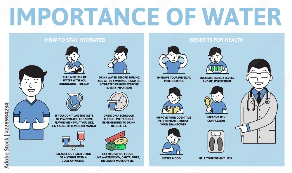
“Drinking water does more than just quench your thirst. It’s essential to keeping your body functioning properly and feeling healthy.

Nearly all of your body’s major systems depend on water to function and survive. With water making up about 60% of your body weight, it’s no surprise what staying hydrated can do for you.

Some ways the water works in our bodies is the following:

  • Regulates body temperature
  • Moistens tissues in the eyes, nose, mouth and other areas
  • Protects body organs and tissues
  • Carries nutrients and oxygen to cells.”

MAYO CLINIC (Water: Essential for your body – Mayo Clinic Health System)


Why water is so vital to humans!

You get most of your water from drinking beverages, but food also contributes a small amount to your daily water intake.

It’s common to hear that water is essential for your health. But why?

Here are the reasons why:

1. It helps create saliva

Water is a main component of saliva. Saliva also includes small amounts of electrolytes, mucus, and enzymes. It’s essential for breaking down solid food and keeping your mouth healthy.

Your body generally produces enough saliva with regular fluid intake. However, your saliva production may decrease as a result of age or certain medications or therapies.

If your mouth is drier than usual and increasing your water intake isn’t helping, see your doctor.

2. It regulates your body temperature

Staying hydrated is crucial to maintaining your body temperature. Your body loses water through sweat during physical activity and in hot environments.

Your sweat keeps your body cool, but your body temperature will rise if you don’t replenish the water you lose. That’s because your body loses electrolytes and plasma when it’s dehydrated.

If you’re sweating more than usual, make sure you drink plenty of water to avoid dehydration.

3. It protects your tissues, spinal cord, and joints

Water consumption helps lubricate and cushion your joints, spinal cord, and tissues. This will help you enjoy physical activity and lessen discomfort caused by conditions like arthritis.

4. It helps excrete waste through perspiration, urination, and defecation

Your body uses water to sweat, urinate, and have bowel movements.

Sweat regulates body temperature when you’re exercising or in warm temperatures. You need water to replenish the lost fluid from sweat.

You also need enough water in your system to have healthy stool and avoid constipation.

Your kidneys are also important for filtering out waste through urination. Adequate water intake helps your kidneys work more efficiently and helps to prevent kidney stones.

QUOTE FOR THE WEEKEND:

“No matter what stung you, you’re likely to feel some pain, throbbing and stinging, along with redness in the affected area. Unless you have an allergic reaction, these symptoms shouldn’t stick around for too long.

“Redness and swelling at the site of the sting is a normal reaction and will subside over time, usually in one to two days,” says Dr. Fertel. In terms of treatment, bee, wasp and hornet stings can be treated the same — unless the person has a specific allergy.

You may have heard that you should swipe the stinger out with a credit card or your fingernail rather than pinching and pulling it out. But, says Dr. Fertel, there’s no evidence that the technique makes a difference. What does matter is how quickly you remove the stinger. So, don’t stress about how it’s done — just get it over with fast. This is because the quicker you’re able to remove the stinger, the less venom will be pumped into the wound. This will help decrease the side effects of the sting and reduce the risk for anaphylactic shock if you’re allergic.”

Cleveland Clinic (Bee Sting Treatment and Prevention)

What to do if you or the family gets stung by a wasp or a bee.

What you should do if you or your child or family gets stung is:

One get into a safe area and away from a hive or outside where more stinging insects can come and attack you.

Two look at the area and if you see the stinger DO NOT SQUEEZE IT OUT since you will squeeze out more venom from the stinger but what you can do is get a tweezer and pull it our or if not available you can attempt to scratch it out with a nail (like if you are out camping and have no tweezers for example).

Three than wash the area out with soap and apply ice if the area is in pain to give the numbing affect to the area and decrease the pain with decreasing the venom from spreading.

If the area is itching apply oatmeal or a antihistamine cream to the area to decrease the itching or maybe even a cool bath and if you are not allergic (the majority of us are not) but you DO get stung by a bee outside hiking look for some plantain – chew it up a bit at the front of your mouth – and then spit the chewed up leaf and saliva on the sting.

Most stings will cause a small red bump to the area that got stung. For most part they can be treated at home depending on the area that was stung (Foot vs EYE for example). It would also include the reaction the individual has (LOCAL vs SYSTEMIC or even ANAPHYLACTIC=An allergic reaction that needs to be treated immediately or fatal, usually with epinephrine injection.).

Stung in the eye it will get swollen and shut and immediate evaluation from a MD is needed to make sure there is no other injury to the eye or that they didn’t even actually get stung in the eye itself.

If you show hives with DIFFICULTY BREATHING or DIFFICULTY SWALLOWING you NEED TO CALL 911 IMMEDIATELY since this is indicating a ANAPHYLACTIC REACTION most likely that needs treatment ASAP!! Since this can lead to shock or unconsciousness.

If you have reason to think you may be seriously allergic to bee venom, you should carry an Epipen (further discussed below).

How to determine if your even allergic to stings:

The diagnosis is made by a specialist, an allergist, by interviewing the patient and doing special allergy tests. If someone has had what is described as a systemic reaction, they should have venom skin tests done by an allergist to identify which venoms they are allergic to. The allergist can then recommend, based on the kind of reaction that the patient had, what kind of prevention would be the best idea for that person. For some people, it might be enough to be careful and carry an EpiPen, but for most people with insect skin allergy the best recommendation is to be immunized with venom treatment, because the allergy shots are highly effective to prevent dangerous reactions. This would all be done after any serious reactions were first taken care of in the ER if you had to call 911.

If you have reason to think you may be seriously allergic to bee venom, you should carry an Epipen (further discussed below). What it this exactly? An EpiPen is one kind of injector to deliver epinephrine, also known as adrenaline. It is a spring-loaded injector that makes it easy for somebody to give themselves an emergency injection that can be life-saving when there’s a severe allergic reaction. An EpiPen is useful for someone to carry if they have had a severe allergic reaction in the past. This is true for insect sting allergy and for some food allergies or other causes of anaphylaxis.

Let me point outthat there is no other medicine that can counteract a severe allergic reaction, but sometimes even the EpiPen isn’t enough; so when someone needs to use an EpiPen they should call 911, because they may need intravenous fluids or oxygen or other medicines.

BE SAFE RATHER THAN SORRY!

So let us remember it is coming onto summer and their BACK AGAIN!

 References

1-Read more: http://www.ehow.com/how 2-NEWS4JAX.com Published On: May 30 2014 09:38:22 AM EDT

3-http//beestrawbridge.blogspot.com/2013/03/which- bees-sting and which-don’t.html with Phil Chandler of Biobees.

4-Wikipedia-2013 published Bees

5-MedicineNet.com Bee and Wasp Sting 12/11/2013

6-See more at: http://www.about-bees.com/carpenter-bees.html#sthash.NywAhKk2.dpuf


QUOTE FOR FRIDAY:

“You can do a few things to minimize your sun exposure and reduce your risk of skin damage.

  • Try to stay in the shade as much as possible. If you are in the sun, wear protective clothing such as a long-sleeved shirt and pants.
  • You should also apply sunscreen with a high SPF rating to exposed skin.
  • Avoid being in the sun during peak hours, typically between 10 am and 4 pm.

By taking these precautions, you can help to protect your skin from the harmful effects of UV radiation.

UV rays are just as harmful on cloudy days as on sunny days.

While it’s true that people with darker skin have a higher natural SPF than those with lighter skin, this does not mean that they are immune to the effects of UV radiation.

While sunscreen is a vital part of any sun safety plan, it’s not the only thing you need to do to protect yourself from UV radiation. You should also take steps to avoid excessive sun exposure and wear protective clothing when you are in the sun.

Skin and Cancer Institute (July is UV Safety Month | Skin And Cancer Institute)