Archive | August 2022

QUOTE FOR MONDAY:

“Chest surgery at Mayo Clinic (also known as thoracic surgery) involves the organs of the chest, but extends to the esophagus (tube between mouth and stomach), the trachea (airway) and the chest wall (rib cage and breastbone).  From some chest traumas that happen a chest tube maybe needed.  A flexible chest tube is inserted into the air-filled space and may be attached to a one-way valve device that continuously removes air from the chest cavity until your lung is re-expanded and healed.  The chest tube has different functions as well.”

MAYO CLINIC

What Thoracic Surgery and what is a chest tube including why they maybe ordered!

Thoracotomy versus Thoracostomy:

Thoracotomy is surgery that makes an incision to access the chest. It’s often done to remove part or all of a lung in people with lung cancer. Thoracostomy is a procedure that places a tube in the space between your lungs and chest wall (pleural space).

Thoracostomy – the procedure to put a chest tube in places.

Thoracostomy inserts a thin plastic tube into the space between the lungs and the chest wall. The doctor may attach the tube to a suction device to remove excess fluid or air. Or, the doctor may use it to deliver medication into the space to decrease the likelihood that fluid will accumulate. This is called pleurodesis.

Your doctor may use thoracostomy to treat pneumothorax, also known as collapsed lung.

Your doctor will tell you how to prepare if this is not an emergency, including any changes to your medication schedule. Tell them if there’s a possibility you are pregnant and discuss any recent illnesses, medical conditions, allergies, and medications you’re taking, including herbal supplements and aspirin. They may tell you to stop taking aspirin, nonsteroidal anti-inflammatory drugs (NSAIDs) or blood thinners several days prior to your procedure. Leave jewelry at home and wear loose, comfortable clothing. You may most likely need to change into a gown for the procedure.

Know the medical difference in the ending of these 2 terminologies above:  ostomy or -stomy : surgically creating a hole (a new “mouth” or “stoma”, see List of -ostomies) -otomy or -tomy : surgical incision (see List of -otomies) -pexy : to fix or secure. -plasty : to modify or reshape (sometimes entails replacement with a prosthesis).

Chest Tube (s):


How a chest tube removes fluid, blood or air:

Fluid in the intrapleural space is affected by gravity and localizes in the lower portion of the lung cavity. Tubes placed in these positions drain blood and fluid. Frequently applying suction helps with this drainage.


Pneumothorax = Air in the lung through a hole

Hemothorax = Blood in the lung.

They are even used on animals:


Risk Factors getting a Chest Tube:

  • Pain during insertion and after placement of chest tube: Although pain during insertion and mild discomfort after placement are common, your healthcare provider can help minimize these effects with pain medicines.
  • Infection
  • Improper placement
  • Dislodged chest tube
  • Collapsed lung after removal of chest tube
  • Bleeding at the site of insertion


WHAT IS A CARDIAC ARREST VS HEART ATTACK.


Cardiac arrest is the abrupt loss of heart function in a person who may or may not have diagnosed heart disease. The time and mode of death are unexpected. It occurs instantly or shortly after symptoms appear.

Each year, more than 420,000 emergency medical services-assessed out-of-hospital cardiac arrests occur in the United States.

No. The term “heart attack” is often mistakenly used to describe cardiac arrest. While a heart attack may cause cardiac arrest and sudden death, the terms don’t mean the same thing. Heart attacks are caused by a blockage that stops blood flow to the heart. A heart attack (or myocardial infarction) refers to death of heart muscle tissue due to the loss of blood supply, not necessarily resulting in the death of the heart attack victim.

Cardiac arrest is caused when the heart’s electrical system malfunctions. In cardiac arrest death results when the heart suddenly stops working properly. This may be caused by abnormal, or irregular, heart rhythms (called arrhythmias). A common arrhythmia in cardiac arrest is ventricular fibrillation. This is when the heart’s lower chambers suddenly start beating chaotically and don’t pump blood. Death occurs within minutes after the heart stops. Cardiac arrest may be reversed if CPR (cardiopulmonary resuscitation) is performed and a defibrillator is used to shock the heart and restore a normal heart rhythm within a few minutes.

Heart Attack and Cardiac Arrest

People often use these terms interchangeably, but they are not synonyms. A heart attack is when blood flow to the heart is blocked, and sudden cardiac arrest is when the heart malfunctions and suddenly stops beating unexpectedly. A heart attack is a “circulation” problem and sudden cardiac arrest is an “electrical” problem.

What is a heart attack? A heart attack occurs when a blocked artery prevents oxygen-rich blood from reaching a section of the heart. If the blocked artery is not reopened quickly, the part of the heart normally nourished by that artery begins to die. The longer a person goes without treatment, the greater the damage. Symptoms of a heart attack may be immediate and intense. More often, though, symptoms start slowly and persist for hours, days or weeks before a heart attack. Unlike with sudden cardiac arrest, the heart usually does not stop beating during a heart attack. The heart attack symptoms in women can be different than men.  A heart attack actually caused scarring to the heart since it causes damaging to the heart muscle tissue.

What is cardiac arrest? Sudden cardiac arrest occurs suddenly and often without warning. It is triggered by an electrical malfunction in the heart that causes an irregular heartbeat (arrhythmia). With its pumping action disrupted, the heart cannot pump blood to the brain, lungs and other organs. Seconds later, a person loses consciousness and has no pulse. Death occurs within minutes if the victim does not receive treatment.

Fast action can save lives. Find out what to do if someone experiences a heart attack or cardiac arrestFast action can save lives. Find out what to do if someone experiences a heart attack or cardiac arrest.Fast action can save lives. Find out what to do if someone experiences a heart attack or cardiac arrest.

What to do: Heart Attack Even if you’re not sure it’s a heart attack, don’t wait more than five minutes to call 9-1-1 or your emergency response number. Every minute matters! It’s best to call EMS to get to the emergency room right away. Emergency medical services staff can begin treatment when they arrive — up to an hour sooner than if someone gets to the hospital by car. EMS staff are also trained to revive someone whose heart has stopped. Patients with chest pain who arrive by ambulance usually receive faster treatment at the hospital, too.

What to do: Sudden Cardiac Arrest Cardiac arrest is reversible in most victims if it’s treated within a few minutes. First, call 9-1-1 for emergency medical services. Then get an automated external defibrillator if one is available and use it as soon as it arrives. Begin CPR immediately and continue until professional emergency medical services arrive. If two people are available to help, one should begin CPR immediately while the other calls 9-1-1 and finds an AED.  Learn CPR  you may just save someone one day being at the right place at the right time.

Sudden cardiac arrest is a leading cause of death unfortunately – nearly 400,000 out-of-hospital cardiac arrests occur annually in the United States. By performing Hands-Only CPR to the beat of the classic disco song “Stayin’ Alive,” you can double or even triple a victim’s chance of survival.

Learn the two easy steps to save a life CPR, go to heart.org/handsonlycpr.


QUOTE FOR FRIDAY:

“Sepsis arises when the body’s response to an infection injures its own tissues and organs. It may lead to shock, multi-organ failure, and death – especially if not recognized early and treated promptly. Sepsis is the final common pathway to death from most infectious diseases worldwide, including viral infections.   Sepsis is a global health crisis.  It affects between 47 and 50 million people every year, at least 11 million die – one death every 2.8 seconds.  Depending on country, mortality varies between 15 and more than 50 %.  Many surviving patients suffer from the consequences from the Sepsis.

Severe acute respiratory syndrome (SARS) is a rapidly spreading, potentially fatal infectious viral disease.  The viral disease first appeared in Southern China in November, 2002 and spread to more than 24 countries including North America.”.

John Hopkins Medicine (https://www.hopkinsmedicine.org)

What is sepsis/SIRS actually?


Sepsis is a potentially dangerous or life-threatening medical condition, found in association with a known or suspected infection (usually caused by but not limited to bacteria).

     1.What causes sepsis?

In sepsis the infection has spread from a local area of the body with spreading into a systemic infection, this means the infection is in the blood stream now.  So you have gone from a infection in one spot (local) now in another area of the body, the blood stream (now a systemic infection), that goes to every tissue of our body bringing the infection to effect anywhere in our body from 2 or several organs in the body to death if not treated.

Bacterial infections are the most common cause of sepsis. Sepsis can also be caused by fungal, parasitic, or viral infections. The source of the infection can be any of a number of places throughout the body. Common sites and types of infection that can lead to sepsis include:

  • The abdomen—An inflammation of the appendix (appendicitis), bowel problems, infection of the abdominal cavity (peritonitis), and gallbladder or liver infections.  If spreads the peritoneum (The serous membrane lining the cavity of the abdomen and covering the abdominal organs.)  Through the membrane the infection spreads to a abdominal organ or organs.
  • The central nervous system—Inflammation or infections of the brain or the spinal cord, easy to get into the bloodstream.
  • The lungs—Infections such as pneumonia, our lungs give our blood oxygen so easy for the infection to spread into our bloodstream.
  • The skin—Bacteria can enter skin through wounds or skin inflammations, or through the openings made with intravenous (IV) catheters (tubes inserted into the body to administer or drain fluids). Conditions such as cellulitis (inflammation of the skin’s connective tissue) can cause sepsis.
  • The urinary tract (kidneys or bladder)—Urinary tract infections are especially likely if the patient has a urinary catheter to drain urine.

    Sepsis can strike anyone, but their are those at particular risk.

Sepsis has to show signs and symptoms to fulfill at least two of the following criteria of a systemic inflammatory response syndrome (SIRS):

  • elevated heart rate (tachycardia) >90 beats per minute at rest, your heart is fighting this infection.
  • body temperature either high (>100.4 F or 38 C) usually the case at first or low (<96.8 F or 36 C), the body now
  • increased respiratory rate of >20 breaths per minute or a reduced PaCO2 (partial pressure of carbon dioxide in arterial blood level)
  • abnormal white blood cell count (>12,000 cells/µL or <4,000 cells/µL or >10% bands [an immature type of white blood cell])

     2.Who is at risk for sepsis?

  • People with weakened immune systems
  • Patients who are in the hospital
  • People with pre-existing infections or medical conditions
  • People with severe injuries, such as large burns or bullet wounds
  • People with a genetic tendency for sepsis
  • The very old or very young.

    3.What are the symptoms of sepsis?

  • Decreased urine output
  • Fast heart rate
  • Fever
  • Or the opposite Hypothermia (very low body temperature)
  • Shaking
  • Chills
  • Warm skin or a skin rash
  • Confusion or delirium
  • Hyperventilation (rapid breathing)A person may have sepsis if he or she has:

    4.How is sepsis diagnosed?

  • A high or low white blood cell count
  • A low platelet count
  • Acidosis (too much acid in the blood); in the hospital what is checked is lactic acid blood level.
  • A blood culture that is positive for bacteria
  • Abnormal kidney or liver functionm

     5.TREATMENT:

  • Patients who meet the above criteria on symtoms have sepsis and are also termed septic.  In acute hospitals if 2 of these infections are present in the patient a “Septic Code” is called to get pt on antibiotics 2 usually that include Vancomycin, with IVFs started continuously, blood tests including bacterial culture x2, to the ICU where closely monitored, on telemetry.  Keep the pt continuously clean.
  • The most important intervention in sepsis is quick diagnosis and prompt treatment. Patients diagnosed with severe sepsis are usually placed in the intensive care unit (ICU) of the hospital for special treatment. The doctor will first try to identify the source and the type of infection, and then administer antibiotics to treat the infection. (Note: antibiotics are ineffective against infections caused by viruses; if anything what is used is antiviral medications.)
  • The doctor also administers IV fluids to prevent blood pressure from dropping too low. In some cases, vasopressor medications (which constrict blood vessels) are needed to achieve an adequate blood pressure. Some patients are given new drug therapies, such as activated protein C (APC). And finally, if organ failures occur, appropriate supportive care is provided (for example, dialysis for kidney failure, mechanical ventilation for respiratory failure, etc.).
  • Commonly what is used when initially sepsis is diagnosed is Vancomycin with other antibiotics like Imipenum, Cefepime, and others depending on what the blood culture results show as the microorganism.  Antibiotics with Sepsis and SIRS is caused by a bacterial infection (many times it is).

SIRS –

SIRS with a suspected source of infection is termed sepsis. Confirmation of infection with positive cultures is therefore not mandatory, at least in the early stages. Sepsis with one or more end-organ failures is called severe sepsis, and hemodynamic instability despite intravascular volume repletion is called septic shock.  Together they represent a physiologic continuum with progressively worsening balance between pro and anti-inflammatory responses of the body.

The American College of Chest Physicians/Society of Critical Care Medicine-sponsored sepsis definitions consensus conference also identified the entity of multiple organ dysfunction syndrome (MODS) as the presence of altered organ function in acutely ill septic patients such that homeostasis is not maintainable without intervention.

Objectively, SIRS is defined by the satisfaction of any two of the criteria below:

  • Body temperature over 38 or under 36 degrees Celsius. (100.4 Farenheit down to 96.8° Fahrenheit)
  • Heart rate greater than 90 beats/minute
  • Respiratory rate greater than 20 breaths/minute or partial pressure of CO2 less than 32 mmHg
  • Leukocyte (white blood cell) count greater than 12000 or less than 4000 /microliters or over 10% immature forms or bands.

Treatment:

Treatment focuses on the underlying cause. Early care to treat for septic shock is critical in patients suspected of infection and hemodynamic instability.

Routine prophylaxis, including deep vein thrombosis (DVT) and stress ulcer prophylaxis, should be initiated when clinically indicated in severely ill bed-ridden patients, especially if they require mechanical ventilation. Long-term antibiotics, when clinically indicated, should be as narrow spectrum as possible to limit the potential for superinfection (suggested by a new fever, a change in the white blood cell [WBC] count, or clinical deterioration). Unnecessary vascular catheters and Foley catheters should be removed as soon as possible.

SIRS is a syndrome, not a disease. Treatment of SIRS should focus on possible inciting causes. As the causes of SIRS include a wide range of disorders (eg, acute myocardial infarction, community-acquired pneumonia, [9] pancreatitis), the appropriate interventions will likewise differ from patient to patient.

QUOTE FOR THURSDAY:

“A virus is an infectious microbe consisting of a segment of nucleic acid (either DNA or RNA) surrounded by a protein coat. A virus cannot replicate alone; instead, it must infect cells and use components of the host cell to make copies of itself. Often, a virus ends up killing the host cell in the process, causing damage to the host organism. Well-known examples of viruses causing human disease include AIDS, COVID-19, measles and smallpox. “.

NIH National Human Genome Research Institute (https://www.genome.gov/genetics-glossary/Virus)

Know the difference between Adeno Virus and Arena Virus

 Adeno Viruse

Colorful diagram of Arena virus particle structure with annotations on white background. Vector illustration


1.) Adenovirus:

Adenoviruses are common viruses that typically cause mild cold- or flu-like illness.  Adenoviruses can cause illness in people of all ages any time of year. You can protect yourself and others from adenovirus infection: wash your hands often with soap and water for 20 seconds.

2.) Arenavirus:

Viruses in the family Arenaviridae are generally spread by rodents, with each virus associated with one, or a few, closely related rodent species that serve as the virus’ natural reservoir. The types of rodents that spread arenaviruses are located across much of the world, including Europe, Asia, Africa, and the Americas. In some areas of the world, arenavirus infections in people are relatively common and can cause severe disease. 

The first Arenavirus, Lymphocytic choriomeningitis virus (LCMV), was isolated in 1933 during a study of an epidemic of St. Louis encephalitis. Although not the cause of the outbreak, LCMV was found to be a cause of aseptic (nonbacterial) meningitis. By the 1960s, several similar viruses had been discovered and they were classified into the new family Arenaviridae. Since Tacaribe virus was found in 1956, new Arenavirus have been discovered on the average of every one to three years. A number of Arenavirus have been isolated in rodents only, but few cause hemorrhagic disease. Junin virus, isolated in 1958, was the first of these to be recognized. This virus causes Argentine hemorrhagic fever in a limited agrigultural area of the pampas in Argentina. Several years later, in 1963, in the remote savannas of the Beni province of Bolivia, Machupo virus was isolated. The next member of the virus family to be associated with an outbreak of human illness was Lassa virus in Nigeria in 1969. The most recent additions to these human pathogenic viruses were Guanarito detected in Venezuela in 1989, Sabia in Brazil in 1993, Chapare in Bolivia in 2004, and Lujo in South Africa in 2008.

The Arenaviridae are a family of viruses whose members are generally associated with rodent-transmitted diseases in humans. Each virus usually is associated with a particular rodent host species in which it is maintained. Arenavirus infections are relatively common in humans in some areas of the world and can cause severe illnesses.  The virus particles are spherical and have an average diameter of 110-130 nanometers. All are enveloped in a lipid (fat) membrane. Viewed in cross-section, they show grainy particles that are ribosomes acquired from their host cells. It is this characteristic that gave them their name, derived from the Latin “arena”, which means “sandy”. Their genome, or genetic material, is composed of RNA only, and while their replication strategy is not completely understood, we know that new viral particles, called virions, are created by budding from the surface of their hosts’ cells.

Animal hosts

These viruses are zoonotic, meaning that, in nature, they are found in animals. Each virus is associated with either one species or a few closely related rodents, which constitute the virus’ natural reservoir. Tacaribe complex viruses are generally associated with the New World rats and mice (family Muridae, subfamily Sigmodontinae). The LCM/Lassa complex viruses are associated with the Old World rats and mice (family Muridae, subfamily Murinae). Taken together, these types of rodents are located across the greater proportion of the earth’s land mass, including Europe, Asia, Africa, and the Americas. One notable exception is Tacaribe virus, found in Trinidad, which was isolated from bats and mosquitoes.

Spreading Arenavirus infections

The rodent hosts of Arenavirus species are chronically infected with virus; however, the viruses do not appear to cause obvious illness in them. Some Arenavirus types appear to be passed from mother rodents to their offspring during pregnancy, and thus remain in the rodent population generation after generation. Most infections are transmitted among adult rodents, likely via fighting and inflicting bites. Only a portion of the rodents in each host species is infected at any one time, and in many cases only in a limited portion of the host’s geographical range. Viruses are shed into the environment in the urine or droppings of the infected hosts.

Human infection with an Arenavirus is incidental to the natural cycle of the viruses and occurs when an individual comes into contact with the excretions or materials contaminated with the excretions of an infected rodent, such as ingestion of contaminated food, or by direct contact of abraded or broken skin with rodent excrement. Infection can also occur by inhalation of tiny particles soiled with rodent urine or saliva (aerosol transmission). The types of incidental contact depend on the habits of both humans and rodents. For example, where the infected rodent species prefers a field habitat, human infection is associated with agricultural work. In areas where the rodent species’ habitat includes human homes or other buildings, infection occurs in domestic settings.

Some Arenaviruses, such as Lassa, Machupo, and Lujo viruses, are associated with secondary person-to-person and nosocomial (healthcare setting) transmission. This occurs when a person infected by exposure to the virus from the rodent host spreads the virus to other humans. This may occur in a variety of ways. Person-to-person transmission is associated with direct contact with the blood or other body fluids, containing virus particles, of infected individuals. Airborne transmission has also been reported in connection with certain viruses. Contact with objects contaminated with these materials, such as medical equipment, is also associated with transmission. In these situations, use of protective clothing and disinfection procedures (together called barrier nursing) help prevent further spread of illness.

Adenoviruses can cause a wide range of illnesses such as:

  • common cold or flu-like symptoms
  • fever
  • sore throat
  • acute bronchitis (inflammation of the airways of the lungs, sometimes called a “chest cold”)
  • pneumonia (infection of the lungs)
  • pink eye (conjunctivitis)
  • acute gastroenteritis (inflammation of the stomach or intestines causing diarrhea, vomiting, nausea and stomach pain)

Less common symptoms of adenovirus infection include

  • bladder inflammation or infection
  • neurologic disease (conditions that affect the brain and spinal cord)

Treatment:

There is no specific treatment for people with adenovirus infection. Most adenovirus infections are mild and don’t require any medical care; clinical care of adenovirus infections includes treatment of symptoms and complications. Cidofovir has been used to treat severe adenovirus infections in people with immunocompromised systems in specific situations, however there are no FDA-approved antiviral drugs for adenovirus treatment.


With arenavirus infection its different in medical care treatment.

No specific medical care is required for mild infections associated with any of the Arenaviridae. [11] Specific and/or careful symptomatic care is needed in more severe infections associated with those agents linked to hemorrhagic fever.

Lymphocytic choriomeningitis(LCMV) infection requires no more than symptomatic treatment.

QUOTE FOR WEDNESDAY:

“Sometimes when patients are admitted to the hospital, they can get infections. This is a hospital-acquired infection. In the case of either MRSA or VRE, this may mean that symptoms begin 72 hours after admission to the hospital.”

Cornwall Community Hospital (https://www.cornwallhospital.ca)

MRSA and VRE=Types of Hospital Acquired Resistant Infections

1-MRSA Infection

Methicillin-resistant Staphylococcus aureus (MRSA), also known as multidrug resistant S. aureus, includes any strain of S. aureus that has become resistant to the group of antibiotics known as beta-lactam antibiotics. Included in this group are the penicillins (methicillin, amoxicillin, oxacillin) and cephalosporins. Staphylococcus aureus includes gram-positive, nonmotile, non-spore-forming cocci that can be found alone, in pairs, or in grapelike clusters.

Methicillin-resistant Staphylococcus aureus.

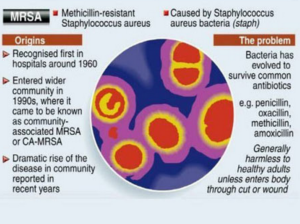
When penicillin was first introduced in the early 1940s, it was considered to be a wonder drug because it reduced the death rate from Staphylococcus infection from 70% to 25%. Unfortunately, by 1944, drug resistance was beginning to occur, so methicillin was synthesized, and, in 1959, it became the world’s first semisynthetic penicillin. Shortly thereafter in 1961, staphylococcal resistance to methicillin began as well, and the name “methicillin-resistant S. aureus” and the acronym MRSA were coined. Although methicillin was discontinued in 1993, the name and acronym have remained because of MRSA history.

MRSA is now the most common drug-resistant infection acquired in healthcare facilities. In addition to becoming more problematic as a top HAI in recent years, transmission of MRSA has also become more common in children, prison inmates, and sports participants. Community-associated MRSA (CA-MRSA) most often presents in the form of skin infections (see Figure 5). Hospital-acquired MRSA (HA-MRSA) infections manifest in various forms, including bloodstream infections, surgical site infections, and pneumonia. Although approximately 25–30% of persons are colonized in the nasal passages with Staphylococcus, less than 2% are colonized with MRSA.

MRSA are extremely resistant and can survive for weeks on environmental surfaces. Transfer of the pathogen can occur directly from patient contact with a contaminated surface or indirectly as healthcare workers touch contaminated surfaces with gloves or hands and then touch a patient.

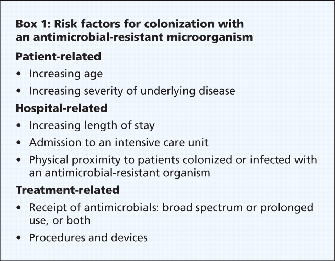
Risk factors for healthcare-acquired MRSA infection include advanced age, young age, use of quinolone antibiotics, and extended stay in a healthcare facility. Those with diabetes, cancer, or a compromised immune system are also at increased risk of infection.

Symptoms of MRSA infection vary depending on the type and stage of infection and the susceptibility of the organism. Skin infections may appear as painful, red, swollen pustules or boils; as cellulitis; or as a spider bite or bump. They can be found in areas where visible skin trauma has occurred or in areas covered by hair. Patients may also have fever, headaches, hypotension, and joint pain. Complications of MRSA-related skin infections include endocarditis, necrotizing fasciitis, osteomyelitis, and sepsis.

Patient history of admission to a healthcare facility is useful in diagnosing HA-MRSA. Definitive diagnosis of MRSA is made by oxacillin/methicillin resistance that is shown by lab culture and susceptibility testing. Specimens submitted for testing vary depending on the site of suspected infection and may include tissue, wound drainage, sputum, respiratory secretions, and blood or urine cultures.

Treatment for MRSA infections varies based on site of infection, stage of infection, and age of the individual. Treatment includes drainage of abscesses, surgical debridement, decolonization strategies, and antimicrobial therapy with antibiotics such as vancomycin #1 in alot of cases, clindamycin, daptomycin, linezolid, rifampin, trimethoprim-sulfamethoxazole (TMP-SMX), quinupristin-dalfopristin, telavancin, and tetracyclines (limited use). MRSA is rapidly becoming resistant to rifampin; therefore, this drug should not be used alone in the treatment of MRSA infections. Consultation with an infectious disease specialist is recommended for treatment of severe MRSA infections.

The CDC recommends healthcare personnel follow these guidelines to help prevent MRSA infections:

  • Follow procedures to recognize previously colonized and infected patients.
  • Follow appropriate hand hygiene practices and isolation precautions (see discussion on hand hygiene and isolation precautions later in this course). The CDC has not made a recommendation on when to discontinue contact precautions. Healthcare workers should check with their individual institution’s infection control policies.
  • Place patients in single rooms, or, if a single room is not available, cohort patients with the same MRSA in the same room or in the same patient care area. If cohorting patients with the same MRSA is not possible, place MRSA patients in rooms with patients who are at low risk for acquisition of MRSA and are likely to have short lengths of stay.
  • Keep skin wounds of MRSA patients clean and covered until healed.
  • Handle equipment and instruments/devices used for MRSA patients appropriately, with care and attention to disinfection according to institution infection control policy. Ensure that equipment is properly cleaned and disinfected before being used with another patient.

2-VancoResistantEnterococci


Vancomycin-Resistant Enterococci Infection (VRE)

Enterococci (formerly known as Group D streptococci) are non-spore-forming, gram-positive cocci that exist in either pairs or short chains. They are commonly found in the human intestine or the female genital tract. The most common organism associated with vancomycin-ressistant enterococci (VRE) infection in hospitals is Enterococcus faecium. Enterococcus faecalis is also a cause of human disease. VRE infections can occur in the urinary tract, in wounds associated with catheters, in the bloodstream, and in surgical sites. Enterococci are a common cause of endocarditis, intra-abdominal infections, and pelvic infections.

VRE was first reported in Europe in 1986, followed in 1989 by the first report in the United States. Since then it has spread rapidly. Between 1990 and 1997, the prevalence of VRE in hospital patients increased from less than 1% to 15%.

VRE, which is found predominantly in hospitalized or recently hospitalized patients, are difficult to eliminate because they are able to withstand extreme temperatures, can survive for long periods on environmental surfaces, and are resistant to vancomycin. Transmission of VRE occurs most commonly in the form of person-to-person contact by the hands of healthcare workers after contact with the blood, urine, or feces an infected individual. VRE is also spread from contact with environmental surfaces, or through contact with the open wound of an infected person.

People most at risk for infection with VRE include the elderly and those with diabetes, those with compromised immune systems, and those who are already colonized with the bacteria. Prolonged hospitalization, catheterization (urinary and intravenous), and long-term use of vancomycin or other antibiotics also increase a person’s risk of infection.

Symptoms of VRE infection vary depending on the site of infection and may include erythema, warmth, edema, fever, abdominal pain, pelvic pain, and organ pain. Definitive diagnosis is made by culture and susceptibility testing with specimens obtained from suspected sites of infection. Treatment of VRE infection may include drainage of abscesses; removal of prosthetic devices, IV lines, or catheters; and antibiotic therapy with one or more appropriate antibiotics that show activity against VRE. Consultation with an infectious disease specialist is recommended for treatment of patients with serious infections or VRE that is resistant to other antibiotics.

To prevent infection from VRE, the CDC recommends healthcare professionals use vancomycin prudently and promptly detect and report VRE infections. Healthcare providers in direct contact with patients should follow steps for proper hand hygiene and contact precautions.