Archive | September 2018

QUOTE FOR WEDNESDAY:

“Affects approximately 30,000 children in the U.S. if all forms are considered. This is comparable to the number of people affected with cystic fibrosis in the U.S. According to the Pediatric Cardiomyopathy Registry, one in every 100,000 children are diagnosed with symptoms each year. This number is conservative though, and for every diagnosed child there is another undiagnosed child unknowingly at risk of premature death.”

Children’s Cardiomyopathy Foundation (http://childrenscardiomyopathy.org/)

Cardiomyopathy in Children


C

Heart Enlarged


Cardiomyopathy is cardiomyopathy, meaning rather in adult or child the same problems in each age group occur but degree of the disease varies for all, it is case by case.  What is cardiomyopathy?

Cardiomyopathy is a disease of the heart muscle characterized by an abnormally large, thick or stiff heart muscle. It may affect only the heart’s lower chambers (ventricles) or both the lower and upper chambers (atria).  For an adult there just more wear and tear on the organ due to age and severity of the disease.

Cardiomyopathy causes damage to tissue around the heart, as well as heart muscle cells. In severe cases, the heart becomes so weak that it can’t pump blood properly. This can lead to heart failure or irregular heartbeats (arrhythmias). In some cases, cardiomyopathy also involves a buildup of scar tissue or fat within the heart muscle. In rare cases, the heart muscle can’t relax and blood can’t fill the heart properly.

Remember the natural pacemaker in humans is located in the upper right chamber (Rt. atrium) in the upper right region called the sinus node.  The sinus node initiates our impulses from that right atrium across to the Lt. atrium and works the impulse sensation down thru the A-V node-atrioventricular node (site between upper and lower chambers in the heart located at the inferior end of the interatrial septum ending at the top of the ventricles in the heart)).   From the A-V node the impulse sensation continues on from A-V node to the end of the lower right and left lower chambers (called the ventricles) of the heart that continues up the ventricles and passing through the bundle of His to the bundle branches/Purkinje fibers (The bundle of His is an important part of the electrical conduction system of the heart, as it transmits impulses from the A-V node, located at the inferior end of the interatrial septum, to the ventricles of the heart).   Than the impulse sensation goes located in the inner ventricular walls of the heart radiating over the outside of the ventricles = Purkinje Fibers. This whole process allows the heart beat to occur sounding “LubDub”.

This entire impulse conduction process allows the heart to fill up the chambers with blood first in upper chambers to lower ones to releasing the blood in the ventricles (the Rt Ventricle pumps blood to the heart to get more oxygen from lungs that sends this fresh oxygenated blood to the left side of the heart.  The left ventricle pumps the filling of its blood into our blood stream called Cardiac Output in creating the heart to beat.  Stroke Volume x Heart Rate=our Cardiac Output (SVxHR=CO).  Stroke volume is the amount of blood circulated by the heart with each beat  x HR = beat in 60 secs.

Cardiomyopathy is very often a “time-will-tell” disease. Symptoms can vary and the progression of the disease can be unpredictable.  There are many forms of cardiomyopathy; just like in the adult.

What are the types of cardiomyopathy?

The main types include:

Dilated cardiomyopathy (DCM)

DCM is the most common type and occurs when the main pumping chamber of the heart muscle is too stretched out (dilated). Dilated cardiomyopathy makes the heart unable to pump blood effectively.

Hypertrophic cardiomyopathy (HCM)

HCM makes the heart muscle too thick. Usually, the thickening occurs in the muscle of the left ventricle in the heart, often involving the wall between the heart’s two ventricles.

Restrictive cardiomyopathy

Restrictive cardiomyopathy is a rare type of cardiomyopathy that causes the heart muscle to become very rigid or stiff. This makes it difficult for the ventricles of the heart to properly fill with blood.

Arrhythmogenic right ventricular cardiomyopathy (ARVC)

ARVC is a rare form of cardiomyopathy that affects only one in 5,000 people. It occurs when the muscle of the heart’s right ventricle is replaced by thick or fatty scar tissue. The scarring “scrambles” electrical signals within the heart and can make it difficult for the heart to pump blood.

Remember the sinus node (natural pacemaker) of the heart.

How Cardiomyopathy is diagnosed:

Because the symptoms of cardiomyopathy can be diverse, it is often misdiagnosed as asthma, an infection or a gastrointestinal problem but the tests similar to adults tests being ruled out for this disease.  Remember its the same problem at a different age.

In children who have no symptoms, cardiomyopathy is sometimes diagnosed when the child has a chest x-ray that shows an enlarged heart or an echocardiogram for another reason.

If your child’s doctor suspects cardiomyopathy, he or she may order one or more of the following tests, from least invasive to most invasive tests:

  • blood tests
  • chest x-rays
  • electrocardiogram  (EKG or ECG)
  • echocardiogram (cardiac ultrasound)
  • cardiac magnetic resonance imaging (MRI)
  • stress echocardiography, which uses ultrasound and heart-rate monitoring to assess your child’s heart function just before and just after exercise
  • stress test, or exercise test
  • cardiac catheterization
  • coronary angiography

What are the treatment (Rx) options for cardiomyopathy, again similar to adults Rx.?

Your child’s treatment options will be determined by the type of cardiomyopathy he or she has, as well as the specific symptoms. A child with no symptoms might not need medication or other treatment right away. Instead, the cardiologist will monitor your child to gauge the progression of the disease.

A child with more serious symptoms may need additional tests to give the treatment team more detailed information about how the cardiomyopathy is affecting the heart and the rest of the body. .

Many children with cardiomyopathy do well with medication alone. Others need surgery to improve the function of valves, and in the most severe cases, some children need a heart transplant.

Medication for cardiomyopathy

There are several different types of medications for cardiomyopathy, depending on which type your child has and the symptoms.

  • Angiotestin converting enzyme (ACE) inhibitors are drugs that dilate blood vessels in the body, fighting the constricting effect caused by heart failure.
  • Antiarrhythmic medications combat the abnormal heart rhythms caused by irregular electrical activity within the heart.
  • Beta blockers block certain chemicals from binding to nerve receptors in the heart, slowing the heart rate and lowering blood pressure.
  • Blood thinners or anticoagulants help prevent the formation of blood clots, especially in children with the dilated form of cardiomyopathy.
  • Diuretics prevent the buildup of fluid in the body and can help breathing by reducing fluid in the lungs. These drugs may also be helpful in treating scar tissue on the heart.

Surgical treatments for cardiomyopathy

There are several options for treating cardiomyopathy using surgery or minimally-invasive procedures.

Defibrillators

For some children with cardiomyopathy — particularly HCM — an implantable cardioverter-defibrillator (ICD) can be a life-saving option. This tiny instrument, about the size of a deck of cards, is placed in the chest to monitor the child’s heartbeat. If the child has an arrhythmia, the defibrillator will administer a precise electrical pulse to restore normal heart rhythm.

Pacemakers

Pacemaker implantation is minimally invasive option for some children with cardiomyopathy. The pacemaker — a small electronic device — is inserted directly under the skin, where it sends electric signals to the child’s heart, controlling and monitoring the heart rate. The procedure can be performed under local anesthesia in a matter of hours.

Radiofrequency ablation

Radiofrequency ablation is another type of minimally invasive treatment that can be very effective for some children with cardiomyopathy due to arrhythmia. A small, needle-like probe is inserted into the scarred tissue of the heart muscle, sending out radiofrequency waves that burns away the scar tissue and the arrhythmia.

Surgical removal of some heart muscle

In serious cases of hypertrophic cardiomyopathy, the treatment team may perform a surgery called a septal myectomy or septal myomectomy. This procedure involves the removal of a portion of the thickened muscle in the heart, widening the channel in the heart’s ventricle that leads to the aortic valve. The procedure has a very good success rate, and most children have improved blood flow throughout the heart and body.

Ventricular assist device

In certain types of cardiomyopathy, a ventricular-assist device (VAD) can be used to help recover the heart and normalize the heart’s function. After the heart’s function has returned to normal, the VAD will be removed and the child will be monitored for any further heart problems. This approach using the VAD is called a “bridge to recovery.”

Heart transplant

Children with the most severe cases of cardiomyopathy may need a heart transplant if other methods don’t manage symptoms. While your child is waiting for an available heart, a VAD may be used to support the heart. In many cases, children can return to school and other activities while waiting for a transplant.

So similar to how adults are treated also with cardiomyopathy.  The age is the pretty much the difference.


QUOTE FOR TUESDAY:

“If you have a bad case of acne, you may feel like you are the only one. But many people have acne. It is the most common skin problem in the United States. About 40 to 50 million Americans have acne at any one time.”

The American Academy of Dermatology AAD


QUOTE FOR MONDAY:

“In years past we used to call ovarian cancer the silent killer but it’s really not completely silent, at least in some patients.”

Dr. Edward Tanner (an assistant professor of gynecology and obstetrics at the Johns Hopkins University School of Medicine in Baltimore, Maryland).

QUOTE FOR THE WEEKEND:

“Peyronie’s disease is penis problem caused by scar tissue, called plaque, that forms inside the penis. It can result in a bent, rather than straight, erect penis. Most men with Peyronie’s disease can still have sex. But for some, it can be painful and cause erectile dysfunction.”

MAYO CLINIC

QUOTE FOR FRIDAY:

“Sept. 12, 2003 — Popular TV and movie star John Ritter has died from an undetected flaw in his heart called an aortic dissection.  An aortic aneurysm is an abnormal bulge that occurs in the wall of the major blood vessel (aorta) that carries blood from your heart to your body. Aortic aneurysms can occur anywhere in your aorta and may be tube-shaped (fusiform) or round (saccular). Aortic aneurysms include: Abdominal aortic aneurysm.

MAYO CLINIC


Thoracic Aortic Aneurysm and Aortic Dissection Month

Named for actor John Ritter, who died of a thoracic aortic dissection, Ritter Rules combine knowledge with action. Address urgency, symptoms, who is most at risk and which imaging tests.

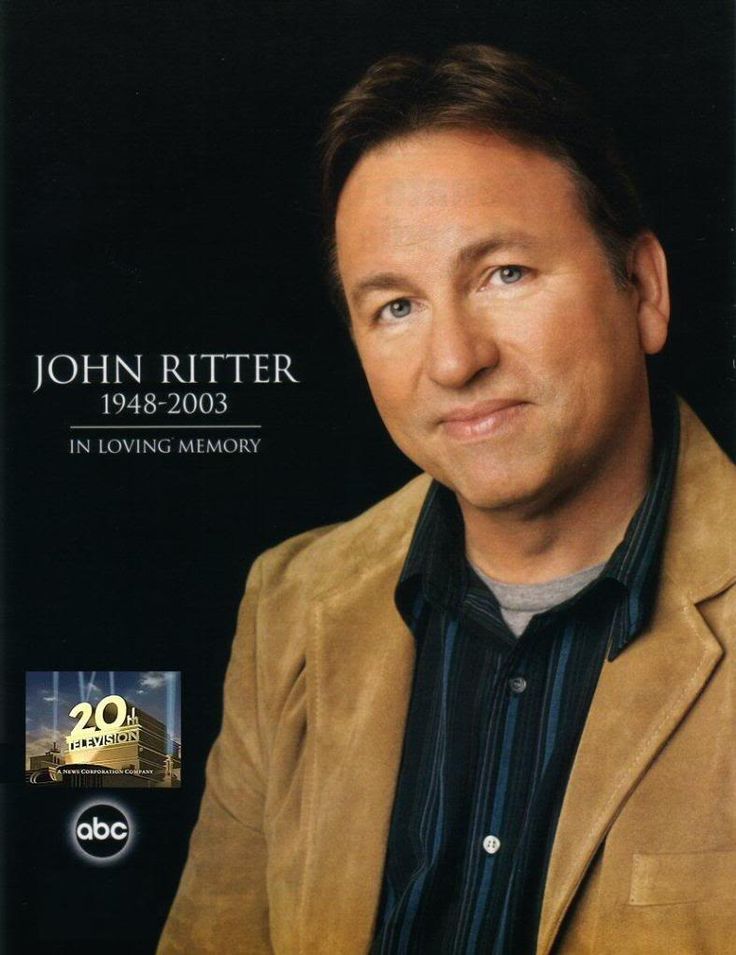
Remember that great comedian and actor in our time John Ritter.  A man non stop in his work and one day on his set doing doing the show “Eight simple rules for dating my daughter.” when becoming severely perspired, weak to nausea/vomiting than laying down in his trailer.  His colleagues knew this was never him and took him to the hospital across the street where he was being treated for a heart attack.  After 4 to 5 hours this occurred he was being rushed to the OR to be possibly saved for Aortic Dissection but like many it was too LATE!  No one knows they have it till in the hospital and hopefully caught in time.  How do you prevent this GO TO A CARDIOLOGIST yearly and be checked after age 50 or much sooner if heredity in the family.  Just like so many other diseases, you don’t know till your diagnosed by a doctor that you have it.  Some are easier in picking up but this problem is sneaks up behind you and may not be reversible for you, like in Ritter’s case.

What is a thoracic aortic aneurysm?

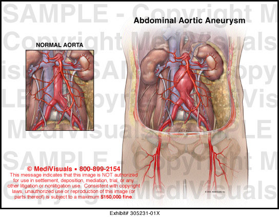
The aorta is the largest blood vessel in the body. It delivers oxygenated blood from the heart to the rest of the body. An aortic aneurysm is a bulging, weakened area in the wall of the aorta. Over time, the blood vessel balloons and is at risk for bursting (rupture) or separating (dissection). This can cause life threatening bleeding and potentially death.

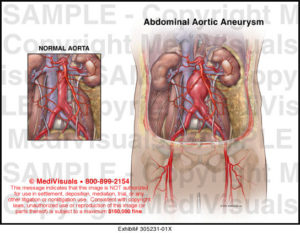
“Thoracic” refers to the part of the aorta that runs through the chest (thoracic aortic aneurysm). Aneurysms occur more often in the portion of the aorta that runs through the abdomen (abdominal aortic aneurysm).

Once formed, an aneurysm will gradually increase in size and get progressively weaker.

Thoracic Aortic Aneurysm and Aortic Dissection describe forms of aortic disease. When the walls of a section of blood vessel become weak and thin, it results in a bulging or ballooning of the vessel that is commonly called an aneurysm (like a thin punching bag). Dissection is the tearing of a blood vessel’s inner lining, causing blood to leak between the layers of the vessel wall and blood only for so long can bleed between those layers till the obvious. An aneurysm may tear (dissect) or completely rupture that area of the aorta. Under certain conditions, a blood vessel may dissect even when there is no aneurysm.

When the aneurysm and/or dissection involve the aorta (the main artery leading away from the heart), the bulging and tearing are considered among the most serious, life-threatening conditions. However, the diseased aorta can be successfully treated, especially when found before an emergency occurs. The US National Center for Health Statistics reports almost 15,000 aorta-related deaths (including both the thoracic and abdominal aorta) each year.

What are the symptoms of a thoracic aortic aneurysm?

Thoracic aortic aneurysms may not cause symptoms. When symptoms do occur, they may be related to the location, size, and how fast the aneurysm is growing.

Sudden, severe pain associated with a thoracic aneurysm may be a sign of a life-threatening medical emergency.

Symptoms of a thoracic aneurysm may include:

  • Pain in the jaw, neck, or upper back-This is why the ER is thinking its a heart attack.

  • Pain in the chest or back-This is why the ER is thinking its a heart attack.

  • Wheezing, coughing, or shortness of breath as a result of pressure on the trachea (windpipe)

  • Hoarseness as a result of pressure on the vocal cords

  • Trouble swallowing due to pressure on the esophagus

The symptoms of a thoracic aortic aneurysm may look like other conditions. See your doctor for a diagnosis.

How is a thoracic aortic aneurysm diagnosed?

Your doctor will do a complete medical history and physical exam. Other possible tests include:

  • Computed tomography scan (also called a CT or CAT scan). This test uses X-rays and computer technology to make horizontal, or axial, images (often called slices) of the body. A CT scan shows detailed images of any part of the body, including the bones, muscles, fat, and organs. CT scans are more detailed than standard X-rays.

  • Magnetic resonance imaging (MRI). This test uses a combination of large magnets, radiofrequencies, and a computer to produce detailed images of organs and structures within the body.

  • Echocardiogram (also called echo). This test evaluates the structure and function of the heart by using sound waves recorded on an electronic sensor that make a moving picture of the heart and heart valves, as well as the structures within the chest, such as the lungs and the area around the lungs and the chest organs.

  • Transesophageal echocardiogram (TEE). This test uses echocardiography to check for aneurysm, the condition of heart valves, or presence of a tear of the lining of the aorta. TEE is done by inserting a probe with a transducer on the end down the throat.

  • Chest X-ray. This test uses invisible electromagnetic energy beams to make images of internal tissues, bones, and organs onto film.

  • Arteriogram (angiogram). This is an X-ray image of the blood vessels that is used to assess conditions such as aneurysm, narrowing of the blood vessel, or blockages. A dye (contrast) will be injected through a thin, flexible tube placed in an artery. The dye makes the blood vessels visible on an X-ray.

What is the treatment for thoracic aortic aneurysm?

Treatment may include:

  • Monitoring with MRI or CT. These tests are done to check the size and rate of growth of the aneurysm.

  • Managing risk factors. Steps, such as quitting smoking, controlling blood sugar if you have diabetes, losing weight if overweight, and eating a healthy diet may help control the progression of the aneurysm.  Reversing these problems always helps, doesn’t cure it.

  • Medicine. Used to control factors such as high cholesterol or high blood pressure.  These medications doing this control of conditions listed helps keep it from getting worse with pt following up with being checked as M.D. prescribes.

  • Surgery Treatment for a thoracic aneurysm may include surgical repair or removal of the aneurysm, or inserting a metal mesh coil (stent) to support the blood vessel and prevent rupture.

    • Thoracic aortic aneurysm open repair. The type of surgery will depend on the location and type of aneurysm, and your overall health. For an ascending or aortic arch aneurysm, a large incision may be made through the breastbone. If an ascending aneurysm involves damage to the aortic valve of the heart, the valve may be repaired or replaced during the procedure. For a descending aneurysm, a large incision may extend from the back under the shoulder blade around the side of the rib cage to just under the breast. This lets the surgeon to see the aorta directly to repair the aneurysm.

    • Endovascular aneurysm repair (EVAR). EVAR requires only small incisions in the groin. Using X-ray guidance and specially-designed instruments, the surgeon can repair the aneurysm by inserting a metal mesh coil, called a stent-graft, inside the aorta. Not all thoracic aneurysms can be repaired by means of EVAR.

A small aneurysm or one that doesn’t cause symptoms may not require surgical treatment until it reaches a certain size or is rapidly increasing in size over a short period of time. Your doctor may recommend “watchful waiting.” This may include a CT scan or MRI scan every 6 months to closely monitor the aneurysm, and blood pressure medicine may be used to control high blood pressure.

Your doctor may recommend surgery if the aneurysm is causing symptoms or is large.

What is aortic dissection?

An aortic dissection starts with a tear in the inner layer of the aortic wall of the thoracic aorta. The aortic wall is made up of 3 layers of tissue. When a tear occurs in the innermost layer of the aortic wall, blood is then channeled into the wall of the aorta separating the layers of tissues. This generates a weakening in the aortic wall with a potential for rupture. Aortic dissection can be a life-threatening emergency, like in John Ritter’s situation Sept 11, 2003 which led him to death in the ER and attempt to save him in the OR but too late (Died at 54 y/o. missing his next birthday Sept 17). The most commonly reported symptom of an acute aortic dissection is severe, constant chest or upper back pain, sometimes described as “ripping” or “tearing.” The pain may move from one place to another.

When a diagnosis of aortic dissection is confirmed, immediate surgery or stenting is usually done.

For a thoracic aneurysm may include surgical repair or removal of the aneurysm, or inserting a metal mesh coil (stent) to support the blood vessel and prevent rupture.  If not treated the vessel continues to weaken getting thinner.

PREVENTION is the key = seeing your cardiologist every 6 months to yearly not for anything overlooked by the expert on our engine of the body (the heart) and diseases of cardiology.  Go to the expert over a general doctor so nothing is missed!

QUOTE FOR THURSDAY:

“Stroke is the greatest risk for the patient with atrial fibrillation. Some patients are not aware of atrial fibrillation and in them, the risk remains unrecognized. In these patients, the first indication of A Fib may be admission to a hospital with a stroke.  If the heartbeat is very fast for a long period of time, it can also lead to heart failure.”

Heart Rhythm Society

QUOTE FOR WEDNESDAY:

“Atrial Fibrillation (AFib) is caused when the two upper chambers of the heart beat unpredictably and sometimes rapidly, in a fibrillation. These irregular heartbeats can cause blood to collect in the heart and potentially form a clot, which can travel to a person’s brain and cause a stroke. While AFib can occur any age, it is more common in people 65 years and older. AFib is more common in people with high blood pressure, heart disease, or diabetes.”

American Heart Association

QUOTE FOR TUESDAY:

“A month after 9/11, nearly 50% of people living near the WTC site who participated in a survey about their health needs and concerns said they had nose, throat or eye irritation.  Respiratory hospital admissions increased significantly among lower Manhattan residents the first week after 9/11 compared with a similar demographic in Queens. Cardiovascular and cerebrovascular disease admissions also increased two to three weeks after 9/11, especially among women and those over 65 years of age.”

NYC 9/11 Health /www1.nyc.gov/