Part I What actually is Chronic Obstructive Pulmonary Disease (COPD)?

COPD2  COPD3 Usually due to smoking

This is Healthy Lung Month covering COPD.

What is Chronic Obstructive Pulmonary Disease (COPD)?

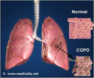
Chronic obstructive pulmonary disease (COPD) is a term that applies to patients with chronic bronchitis, bronchiectasis, emphysema and, to a certain extent, asthma. A brief review of normal functional anatomy will provide a background for the discussion of pathology.

The airway down to the bronchioles normally is lined with ciliated pseudo-stratified columnar cells and goblet cells. Mucus derives from mucus glands that are freely distributed in the walls of the trachea and bronchi. The cilia sweep mucus and minor debris toward the upper airway. Low humidity, anesthesia gases, cigarette smoking and other chemical irritants paralyze the action of these cilia. The mucociliary action starts again after a matter of time. This is why people awaken to “smokers cough.”

“Chronic obstructive pulmonary disease (COPD) is a term that applies to patients with chronic bronchitis, bronchiectasis, emphysema and, to a certain extent, asthma.”

Bronchi run in septal connective tissue, but bronchioles are suspended in lung parenchyma by alveolar elastic tissue. The elastic tissue extends throughout alveolar walls, air passages, and vessels, connecting them in a delicate web. Bronchiolar epithelium is ciliated, single-layered and columnar or cuboidal. Beyond the bronchioles the epithelium is flat and lined with a film of phospholipid (surfactant), which lowers surface tension and thereby helps to keep these air spaces from collapsing. Remember that the phospholipid develops during later gestation in utero. This is the reason why premature infant’s lungs cannot stay inflated without the addition of surfactant therapy. Macrophages are found in alveolar lining. Smooth muscles surround the walls of all bronchi, bronchioles, and alveolar ducts and when stimulated they shorten and narrow the passages. Cartilage lends rigidity and lies in regular horse-shaped rings in the tracheal wall. Cartilage is absent in bronchi less than 1 mm in diameter.

The terminal bronchiole is lined with columnar epithelium and is the last purely conducting airway. An acinus includes a terminal bronchiole and its distal structures. Five to ten acini together constitute a secondary lobule, which is generally 1 to 2 cm in diameter and is partly surrounded by grossly visible fibrous septa. Passages distal to the terminal bronchiole include an average of three but as many as nine generations of respiratory bronchioles lined with both columnar and alveolar epithelium. Each of the last respiratory bronchioles gives rise to about six alveolar ducts, each of these to one or two alveolar sacs, and finally each of the sacs to perhaps seventy-five alveoli. Alveolar pores (pores of Kohn) may connect alveoli in adjacent lobules.

Two different circulations supply the lungs. The pulmonary arteries and veins are involved in gas exchange. The pulmonary arteries branch with the bronchi, dividing into capillaries at the level of the respiratory bronchiole, and supplying these as well as the alveolar ducts and alveoli. In the periphery of the lung, the pulmonary veins lie in the interlobular septa rather than accompanying the arteries and airways. The bronchial arteries are small and arise mostly from the aorta. They accompany the bronchi to supply their walls. In some cases of COPD, like bronchiectasis, extensive anastomoses develop between the pulmonary and bronchial circulations. This can allow major shunting and recirculation of blood, therefore contributing to cardiac overload and failure. Lymphatics run chiefly in bronchial walls and as a fine network in the pleural membrane. The lumina of the capillaries in the alveolar walls are separated from the alveolar lining surfaces by the alveolar-capillary membrane, consisting of thin endothelial and epithelial cells and a minute but expansile interstitial space. This interface between air and blood, only 2 microns in thickness, is the only place where gases may be exchanged effectively.

Disease Specific Review

Chronic Bronchitis

Chronic bronchitis is a clinical disorder characterized by excessive mucus secretion in the bronchi. It was traditionally defined by chronic or recurrent productive cough lasting for a minimum of three months per year and for at least two consecutive years, in which all other causes for the cough have been eliminated. Today’s definition remains more simplistic to include a productive cough progressing over a period of time and lasting longer and longer. Sometimes, chronic bronchitis is broken down into three types: simple, mucopurulent or obstructive. The pathologic changes consist of inflammation, primarily mononuclear, infiltrate in the bronchial wall, hypertrophy and hyperplasia of the mucus-secreting bronchial glands and mucosal goblet cells, metaplasia of bronchial and bronchiolar epithelium, and loss of cilia. Eventually, there may be distortion and scarring of the bronchial wall.

Asthma

Asthma is a disease characterized by increased responsiveness of the trachea and bronchi to various stimuli (intrinsic or extrinsic), causing difficulty in breathing due to narrowing airways. The narrowing is dynamic and changes in degree. It occurs either spontaneously or because of therapy. The basic defect appears to be an altered state of the host, which periodically produces a hyperirritable contraction of smooth muscle and hypersecretion of bronchial mucus. This mucus is abnormally sticky and therefore obstructive. In some instances, the illness seems related to an altered immunologic state.

Histological changes of asthma include an increase in the size and number of the mucosal goblet cells and submucosal mucus glands. There is marked thickening of the bronchial basement membrane and hypertrophy of bronchial and bronchiolar smooth muscle tissue. A submucosal infiltration of mononuclear inflammatory cells, eosinophils and plugs of mucus blocks small airways. Patients who have had asthma for many years may develop cor pulmonale and emphysema.

Emphysema

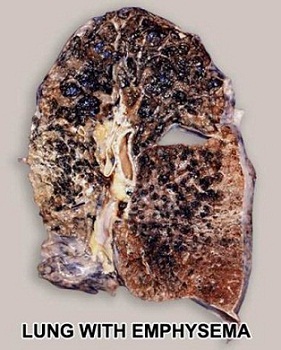
Pulmonary emphysema is described in clinical, radiological and physiologic terms, but the condition is best defined morphologically. It is an enlargement of the air spaces distal to the terminal non-respiratory bronchiole, with destruction of alveolar walls.

Although the normal lung has about 35,000 terminal bronchioles and their total internal cross-sectional area is at least 40 times as great as that of the lobar bronchi, the bronchioles are more delicate and vulnerable. Bronchioles may be obstructed partially or completely, temporarily or permanently, by thickening of their walls, by collapse due to loss of elasticity of the surrounding parenchyma, or by influx of exudate. In advanced emphysema, the lungs are large, pale, and relatively bloodless. They do not readily collapse. They many contain many superficial blebs or bullae, which occasionally are huge. The right ventricle of the heart is often enlarged (cor pulmonale), reflecting pulmonary arterial hypertension. Right ventricular enlargement is found in about 40% of autopsies of patients with severe emphysema. The distal air spaces are distended and disrupted, thus excessively confluent and reduced in number. There may be marked decrease in the number and size of the smaller vascular channels. The decrease in alveolar-capillary membrane surface area may be critical. Death may result from infection that obliterates the small bronchi and bronchioles. There is often organized pneumonia or scarring of the lung parenchyma due to previous infections.

Classification of emphysema relies on descriptive morphology, requiring the study of inflated lungs. The two principal types are centrilobular and panlobular emphysema. The two types may coexist in the same lung or lobe.

Centrilobular emphysema (CLE) or centriacinar emphysema affects respiratory bronchioles selectively. Fenestrations develop in the walls, enlarge, become confluent, and tend to form a single space as the walls disintegrate. There is often bronchiolitis with narrowing of lumina. The more distal parenchyma (alveolar ducts and sacs and alveoli) is initially preserved, then similarly destroyed as fenestrations develop and progress.

The disease commonly affects the upper portions of the lung more severely, but it tends to be unevenly distributed. The walls of the emphysematous spaces may be deeply pigmented. This discoloration may represent failure of clearance mechanisms to remove dust particles, or perhaps the pigment plays an active role in lung destruction. CLE is much more prevalent in males than in females. It is usually associated with chronic bronchitis and is seldom found in nonsmokers.

Panlobular emphysema (PLE) or panacinar emphysema is a nearly uniform enlargement and destruction of the alveoli in the pulmonary acinus. As the disease progresses, there is gradual loss of all components of the acinus until only a few strands of tissue, which are usually blood vessels, remain. PLE is usually diffuse, but is more severe in the lower lung areas. It is often found to some degree in older people, who do not have chronic bronchitis or clinical impairment of lung function. The term senile emphysema was formerly applied to this condition. PLE occurs as commonly in women and men, but is less frequent than CLE. It is a characteristic finding in those with homozygous deficiency of serum alpha-1 antitrypsin. It has also been found that certain populations of IV Ritalin abusers show PLE.

Bullae are common in both CLE and PLE, but may exist in the absence of either. Air-filled spaces in the visceral pleura are commonly termed blebs, and those in the parenchyma greater than 1 cm in diameter are called bullae. A valve mechanism in the bronchial communication of a bulla permits air trapping and enlargement of the air space. This scenario may compress the surrounding normal lung. Blebs may rupture into the pleural cavity causing a pneumothorax, and through a valve mechanism in the bronchopleural fistula a tension pneumothorax may develop.

Paracicatricial emphysema occurring adjacent to pulmonary scars represents another type of localized emphysema. When the air spaces distal to terminal bronchioles are increased beyond the normal size but do not show destructive changes of the alveolar walls, the condition is called pulmonary overinflation. This condition may be obstructive, because of air trapping beyond an incomplete bronchial obstruction due to a foreign body or a neoplasm. Many lung lobules may be simultaneously affected as a result of many check-valve obstructions, as in bronchial asthma. Pulmonary overinflation may also be nonobstructive, less properly called “compensatory emphysema”, when associated with atelectasis or resection of other areas of the lung.

Bronchiectasis

Bronchiectasis means irreversible dilation and distortion of the bronchi and bronchioles. Saccular bronchiectasis is the classic advanced form characterized by irregular dilatations and narrowing. The term cystic is used when the dilatations are especially large and numerous. Cystic bronchiectasis can be further classified as fusiform or varicose.

Tubular bronchiectasis is simply the absence of normal bronchial tapering and is usually a manifestation of severe chronic bronchitis rather than of true bronchial wall destruction.

Repeated or prolonged episodes of pneumonitis, inhaled foreign objects or neoplasms have been known to cause bronchiectasis. When the bronchiectatic process involves most or all of the bronchial tree, whether in one or both lungs, it is believed to be genetic or developmental in origin.

Mucoviscidosis, Kartagener’s syndrome (bronchiectasis with dextrocardia and paranasal sinusitis), and agammaglobulinemia are all examples of inherited or developmental diseases associated with bronchiectasis. The term pseudobronchiectasis is applied to cylindrical bronchial widening, which may complicate a pneumonitis but which disappears after a few months. Bronchiectasis is true saccular bronchiectasis but without cough or expectoration. It is located especially in the upper lobes where good dependent drainage is available. A proximal form of bronchiectasis (with normal distal airways) complicates aspergillus mucus plugging.

Advanced bronchiectasis is often accompanied by anastomoses between the bronchial and pulmonary vessels. These cause right-to-left shunts, with resulting hypoxemia, pulmonary hypertension and cor pulmonale.

Keeping a healthy lung prevents emphysema.  So for starters don’t smoke and exercise; which includes don’t be exposed to smoke frequently!
QUOTE FOR FRIDAY:

Overall numbers the CDC reports in statistics up to 2021:

“OVERALL NUMBERS:

  • Prevalence: In 2021, 38.4 million Americans, or 11.6% of the population, had diabetes.
    • 2 million Americans have type 1 diabetes, including about 304,000 children and adolescents
  • Diagnosed and undiagnosed: Of the 38.4 million adults with diabetes, 29.7 million were diagnosed, and 8.7 million were undiagnosed.
  • Prevalence in seniors: The percentage of Americans age 65 and older remains high, at 29.2%, or 16.5 million seniors (diagnosed and undiagnosed).
  • New cases: 1.2 million Americans are diagnosed with diabetes every year.
  • Prediabetes: In 2021, 97.6 million Americans age 18 and older had prediabetes.”

“Diabetes was the eighth leading cause of death in the United States in 2021 based on the 103,294 death certificates in which diabetes was listed as the underlying cause of death. In 2021, diabetes was mentioned as a cause of death in a total of 399,401 certificates.

Cost of diabetes

Updated November 2, 2023

$412.9 billion: Total cost of diagnosed diabetes in the United States in 2022

$306.6 billion was for direct medical costs

$106.3 billion was in indirect costs

After adjusting for population age and sex differences, average medical expenditures among people with diagnosed diabetes were 2.6 times higher than what expenditures would be in the absence of diabetes.”

American Diabetes Association (https://diabetes.org/about-diabetes/statistics/about-diabetes)


QUOTE FOR THURSDAY:

“Natural insulin (i.e. insulin released from your pancreas) keeps your blood sugar in a very narrow range. Overnight and between meals, the normal, non-diabetic blood sugar ranges between 60-100mg/dl and 140 mg/dl or less after meals and snacks.  To keep the blood sugar controlled overnight, fasting and between meals, your body releases a low, background level of insulin. When you eat, there is a large burst of insulin. This surge of insulin is needed to dispose of all the carbohydrate or sugar that is getting absorbed from your meal. All of this happens automatically!  Insulin is continuously released from the pancreas into the blood stream. Although the insulin is quickly destroyed (5-6 minutes) the effect on cells may last 1-1/2 hours. When your body needs more insulin, the blood levels quickly rise, and, the converse – when you need less, the blood levels rapidly fall —The situation is different when you have diabetes and are getting insulin replacement therapy. Once you have injected a dose of insulin, it is going to get absorbed into your bloodstream whether you need it or not.

Insulin pump therapy is increasingly popular. Because insulin pumps more closely mimic what your body does naturally, you can improve your blood sugar control. With that control comes a more flexible lifestyle. Remember, though, that the pumps still require a lot of input from users.

Type 1 diabetes is caused by a loss or malfunction of the insulin producing cells, called pancreatic beta cells. Damage to beta cells results in an absence or insufficient production of insulin produced by the body.

You have Type 2 diabetes if your tissues are resistant to insulin, and if you lack enough insulin to overcome this resistance. Type 2 diabetes is the most common form of diabetes of diabetes worldwide and accounts for 90-95% of cases.”

University of California (https://dtc.ucsf.edu)

QUOTE FOR WEDNESDAY:

“Research shows that type 2 diabetes increases a person’s risk of developing dementia. Dementia risk also increases with the length of time someone has diabetes and how severe it is. However, it is important to note that diabetes is only a risk factor and does not mean that a person with diabetes will go on to develop dementia.

In people with type 1 diabetes. severe blood sugar highs and lows are also associated with increased risk of developing dementia.As you get older, you are more likely to develop certain health conditions, including diabetes.

To manage this, speak to your GP about going for a health check.

Eating a healthy, balanced diet may reduce your risk of type 2 diabetes. No single ingredient, nutrient or food can improve health by itself. Instead, eating a range of different foods in the right proportions is what makes a difference. This is known as a ‘balanced’ diet.

By eating a balanced diet you are more likely to get all the nutrients you need to stay healthy.”

Alzheimer’s Society (https://www.alzheimers.org.uk/about-dementia/managing-the-risk-of-dementia/reduce-your-risk-of-dementia/diabetes)


QUOTE FOR TUESDAY:

“Diabetes is a condition that happens when your blood sugar (glucose) is too high. It develops when your pancreas doesn’t make enough insulin or any at all, or when your body isn’t responding to the effects of insulin properly. Diabetes affects people of all ages. Most forms of diabetes are chronic (lifelong), and all forms are manageable with medications and/or lifestyle changes.”

Cleveland Clinic (https://my.clevelandclinic.org/health/diseases/7104-diabetes)

Part II Diabetes Awareness Month – Symptoms & Complications of Diabetes!


 ADA_STOP_LockUp_4C

Diabetes is becoming more common in the United States.  From 1980 through 2011, the number of Americans with diagnosed diabetes has than tripled (from 5.6 million to 20.9 million).

DIABETES: The Signs & Symptom and How to control the them:

The signs and symptoms of Diabetes 1 or 2 with hyperglycemia (HIGH GLUCOSE LEVELS):

THINK OF THE 3 P’s=

1.)Polyuria-When all of sudden you are voiding urine.  Poly ,meaning alot, uria, meaning urine,; so a lot of urinating due to your body trying to void out of the body excess glucose in your urine can be a symptom of diabetes. This is a common symptom that causes the next symptoms due to your voiding a lot of urine which causes your body to lose fluid, being water with alot of glucose in the urine, and in return you become very thirsty with hungry. This gives you:

2.)Polydipsia= very thirsty

3.)Polyphagia=very hungry

This should be a red light for a diabetic with these one or all 3 symptoms to finger stick or glucose test themselves.   See where your glucose level is at and if over 200 this is why you have one or all of the “P” symptoms (listed above).

Other s/s of Diabetes consist of:

– Tingling / Numbness in the hands and feet (diabetic neuropathy)

-Very tired and fatigued

-Weight Loss (more common to see in Diabetes 1; most of the time Type II DM is due to obesity and noncompliance of a diabetic )

-Blurred Vision.

-Sores or diabetic ulcers especially in the lower extremities that do not heal; and if not healed, this can cause in time a severe condition.

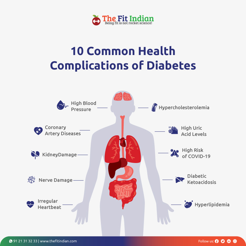
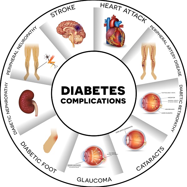
Complications that can come about due to DIABETES:

Dental Disease – Diabetes can lead to problems with teeth and gums, called gingivitis and periodontitis.

Heart Disease – People with diabetes have a higher risk for HTN, heart attack and stroke.

Eye Complications – People with diabetes have a higher risk of blindness and other vision problems.

Kidney Disease – Diabetes can damage the kidneys and may lead all the way up to kidney failure.

Nerve Damage (neuropathy) – Diabetes can cause damage to the nerves that run through the body.  Particularly neuropathy can occur leading to no feeling to other complications occuring (Example diabetic with neuropathy keeps stepping on sharp items not feeling them making a wound develop causing a sore not to heal that leads into a diabetic ulcer that doesn’t heel leading to a foot amputation or worse below or above knee amputation it leads to in time).

Foot Problems – Nerve damage, infections of the feet, and problems with blood flow to the feet can be caused by diabetes.

Skin Complications – Diabetes can cause skin problems, such as infections, sores, and itching. Skin problems are sometimes a first sign that someone has diabetes. Sores that cannot heal due to constant high glucose in the body can lead into a severe condition=AMPUTATION of the foot or leg.

**. (At least 15 % of all people with diabetes eventually have a foot ulcer, and 6 out of every 1000 people with Diabetes have an AMPUTATION. Possibly first surgery with bypassing the blood can resolve the problem 100% or like many only temporary. It is based on your other medical history with how brittle the diabetes and how compliant you are in taking care of yourself with diabetes.   This is why you see with some diabetics amputations of the lower extremities, hardly ever a upper amputation which is usually due to trauma or smoking.***

All these complications are effected by hyperglycemia and in playing a part in the blood circulation of our body. Ending line the person is getting bad oxygenated blood supply sent to the lower extremities when the glucose is poorly controlled over a long time. Based on the principle of gravity; what happens here is the heart pumps our blood throughout our body and when it gets difficult for the organ to do its job due to thick high glucose blood than it has to compensate at some point. Simply a narrowing to a blockage is occurring in that lower extremity and the reason for this is it’s the furthest area from the heart=FEET/LEGS.

This can be caused by just thick high glucose blood flowing throughout the body making it difficult for the heart to pump as effectively as opposed to someone that doesn’t have hyperglycemia which over time leads to further complications (listed above).

Diabetes with constant high glucose blood levels can leaded into poor circulation causes the feet and lower leg to first become cool to cold to changing colors of pale to cyanotic (purple) which takes over weeks to months to years, depending on the patient. Then the tissue gets necrotic (black=dead tissue) and an amputation has to be done to save the person or else this will get infected locally, at first, going into a systemic infection causing the person to go into septicemia and expire.


REFERENCES for Part 1, Part 2 & 3:

1.)  Center for Disease (CDC) – “National Diabetes Fact Sheet”

2.)  NYS Dept. of Health –Diabetes

3.)  Diabetic Neuropathy.org “All about diabetic neuropathy and nerve damage caused by Diabetes.”

4.)  NIDDK “National Institute of Diabetes and Digestive and Kidney Diseases.

5.)  National Diabetes Information Clearinghouse (NIDC) – U.S. Department of Health and Human Services.       “Preventing Diabetes Problems: What you need to know”


QUOTE FOR MONDAY:

“Among the U.S. population overall, crude estimates for 2021 were:

  • 38.4 million people of all ages—or 11.6% of the U.S. population—had diabetes.
  • 38.1 million adults aged 18 years or older—or 14.7% of all U.S. adults—had diabetes (Table 1a; Table 1b).
  • 8.7 million adults aged 18 years or older who met laboratory criteria for diabetes were not aware of or did not report having diabetes (undiagnosed diabetes, Table 1b). This number represents 3.4% of all U.S. adults (Table 1a) and 22.8% of all U.S. adults with diabetes.
  • The percentage of adults with diabetes increased with age, reaching 29.2% among those aged 65 years or older (Table 1a).”

Center for Disease Control and Prevention – CDC (https://www.cdc.gov/diabetes/php/data-research/index.html)

QUOTE FOR THE WEEKEND:

“There are two main types of seizures: generalized and focal seizures.

These types describe where a seizure starts in the brain and how it may affect a person.

Call 911 if a seizure (of any type) lasts more than 5 minutes or if the person does not wake up fully between seizures.”

Centers for Disease Control and Prevention (https://www.cdc.gov/epilepsy/about/types-of-seizures.html)


Part III National Epilepsy Awareness Month: Types of Seizures, and Types of Treatments for Epilepsy/Seizures!

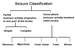
Old Lists Below on Seizure Classification:

Most Updated List on Classifications of Seizures by the Epilepsy Foundation:

Expanded Seizure Classifications


Types of seizures whether with a etiology or unknown:

I-Partial seizures (seizures beginning local)

1-simple partial seizures-(the person is conscious and not impaired).  With motor symptoms, autonomic symptoms and even psychic symptoms.

2.)-Complex partial seizures-(the person is with impairment of consciousness)

II-Generalized seizures-(bilaterally symmetrical and without local onset).

3.) Tonic clonic seizures – Grand Mal

See Above the most updated,being 2017, on classifications of seizures list by the Epilepsy Foundation.

Treatment:

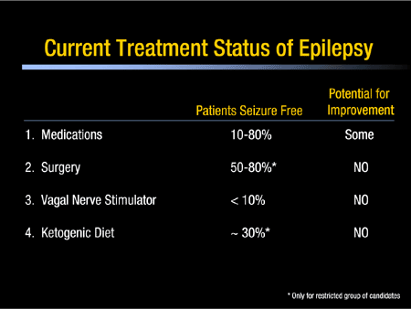
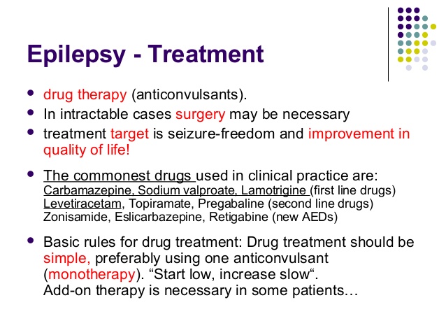
1-Epilepsy is sometimes referred to as a long-term condition, as people often live with it for many years, or for life. Although generally epilepsy cannot be ‘cured’, for most people, seizures can be ‘controlled’ (stopped) so that epilepsy has little or no impact on their lives. So treatment is often about managing seizures in the long-term.

Most people with epilepsy take anti-epileptic drugs (AEDs) to stop their seizures from happening. However, there are other treatment options for people whose seizures are not controlled by anti-epileptic drugs (AEDs).

2-The ketogenic diet is one treatment option for children with epilepsy whose seizures are not controlled with AEDs. The diet may help to reduce the number or severity of seizures and can often have positive effects on behaviour.

3-Vagus nerve stimulation therapy is a treatment for epilepsy that involves a stimulator (or ‘pulse generator’) which is connected, inside the body, to the left vagus nerve in the neck. The stimulator send regular, mild electrical stimulations through this nerve to help calm down the irregular electrical brain activity that leads to seizures.

There are several ways to treat epilepsy. How well each treatment works varies from one person to another. Vagus nerve stimulation therapy is a form of treatment for people with epilepsy whose seizures are not controlled with medication.

4-There are different kinds of epilepsy surgery. One kind of surgery involves removing a specific area of the brain which is thought to be causing the seizures. Another kind involves separating the part of the brain that is causing seizures from the rest of the brain.

Surgery may be possible for both adults and children, and might be considered if:

  • you have tried several AEDs and none of them have stopped or significantly reduced your seizures; and
  • a cause for your epilepsy can be found in a specific area of your brain, and this is an area where surgery is possible.

Whether you are suitable for surgery is something that you may like to talk about with your GP or neurologist. If you meet these criteria and are considered for surgery, you will need to have further tests before you can have the surgery.

If you are referred for surgery you will probably go to a specialist centre for tests. There are many different pre-surgical tests you might have before you can be given the go-ahead for surgery. This could include further MRI scans, an EEG (electroencephalogram) and video telemetry (an EEG while also being filmed). Other types of scans may also be done, which trace a chemical injected into the body. This can show detailed information about where seizures start in the brain.

Memory and psychological tests are also used to see how your memory and lifestyle might be affected after the surgery. These types of tests also help the doctors to see how you are likely to cope with the impact of having this type of surgery.

The tests will confirm whether:

  • the surgeons can reach the epileptogenic lesion during surgery and can remove it safely without causing new problems;
  • other parts of your brain could be affected by the surgery, for example the parts that control your speech, sight, movement or hearing;
  • you have a good chance of having your seizures stopped by the surgery; and
  • you have any other medical conditions that would stop you from having this kind of surgery.

The results from the pre-surgical tests will help you and your neurologist decide whether surgery is an option for you, and what the result of the surgery might be.

Your specialist will also talk with you about the possible risks and benefits of having surgery.

For many people the results show that surgery is not an option: the majority of people who are recommended for surgery, and have these tests carried out, are unable to have surgery.

Take the action and make your life one without seizures occurring putting your life on HOLD you need to TAKE CARE OF YOURSELF!    That is all up to you, the patient diagnosed with it or questioning if they have seizures.

Part II National Epilepsy Awareness Month-Causes, how its diagnosed, and what to know before, during,, and after the seizure!


Possible causes of Epilepsy:

Their epilepsy that is diagnosed with a IDIOPATHIC cause – meaning unknown cause and the patient could grow out of it in childhood in some cases (not all) depending on the type of seizure disorder and if the child doesn’t grow out of it the condition becomes chronic (for life).

Genetic influence (heredity). Some types of epilepsy run in families. In these instances, it’s likely that there’s a genetic influence. Researchers have linked some types of epilepsy to specific genes. But some people have genetic epilepsy that isn’t hereditary. Genetic changes can occur in a child without being passed down from a parent.For most people, genes are only part of the cause of epilepsy. Certain genes may make a person more sensitive to environmental conditions that trigger seizures.

  • A genetic tendency, passed down from one or both parents (inherited).
  • A genetic tendency that is not inherited, but is a new change in the person’s genes.

Although heredity has been known since antiquity to cause epilepsy, the progress to date in identifying the genetic basis of epilepsy has been limited primarily to the discovery of single gene mutations that cause epilepsy in relatively rare families. For the more common types of epilepsy, heredity plays a subtler role, and it is thought that a combination of mutations in multiple genes likely determine an individual’s susceptibility to seizures, as well as the responsiveness to antiepileptic medications.

Epilepsy can be caused by genetic factors (inherited) or acquired (a etiology—cause) , although in most cases it arises in part from both. The neurology and neurological sciences of Stanford Epilepsy Center Dr. Robert S. Fischer Ph D. presents in the article Genetic Causes of Epilepsy.

He also presents in this article our genes are the instruction set for building the human body. Genes reside on chromosomes.

Going to the basics is every person has 46 chromosomes, carrying a total of about 30,000 genes. We get half our chromosomes from our mother and half from our father. While genes determine the structure of our body, they also control the excitability of our brain cells. Defective genes can make hyperexcitable brain cells, which are prone to seizures.

In recent years, several epilepsy conditions have been linked to mutations in genes, but the matter is complicated by the fact that different genes may be involved in different circumstances.

In general, the most common epilepsy conditions, including partial seizures, seem to be more acquired than genetic.

Gene testing will soon be able to identify predispositions to epilepsy, allowing doctors to help a patient get treatment and to assist with family counseling. One day, doctors may simply be able to swap a patient’s cheek, test his or her genes, and predict response to various epilepsy medicines, eliminating much of the trial and error in medication choice that goes on today. Eventually, we may even be able to repair or replace defective genes that predispose a person to epilepsy, a process called gene therapy.

Lastly, Dr. Robert Fischer Ph D presented in his article, that I found very interesting, the general population has about a 1% risk of developing epilepsy.  Meanwhile, children of mothers with epilepsy have a 3 to 9% risk of inheriting this disease, while children of fathers have a 1.5 to 3% risk of inheritence. Still, the actual risk is upon the specific type of epilepsy. For example, partial seizures are less likely to run in families than are generalized seizures. In any event, with the usual forms of epilepsy, even if a parent does have the condition, there is more than a 90% chance that their child will not. So most epilepsies are acquired than inherited.

Clearly, genes determine a great deal of who we are, including our possible risk for epilepsy but slim versus a actual cause. But what happens to us in life and what we do is still the larger part of the risk for epilepsy.

A person given this diagnosis in the 1970’s, or before  and even up to the early 1990’s was quiet about ever letting people know about this since in the 1970’s and back with lack of knowledge, information to the public and definitely technology versus now.  Epilepsy is much more an accepted disease in the overall community compared to 20-25 years ago and back.  Heck in the 1970’s and back these patients when having a seizure episode were characterized as “Freaks”.  This was due to ignorance and lack of information to society/community but due to the past 20 to 25 years with the computer used more as a must in our lives with media, television and even our government they all have made it possible for society everywhere in the world to learn and understand diseases with acceptance in wanting to help those, particularly the US, but we still need a healthier America. It will take time to get there with the many multicultural lives that all live in the U.S. which practice differently on how important a healthy diet is with exercise balanced with rest.  Also including stress well controlled is not always in America on their top priority list in living.  Stress can even be a catalyst for a seizure but not the cause.

For a person diagnosed with or without a cause of epilepsy these steps in learning about the disease with higher technology and continuous research with medications over the years has allowed them to be able to live a completely healthy life doing the same things other people do without the disease but only if the patient is UNDER COMPLETE CONTROL  which includes being COMPLIANT with your Rx; this does exist in America.

Compliant meaning taking their medications everyday as ordered by their neurologist with yearly or sooner follow-up visits with blood levels of the anti-seizure medications there on.  This is the only way one with chronic epilepsy is guaranteed that living this way MAY stop the seizures from occurring (inactive epilepsy you can call it — meaning you’ll always have the disease but can put the seizure activity in a remission by medications preventing the seizure.)

Other Etiologies or Causes of Epilepsy:

Epilepsy has no identifiable cause in about half the people with the condition. In the other half, the condition may be traced to various factors, including:

  • Low oxygen during birth.
  • Head trauma. Head trauma as  a result of a car accident or other traumatic injury can cause epilepsy.
  • Factors in the brain. Brain tumors can cause epilepsy. Epilepsy also may be caused by the way blood vessels form in the brain. People with blood vessel conditions such as arteriovenous malformations and cavernous malformations can have seizures. And in adults older than age 35, stroke is a leading cause of epilepsy.
  • Infections. Meningitis, HIV, viral encephalitis and some parasitic infections can cause epilepsy.
  • Injury before birth. Before they’re born, babies are sensitive to brain damage that could be caused by several factors. They might include an infection in the mother, poor nutrition or not enough oxygen. This brain damage can result in epilepsy or cerebral palsy.

Head trauma/Degenerative Disease like Alzheimer’s or Creutfeldz-Jacob or Huntington’s Chorea or Multiple Sclerosis or Pick’s Disease. There is also tumors or genetic disease or Stroke or Infections or Febrile seizures.

Different epilepsies are due to many different underlying causes. The causes can be complex, and sometimes hard to identify. A person might start having seizures because they have one or more of the following.

  • A structural (sometimes called ‘symptomatic’) change in the brain, such as the brain not developing properly.
  • A stroke or a tumour. A brain scan, such as Magnetic Resonance Imaging (MRI), may show this.

Some researchers now believe that the chance of developing epilepsy is probably always genetic to some extent, in that any person who starts having seizures has always had some level of genetic likelihood to do so. This level can range from high to low and anywhere in between.

Even if seizures start after a brain injury or other structural change, this may be due to both the structural change and the person’s genetic tendency to seizures, combined. This makes sense if we consider that many people might have a similar brain injury, but not all of them develop epilepsy afterwards.

Metabolic and Systemic Causes of Seizures:

a.) Electrolyte Imbalance=In the blood having acidosis, heavy metal poisoning, Hypocalcemia (low Ca+) , Hypocapnea (low carbon dioxide), Hypoglycemia (low glucose), Hypoxia (low oxygen), Sodium-Potassium imbalance, and than Systemic  diseases (liver, renal failure, etc…).  Then their is also toxemia of pregnancy, and water intoxication.

b.) Infections like meningitis, encephalitis, brain abcess.  Structural changes due to genetic conditions such as tuberous sclerosis, or neurofibromatosis, which can cause growths affecting the brain.

Tuberous sclerosis  – a genetic condition that causes growths in organs including the brain. Tuberous sclerosis can cause epilepsy.

Neurofibromatosis  – a genetic condition that causes benign tumours to grow on the covering of nerves. Neurofibromatosis can cause epilepsy.

c.) Withdrawal of sedative-hypnotic drugs=Alcohol, Antiepileptic drugs, Barbiturates, Benzodiazepines.

d.) Iatrogenic drug overdose=Theopylline, Penicillin.

How Epilepsy is Diagnosed:

The purpose for intial visits is for the Neurologist to determine if the patient is having a seizure or something else and to determine what diagnotic tooling tests to start with to help the doctor to find out the problem.  Apart from the description of the seizure, there are other things that can help to explain why your seizures have happened. Your medical history and any other medical conditions will also be considered as part of your diagnosis.

If you have a seizure you may not remember what has happened. It can be helpful to have a description of what happened from someone who saw your seizure, to pass on to your GP or specialist.

Here are some questions that may help you or someone who witnessed your seizure to record useful information about what happened:

Before the seizure

  • Did anything trigger (set off) the seizure – for example, did you feel tired, hungry, or unwell?
  • Did you have any warning that the seizure was going to happen?
  • Did your mood change – for example, were you excited, anxious or quiet?
  • Did you make any sound, such as crying out or mumbling?
  • Did you notice any unusual sensations, such as an odd smell or taste, or a rising feeling in your stomach?
  • Where were you and what were you doing before the seizure?
  • TIME the seizure when it started and ended to tell the MD if not in the hospital!!

During the seizure

  • Did you appear to be ‘blank’ or stare into space?
  • Did you lose consciousness or become confused?
  • Did you do anything unusual such as mumble, wander about or fiddle with your clothing?
  • Did your colour change (become pale or flushed) and if so, where (face or lips)?
  • Did your breathing change (for example, become noisy or look difficult)?
  • Did any part of your body move, jerk or twitch?
  • Did you fall down, or go stiff or floppy?
  • Did you wet yourself?
  • Did you bite your tongue or cheek?

After the seizure

  • How did you feel after the seizure – did you feel tired, worn out or need to sleep?
  • How long was it before you were able to carry on as normal?
  • Did you notice anything else?

For F/U (follow up) visits is for the neurologist to see how well your seizures are under control by taking drug blood levels of the anti seizure medication your taking to make sure the medication is in a therapeutic drug level and if not he or she will make dose changes in the med(s) your on.  Possible do a EEG (electroencephalogram); the only test to decipher if you have spikes in your brain waves indicating you had a seizure determining from which lobe of the brain is having the seizures (a 26 lead to wires on the brain, which is painless).  Go to the expert for keeping you on the right track.  Its just like based on the principle why a person gets a check up on there car by seeing the mechanic (the car’s doctor) who fixes it.  The expert,  the Neurologist,  fix your seizures or get them under control.