QUOTE FOR FRIDAY:

“APHASIA (Uh-Fay-Zhuh) is a result of a STROKE or BRAIN INJURY, and affects a person’s ability to communicate. It is important to remember that you are still a competent adult, you know what you want to say, you can make your own decisions and you are not deaf. Aphasia usually comes on suddenly from a stroke or brain injury. But in rare cases it may develop gradually. This is called Primary Progressive Aphasia (PPA). If you have aphasia, you may find it hard to: TALK, LISTEN/UNDERSTAND OTHERS WHEN THEY SPEAK, READ, WRITE, USE NUMBERS AND DO CALCULATIONS.”

National Aphasia Association (What is Aphasia? – The National Aphasia Association)

National Aphasia Month-Learn what it is, causes, the types of aphasia and the brain area affected for the particular type.

What is aphasia?

Aphasia is a disorder that results from damage to portions of the brain that are responsible for language. For most people, these areas are on the left side of the brain. Aphasia usually occurs suddenly, often following a stroke or head injury, but it may also develop slowly, as the result of a brain tumor or a progressive neurological disease. The disorder impairs the expression and understanding of language as well as reading and writing. Aphasia may co-occur with speech disorders, such as dysarthria or apraxia of speech, which also result from brain damage.

Who can acquire aphasia?

Most people who have aphasia are middle-aged or older, but anyone can acquire it, including young children. About 1 million people in the United States currently have aphasia, and nearly 180,000 Americans acquire it each year, according to the National Aphasia Association.

What causes aphasia?

Aphasia is caused by damage to one or more of the language areas of the brain. Most often, the cause of the brain injury is a stroke. A stroke occurs when a blood clot or a leaking or burst vessel cuts off blood flow to part of the brain. Brain cells die when they do not receive their normal supply of blood, which carries oxygen and important nutrients. Other causes of brain injury are severe blows to the head, brain tumors, gunshot wounds, brain infections, and progressive neurological disorders, such as Alzheimer’s disease.

Causes of aphasia
  • stroke – the most common cause of aphasia.
  • severe head injury.
  • a brain tumor.
  • progressive neurological conditions – conditions that cause the brain and nervous system to become damaged over time, such as dementia.

Illustration of the brain's left side

Areas of the brain affected by Broca’s and Wernicke’s aphasia

What types of aphasia are there?

There are two broad categories of aphasia: 1-fluent and 2-nonfluent, but know there are several types within these groups.  So the 2 main groups of aphasia are as follows:

1- Fluent aphasia

There is damage to the posterior temporal lobe of the brain.

This may result in Wernicke’s aphasia (see figure above), the most common type of fluent aphasia. People with Wernicke’s aphasia may speak in long, complete sentences that have no meaning, adding unnecessary words and even creating made-up words.

For example, someone with Wernicke’s aphasia may say, “You know that smoodle pinkered and that I want to get him round and take care of him like you want before.”

As a result, it is often difficult to follow what the person is trying to say. People with Wernicke’s aphasia are often unaware of their spoken mistakes. Another hallmark of this type of aphasia is difficulty understanding speech.

2- Nonfluent aphasia

The most common type of nonfluent aphasia is Broca’s aphasia (see figure above).

People with Broca’s aphasia have damage that primarily affects the frontal lobe of the brain.  Damage to a discrete part of the brain in the left frontal lobe (Broca’s area) of the language-dominant hemisphere has been shown to significantly affect the use of spontaneous speech and motor speech control. Words may be uttered very slowly and poorly articulated.

They often have right-sided weakness or paralysis of the arm and leg because the frontal lobe is also important for motor movements. People with Broca’s aphasia may understand speech and know what they want to say, but they frequently speak in short phrases that are produced with great effort. They often omit small words, such as “is,” “and” and “the.”

For example, a person with Broca’s aphasia may say, “Walk dog,” meaning, “I will take the dog for a walk,” or “book book two table,” for “There are two books on the table.” People with Broca’s aphasia typically understand the speech of others fairly well. Because of this, they are often aware of their difficulties and can become easily frustrated.

OTHER TYPES OF APHASIA:

– Global aphasia, results from damage to extensive portions of the language areas of the brain. Individuals with global aphasia have severe communication difficulties and may be extremely limited in their ability to speak or comprehend language.  Damage to the language processing centers in the left hemisphere of your brain, including Wernicke’s and Broca’s areas, can cause global aphasia. These two areas are critical for the production and understanding of language.

This is the most severe form of aphasia. It usually involves the following features.

  • Loss of fluency. People with global aphasia struggle with the physical act of speaking. People with the most severe forms of this might only make small or isolated sounds, or they might not make any sounds at all (mutism). They also may repeat words or simple phrases over and over (this is a problem with fluency, as they’ll still have trouble repeating back words or phrases you say to them).
  • Problems with understanding. People with this struggle to understand what others are saying. They might understand very simple sentences, but the more complex the sentence or phrase, the harder it is to understand.
  • Struggle with repetition. Global aphasia affects repetition, meaning a person with it might struggle to repeat back words or phrases you say to them.
  • Other symptoms: This kind of aphasia happens with conditions that cause severe brain damage, such as major strokes or head injuries. The damage is usually severe and affects multiple parts of the brain, causing other serious symptoms like one-sided paralysis, blindness and more.

Other forms of aphasia

  • Transcortical motor aphasia: This is similar to Broca’s aphasia but usually not as severe. A key difference is that people with this don’t have a problem repeating back phrases or sentences you say to them.
  • Transcortical sensory aphasia: This type is similar to Wernicke’s aphasia but usually not as severe. Like with transcortical motor aphasia above, people with this type don’t have a problem repeating back what you say. This type of aphasia is common with degenerative brain conditions like Alzheimer’s disease.
  • Conduction aphasia: This type of aphasia affects fluency but not understanding. People with this struggle to pronounce words, especially when trying to repeat something you say to them.
  • Mixed transcortical aphasia: This aphasia is like global aphasia, except that people with this can still repeat what people say to them.
  • Anomic aphasia: People with this kind of aphasia struggle to find words, especially names of objects or words that describe actions. To get around this problem, they often use several words to explain what they mean or non-specific words like “thing” instead.


QUOTE FOR THURSDAY:

“The brain is a complex organ that controls thought, memory, emotion, touch, motor skills, vision, breathing, temperature, hunger and every process that regulates our body. Together, the brain and spinal cord that extends from it make up the central nervous system, or CNS.

Weighing about 3 pounds in the average adult, the brain is about 60% fat. The remaining 40% is a combination of water, protein, carbohydrates and salts. The brain itself is a not a muscle. It contains blood vessels and nerves, including neurons and glial cells.

Gray and white matter are two different regions of the central nervous system. In the brain, gray matter refers to the darker, outer portion, while white matter describes the lighter, inner section underneath. In the spinal cord, this order is reversed: The white matter is on the outside, and the gray matter sits within.

The brain sends and receives chemical and electrical signals throughout the body. Different signals control different processes, and your brain interprets each. Some make you feel tired, for example, while others make you feel pain.”

John Hopkins Medicine (Brain Anatomy and How the Brain Works | Johns Hopkins Medicine)


Knowing how the brain functions to understand this month’s awareness on Aphasia!

IIlustration body part,human brain left and right functions


The brain is like a committee of experts. All the parts of the brain work together, but each part has its own special properties. The brain can be divided into three basic units: 1 the forebrain, 2 the midbrain, and 3 the hindbrain.

1-THE CEREBRUM (The Forebrain) AND ITS FUNCTIONS:  Knowing what part of the cerebrum, if the brain injury is their, can explain the reasons for the symptoms the individual is having.

1-The forebrain is the largest and most highly developed part of the human brain: it consists primarily of the cerebrum and the structures hidden beneath it, which is the inner brain.

THE REGIONS (The 4 LOBES) THAT MAKE UP THE CEREBRUM:


The cerebrum, the large, outer part of the brain, controls reading, thinking, learning, speech, emotions and planned muscle movements like walking. It also controls vision, hearing and other senses. The cerebrum is divided two cerebral hemispheres (halves): left and right. The right half controls the left side of the body. The left half controls the right side of the body.

Each hemisphere has four sections, called lobes: frontal, parietal, temporal and occipital.  A lobe simply means a part of an organ (earlobe for example).  Each lobe controls specific functions. For example, the frontal lobe controls personality, decision-making and reasoning, while the temporal lobe controls, memory, speech, and sense of smell.

The frontal lobe is the largest lobe of the brain.  The frontal lobe are the last parts of the brain develop as a person ages and the part of the human brain that is most different from other mammals and primates.  The last part to mature is the prefrontal lobe. This happens during adolescence. Many things affect brain development including genetics, individual and environmental factors.  We learn to become adults in our frontal lobes.   You choose between good and bad actions; override and suppress socially unacceptable responses; and determine similarities and differences between objects or situations. The frontal lobe is considered to be the moral center of the brain because it is responsible for advanced decision making processes. It also plays an important role in retaining emotional memories derived from the limbic system, and modifying those emotions to fit socially accepted norms.  The frontal lobes are considered our emotional control center and home to our personality. There is no other part of the brain where lesions can cause such a wide variety of symptoms (Kolb & Wishaw, 1990). The frontal lobes are involved in motor function, problem solving, spontaneity, memory, language, initiation, judgment, impulse control, and social and sexual behavior. Frontal lobe damage effects one or more of these areas depending on the severity of the damage.  The frontal lobes are extremely vulnerable to injury due to their location at the front of the cranium, proximity to the sphenoid wing and their large size. MRI studies have shown that the frontal area is the most common region of injury following mild to moderate traumatic brain injury.

The parietal lobes can be divided into two functional regions. One involves sensation and perception and the other is concerned with integrating sensory input, primarily with the visual system. The first function integrates sensory information to form a single perception (cognition).  The parietal lobes have an important role in integrating our senses. In most people the left side parietal lobe is thought of as dominant because of the way it structures information to allow us to read & write, make calculations, perceive objects normally and produce language. Damage to the dominant parietal lobe can lead to Gerstmann’s syndrome (e.g. can’t tell left from right, can’t point to named fingers), apraxia and sensory impairment (e.g. touch, pain). Damage to the non-dominant lobe, usually the right side of the brain, will result in different problems. This non-dominant lobe receives information from the occipital lobe and helps provide us with a ‘picture’ of the world around us. Damage may result in an inability to recognize faces, surroundings or objects (visual agnosia). So, someone may recognize your voice, but not your appearance (you sound like my daughter, but you’re not her). Damage to the parietal lobe depends on severity and location of the area. Because this lobe also has a role in helping us locate objects in our personal space, any damage can lead to problems in skilled movements (constructional apraxia) leading to difficulties in drawing or picking objects up.

The temporal lobes they are in the section of the brain located on the sides of the head behind the temples and cheekbones.   It’s responsible for processing auditory information from the ears (hearing).   The temporal lobes play an important role in organizing sensory input, auditory perception, language and speech production, as well as short term memory association and formation. The Temporal Lobe mainly revolves around hearing and selective listening. It receives sensory information such as sounds and speech from the ears. It is also the key to being able to comprehend, or understand meaningful speech. In fact, we would not be able to understand someone talking to us, if it wasn’t for the temporal lobe. This lobe is special because it makes sense of the all the different sounds and pitches (different types of sound) being transmitted from the sensory receptors of the ears. Temporal Lobes Kolb & Wishaw (1990) have identified eight principle symptoms of temporal lobe damage: 1) disturbance of auditory sensation and perception, 2) disturbance of selective attention of auditory and visual input, 3) disorders of visual perception, 4) impaired organization and categorization of verbal material, 5) disturbance of language comprehension, 6) impaired long-term memory, 7) altered personality and affective behavior, 8) altered sexual behavior. These can be due to tumors on the right or left side of the temporal lobe, due to seizures in the temporal lobe and if seizures regularly happen to this individual in the temporal region, which causes lack of oxygen to that area of that area of the brain it will effect one or more of the functions of that lobe which we discussed earlier, listed above.

-The last region or lobe that makes up the cerebrum is the occipital lobe. The occipital lobe is important to being able to correctly understand what our eyes are seeing. These lobes have to be very fast to process the rapid information that our eyes are sending. This is similar to how the temporal lobe makes sense of auditory information, the occipital lobe makes sense of visual information so that we are able to understand it. If our occipital lobe was impaired or injured we would not be able to correctly process visual signals, thus visual confusion would result.

2-Midbrain – The uppermost part of the brainstem is the midbrain, which controls some reflex actions and is part of the circuit involved in the control of eye movements and other voluntary movements.


3-The hindbrain includes the upper part of the spinal cord, the brain stem, and a wrinkled ball of tissue called the cerebellum. The hindbrain controls the body’s vital functions such as respiration and heart rate. The cerebellum coordinates movement and is involved in learned rote movements. Rote means “mechanical or habitual repetition of something to be learned.”. Rote learning is flashcards, times tables, any kind of memorization-based learning. Rote movement applies to activities we do in a mechanical, repetitive way. Running, for example.  When you play the piano or hit a tennis ball you are activating the cerebellum= balance/coordination.


Knowing how the brain functions to understand this month’s awareness Aphasia (which we will discuss tomorrow).

QUOTE FOR WEDNESDAY:

“Allergies and sinus problems are common during the warmer seasons. A reason for this is that plants release pollen in the spring, summer, and fall. People allergic to pollen may experience symptoms similar to a cold during these seasons. However, a cold or flu will usually not last longer than 2 weeks, while allergies can last 6 weeks or longer — depending on the cause of your allergic reaction.

Depending on where you live, another cause of summer sinus problems is the hot and dry air, which can irritate your sinuses and lead to a sinus infection — or sinusitis.”

Medicine Net (Can Hot Weather Cause Sinus Problems?)

Sinusitis and Headaches in hot weather! Learn the difference between viral and bacterial sinus infections (plus the symptoms) and learn some summer health tips even with flying!

During summer and on those very hot days it is marvellous to go to the beach, relax at the pool side, have a barbeque or even picnic out with friends and family but then you may notice that your nose starts to run, you suddenly develop a headache or you feel stuffy.

Sinuses, which are paired air spaces connected to your nose, is lined with very delicate skin called the mucus membrane. The mucus membrane may then become inflamed by many things like bacteria, colds and flu’s, allergies or even a fungus invasion.  We start to feel all those dreadful symptoms when these sinus spaces are blocked, causing mucus build up and pressure.

The most common culprit of summer sinus problems is allergies and hay fever. We don’t hear enough about how pet hair, pollen, dander, dust and dust mites and even milk or how certain food allergies cause sinus problems. But what else about those hot days cause our sinuses to go ballistic?

One often hears about sudden extreme temperature changes causing increased sinus pain and a stuffy or runny nose, but how?

There are millions of tiny hair like structures called cilia that make up the mucus membrane, which lines your nose and sinuses. These cilia work together as a broom, helping to get rid of stagnant mucus that may block the sinuses. Your mucus membranes prefer a moist warm environment as opposed to it being very cold or very hot.

On hot summer days the air is very dry causing the mucus in your nose to become thick and sticky. This makes it difficult for the cilia to move or “sweep” the excess mucus out. This thick mucus then blocks the sinuses. When mucus flow is interrupted or blocked, it often becomes hard in nature and causes pressure and congestion. The pressure and the congestion then cause you to experience pain, stuffiness and headaches.

Know the difference:

Viral sinus infections

They are the most common type of sinusitis. Viruses that cause the common cold are usually the reason for the sinus infection. The symptoms of a viral sinus infection typically last up to seven to ten days.  The first few days your symptoms will worsen and after about the fifth day you may start to see improvement. These types of sinus infections generally go away on their own with rest and vitamin C. The symptoms you may experience while having a viral sinus infection are headaches, congestion, low fever, nasal discharge, and trouble sleeping.

Bacterial Infections

If your cold is lingering around and does not runs its course naturally you may actually have a bacterial sinus infection. Learn the signs that indicate your cold may be a bacterial sinus infection.  Viral sinus infections do not require antibiotics. When you experience sinus infection it will more than likely be a viral sinus infection.

Bacterial sinus infections are less common than viral sinus infections. It may be difficult to tell the difference between bacterial and viral sinus infections because they share common symptoms.

In some cases a viral sinus infection can develop into a bacterial sinus infections. This occurs when bacteria multiplies in fluid-filled sinus pockets. Bacterial sinus infections may also resolve on their own but may require antibiotics to fight off the bacteria.

Know when the signs or symptoms of a viral sinus infection has increased to a bacterial sinus infection are a noticeable increase in the severity of symptoms or failure to get better after seven to ten days.

Some common symptoms of bacterial sinusitis are worsening congestion,  more severe facial pain, and thickening or increase of nasal discharge. Bacterial sinus infections can also return shortly after your symptoms have healed, making you think, why do I keep getting sinus infections? If your nasal discharge or mucus is thick, dark, and/or greenish-yellowish, you may have converted to a bacterial sinus infection. Evaluation by your doctor can determine whether or not you have a bacterial or viral sinus infection. If your symptoms show no sign of improvement after ten days it is safe to say you have developed a bacterial sinus infection.

Summer Sinus Health Tips

  1. Try to Avoid Pollen: Pollen levels are usually highest in the morning so try and stay indoors. Use a HEPA filter, reduce carpeting, and try and vacuum the interior of your home often. Keep your windows closed during especially bad sinus days.

  2. Make sure you always have a Saline Nasal Spray: One of the most helpful precautions to ward off colds are to keep your nasal passages moist. 

  3. Consider Nasal Irrigation: Cleaning out the nasal passages helps remove bacteria, pollutants and other irritants that can lead to sinus pain and infection. The simplest way is to lean over a sink and use a neti pot to run warm saline into and out of your nasal cavity. You can also use a squeeze bottle which is specifically designed to irrigate the nasal passages. 

  4. Avoid nasal irritants: The membranes that line the nostrils and sinuses are very sensitive and can be damaged from an array of irritants. Two common irritants are tobacco smoke and chlorine. Additional items to avoid are exhaust, mold, and smoke of any kind. 

  5. Be prepared when flying: The dry air and poor interior conditions in an airplane cabin are a perfect area for germs and bacteria to collect. Drinking lots of water and sporadically spraying inside your nose with a saline solution can help fight against these conditions.


National Headache & Migraine Month – Tension Headaches vs. Migraines (the difference, the symptoms and treatments.

June is National Migraine & Headache Awareness Month (MHAM), an opportunity to raise awareness about migraine and other headache diseases. Migraine impacts forty million people in the United Statesone billion people across the globe, and is recognized as the #2 cause of disability worldwide. Currently, about 16 million people with migraines in the U.S. are undiagnosed. Approximately 400,000 Americans experience cluster headaches, recognized as one of the most painful diseases a person can have.

The word “headache” is a broad term used to describe pain in the scalp, head and neck. There are many different types of headaches. They may be primary conditions such as tension headaches, migraines and cluster headaches, or they may occur due to underlying health conditions.

Headaches are very common, most people experience them to some degree during their life.

“Headache disorders are amongst the world’s most debilitating conditions globally,” says Susan Broner, M.D., medical director of the Weill Cornell Medicine Headache Program. “In fact, migraine itself is the second most disabling condition in the world in terms of years lost to disability. And if you look at populations of people under 50, it’s the first most disabling condition yet many people go undiagnosed and untreated.”

Severe or recurring headaches of any type can significantly impact daily life. Learning about them can help you communicate your concerns more clearly to your primary care provider.

Tension Headaches:

Tension headaches are the most common type of headache. People often experience occasional tension headaches and don’t seek medical care. However, if you have tension headaches 15 days per month or more, you should consult with your primary care provider.

Causes of Tension Headaches

These headaches are caused by tense muscles around the head and neck, often due to stress, anxiety or depression. Tension headaches may also be triggered by:

  • Alcohol
  • Caffeine or caffeine withdrawal
  • Dental problems such as frequently grinding your teeth or clenching your jaw
  • Eyestrain
  • Keeping your head in one position for a long time
  • Not getting enough sleep

Treating and Preventing Tension Headaches

Occasional tension headaches can often be prevented by:

  • Exercising regularly
  • Getting enough sleep
  • Maintaining good posture while seated and taking breaks from sitting
  • Managing daily stress

Chronic tension-type headaches are typically treated with stress reduction techniques such as meditation or cognitive behavioral therapy and biofeedback.

Over-the-counter pain medications (e.g., ibuprofen or acetaminophen) may be used to decrease pain. Muscle relaxers or prescription antidepressants may also be recommended in some cases.

Migraine Headaches:

Migraines are a severe, recurring type of headache that is often debilitating. About 12% of people in the United States have migraines and they are more common in women.

Causes of Migraines

The exact cause of migraines is unknown. But researchers believe that genetics plays a part. Having certain medical conditions may increase your risk of developing migraines, including:

  • Anxiety
  • Bipolar disorder
  • Depression
  • Epilepsy
  • Sleep disorders

Migraines can be triggered by several factors. Caffeine is one of the more common triggers people encounter.  Caffeine is always a mystery to people because many people find that it helps a headache,

There is currently no cure for migraine, but we are in a renaissance of new treatments, therapies, and approaches to managing the disease.  If you have been struggling in silence, now is the time to speak up, educate yourself, and seek care. You might just change your life for the better.

During the Covid pandemic, there was a significant rise in telemedicine. According to a survey by the Headache and Migraine Policy Forum and MigraineAgain, 78% of migraine and headache patients used telemedicine after the start of the pandemic, in comparison to just 22% before the pandemic. This trend was ushering in a new era of care, where patients could interact with health professionals from the convenience and comfort of their homes. The survey also revealed that there had been a nearly 70% rise in the number of migraine attacks during the pandemic and 84% of people had more stress managing their disease.

On the treatment front, there are new options for patients to explore. “Migraine and headache patients now have more options due to a wave of innovation in acute and preventive care,” noted Dr. William Young, Professor of Neurology, Thomas Jefferson University and Medical Advisor for the Coalition for Headache and Migraine Patients. “These include CGRP monoclonal antibodies, gepants, lasmiditan, and several neuromodulation devices.”

It is so much easier to for many humans to feel since they don’t see anything a migraine or general headache is not so bad but when you experience you may feel different when seeing those people with that symptom.  Headache specialists play a vital role in migraine advocacy and awareness efforts. The symptoms of migraine often cause patients living with the disease to withdraw from their daily lives.. Others may refrain from identifying themselves as a migraine patient, due to stigma surrounding the disease and a lack of compassion surrounding its symptoms. For these patients and so many more, we as healthcare professionals must continue to advocate on their behalf.

A disease awareness month plays a vital role to raise public knowledge, address stigma and build a stronger community of patient advocates.

QUOTE FOR MONDAY:

“First described in 1955, Alice in Wonderland syndrome (AIWS) is a perceptual disorder characterized by distortions of visual perception (metamorphopsias), the body schema, and the experience of time.

Most nonclinical and clinical cases of AIWS are considered benign, in the sense that full remission of the symptoms can often be obtained, sometimes spontaneously and in other cases after proper treatment.”

Neurological Clinical Practice (Alice in Wonderland syndrome | Neurology Clinical Practice)

Part II Alice in Wonderland-prognosis, causes, how its diagnosed and treatment!

Genetic and environmental influences

While there currently is no identified genetic locus/loci associated with Alice in Wonderland Syndrome, observations suggest that a genetic component does exist. AiWS does appear to be passed on from parent to child, with one case study showcasing a grandmother, mother, son, and daughter all with Alice in Wonderland Syndrome. In addition, there is an established hereditary trait of migraines. Examples of environmental influences on the incidence of AiWS include the use of the drug topiramate and potentially the dietary intake of tyramine. Further research is required to establish the genetic and environmental influences on Alice in Wonderland Syndrome.
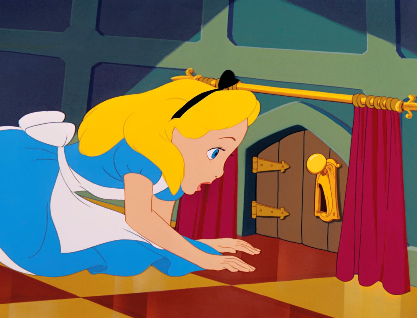
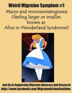
Alice in Wonderland

Alice in Wonderland Syndrome was named after Lewis Carroll’s famous 19th-century novel Alice’s Adventures in Wonderland. In the story, Alice, the title character, experiences numerous situations similar to those of micropsia and macropsia. The thorough descriptions of metamorphosis clearly described in the novel were the first of their kind to depict the bodily distortions associated with the condition. Speculation has arisen that Carroll may have written the story using his own direct experience with episodes of micropsia resulting from the numerous migraines he was known to suffer from. It has also been suggested that Carroll may have suffered from temporal lobe epilepsy.

Gulliver’s Travels

Alice in Wonderland Syndrome’s symptom of micropsia has also been related to Jonathan Swift’s novel Gulliver’s Travels. It has been referred to as “Lilliput sight” and “Lilliputian hallucination”, a term coined by British physician Raoul Leroy in 1909, based on the small people that inhabited the island of Lilliput in the novel.

Etiology

Complete and partial forms of the Alice in Wonderland syndrome exist in a range of disorders, including epilepsy, intoxicants, infectious states, fevers, and brain lesions. Furthermore, the syndrome is commonly associated with migraines, as well as the use of psychoactive drugs. It can also be the initial symptom of the Epstein–Barr virus (see mononucleosis), and a relationship between the syndrome and mononucleosis has been suggested. Epstein-Barr Virus appears to be the most common cause in children, while for adults it is more commonly associated with migraines.

Cerebral hypotheses

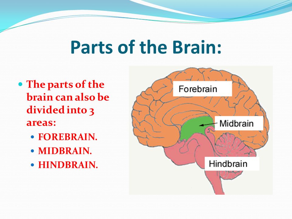
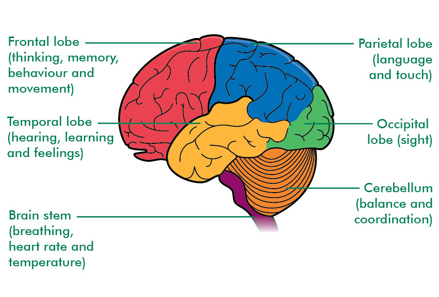
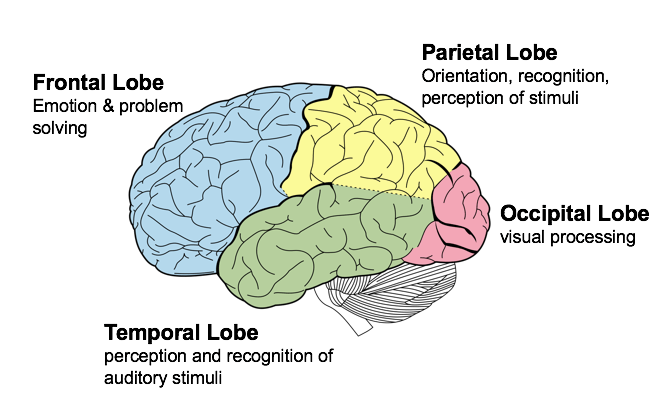
AiWS can be caused by abnormal amounts of electrical activity causing abnormal blood flow in the parts of the brain that process visual perception and texture. Nuclear medical techniques using technetium, performed on patients during episodes of Alice in Wonderland syndrome, have demonstrated that AiWS is associated with reduced cerebral perfusion in various cortical regions (frontal, parietal, temporal and occipital), both in combination and in isolation. It has been hypothesized that any condition resulting in a decrease in perfusion of the visual pathways or visual control centers of the brain may be responsible for the syndrome. For example, one study used single photon emission computed tomography to demonstrate reduced cerebral perfusion in the temporal lobe in patients with AiWS. Other theories exist that suggest the syndrome is a result of unspecific cortical dysfunction (e.g. from encephalitis, epilepsy, decreased cerebral perfusion), or reduced blood flow to other areas of the brain.  Other theories suggest that disordered body image perceptions stem from within the parietal lobe. This has been demonstrated by the production of disturbances of body image through electrical stimulation of the posterior parietal cortex. Other researchers suggest that metamorphopsias may be a result of reduced perfusion of the non-dominant posterior parietal lobe during migraine episodes.

Throughout all the neuroimaging studies, several cortical regions (including the temporoparietal junction within the parietal lobe, and the visual pathway, specifically the occipital lobe) are associated with the development of Alice in Wonderland syndrome symptoms.

Migraines

The role of migraines in Alice in Wonderland syndrome is still not understood, but both vascular and electrical theories have been suggested. For example, visual distortions may be a result of transient, localized ischaemia (an inadequate blood supply to an organ or part of the body) in areas of the visual pathway during migraine attacks. In addition, a spreading wave of depolarization of cells (particularly glial cells) in the cerebral cortex during migraine attacks can eventually activate the trigeminal nerve’s regulation of the vascular system. The intense cranial pain during migraines is due to the connection of the trigeminal nerve with the thalamus and thalamic projections onto the sensory cortex. Alice in Wonderland syndrome symptoms can precede, accompany, or replace the typical migraine symptoms.

Diagnosis

Alice in Wonderland syndrome is a disturbance of perception rather than a specific physiological change to the body’s systems. The diagnosis can be presumed when other causes have been ruled out and if the patient presents symptoms along with migraines and complains of onset during the day (although it can also occur at night). As there are no established diagnostic criteria for Alice in Wonderland syndrome, there is likely to be a large degree of variability in the diagnostic process and thus it is likely to be poorly diagnosed.

Prognosis

Whatever the cause, the bodily related distortions can recur several times a day and may take some time to abate. Understandably, the person can become alarmed, frightened, and panic-stricken throughout the course of the hallucinations—maybe even hurt themselves or others around them. The symptoms of the syndrome themselves are not harmful and are likely to disappear with time. The outcome is typically not harmful, especially in children, and most patients outgrow these episodes. The long-term prognosis typically depends on the root cause of the syndrome, and it is the underlying condition which must be evaluated and treated. Often, the difficulty lies within the patient’s reluctance to describe their symptoms out of fear of being labeled with a psychiatric disorder.

Treatment

At present, Alice in Wonderland syndrome has no standardized treatment plan. Often, treatment methods revolve around migraine prophylaxis, as well as the promotion of a low tyramine diet. Drugs that may be used to prevent migraines include: anticonvulsants, antidepressants, calcium channel blockers, and beta blockers. Other treatments that have been explored include repetitive transcranial magnetic stimulation (rTMS). Further research is required to establish an effective treatment regime.

Epidemiology

The lack of established diagnostic criteria or large-scale epidemiological studies on Alice in Wonderland syndrome means that the exact prevalence of the syndrome is currently unknown. One study on 3,224 adolescents in Japan demonstrated the occurrence of macropsia and micropsia to be 6.5% in boys and 7.3% in girls, suggesting that the symptoms of AiWS may not be so rare.

It appears that the male/female ratio is dependent on the age range being observed. Studies showed that younger males (age range of 5 to 14 years) were 2.69 times more likely to experience AiWS than girls of the same age, while there were no significant differences between students of 13 to 15 years of age. Conversely, female students (16- to 18-year-olds) showed a significantly greater prevalence.

The average age of the start of Alice in Wonderland syndrome is six but it is very normal for some to experience the syndrome from childhood to their late 20’s. It is also thought that this syndrome is hereditary because many parents who have AiWS report their children having it as well.