QUOTES FOR THURSDAY:

“In the United States, for equine encephalitides for which vaccines are available include eastern equine encephalomyelitis (EEE), western equine encephalomyelitis (WEE), Venezuelan equine encephalomyelitis (VEE) and West Nile Virus encephalomyelitis. The availability of licensed vaccine products combined with an inability to completely eliminate risk of exposure justifies immunization against EEE and WEE as core prophylaxis for all horses residing in or traveling to North America and any other geographic areas where EEE and/or WEE is endemic.  Adult horses previously vaccinated against EEE/WEE: Annual revaccination must be completed prior to vector season in the spring. In animals of high risk or with limited immunity, more frequent vaccination or appropriately timed vaccination is recommended in order to induce protective immunity during periods of likely exposure.”

American Association of  Equine Practioners

“Know there’s no cure for Eastern equine encephalitis, or EEE, but there is a vaccine for the mosquito-borne illness. It’s just not commercially available for humans.The United States military developed it in the 1980s as part of a vaccine program to protect military personnel from dangerous pathogens, says Sam Telford, an epidemiologist at Tufts University.”

Common Health


Part II What is Eastern Equine Encephalitis?

eee_cases_by_year-2018

Eastern equine encephalitis virus (EEEV) is a zoonotic alphavirus and arbovirus, and was first recognized in horses in 1831 in Massachusetts. The first confirmed human cases were identified in New England in 1938. EEEV is present today in North, Central and South America, and the Caribbean. In rare cases, those that contract the virus will develop the serious neuroinvasive disease, Eastern equine encephalitis (EEE). From 2009 to 2018, between three (3) and fifteen (15) cases of EEE were reported annually in the U.S. EEE may also be commonly referred to as Triple E or sleeping sickness. EEEV is a vector-borne disease that is transmitted to humans through the bite of an infected mosquito. Culiseta melanura is the primary vector among birds, but this mosquito species does not typically feed on humans. It is believed that EEEV is mainly transmitted to humans and horses by bridge vectors that have contracted the virus by feeding on infected birds. Bridge vector species of mosquitoes may include Coquillettidia pertubans, Aedes sollicitans, and Ochlerotatus canadensis. The risk of contracting the EEE virus is highest during the summer months, and those who live and work near wetland and swamp areas are at higher risk of infection. EEEV is only spread to humans via mosquito bite, and cannot be transmitted directly by other humans or horses. There is an EEEV vaccine available for horses, and owners are encouraged to discuss vaccination with their veterinarian.

A Global View of Eastern Equine Encephalitis

EEE affects areas throughout North and South America, with outbreaks occurring mainly in the eastern coastal areas of the United States and Canada, the Caribbean, and Argentina.

Know Your Mosquitoes

In the U.S., Culiseta melanura is the mosquito responsible for the spread of EEEV in the mosquito-bird-mosquito cycle. Known as the black-tailed mosquito, Cs. melanura can be found in swamps from the Great Lakes and Maine to southern Florida and southeastern Texas. It is distinguished by its unusually long, curved dark-scaled proboscis. This mosquito is also unique because it overwinters as larvae, as opposed to most mosquito species that overwinter as adults or eggs.

EEEV is mainly transmitted to humans by bridge vectors that contract the virus by feeding on infected birds. Bridge vectors may include Aedes, Coquillettidia, and Culex species.

Aedes mosquitoes have distinct black and white markings on their body and legs. They bite during the daytime only, with the highest levels of activity occur in the early morning and evening hours. Members of the Aedes genus are known vectors of EEE, Zika virus, dengue, yellow fever, West Nile virus, and chikungunya.

Coquillettidia mosquitoes have slender bodies and long legs. They are commonly found in humid, low-lying areas that have warm summer and lots of vegetation. In addition to acting as vectors for EEE, Coquillettidia mosquitoes are also known to transmit West Nile virus to humans.

Culex mosquitoes are brown with whitish markings on the abdomen. They typically bite at dusk and at night, and are known to vector several diseases including EEE, West Nile virus, Japanese encephalitis, St. Louis encephalitis, and avian malaria.


QUOTE FOR WEDNESDAY:

“We don’t know some of the basic details about these [mosquito-transmitted] diseases, unfortunately.  The ideal is to anticipate outbreaks, which is very, very difficult. But we need to be prepared for an outbreak when it comes.”

Stephen Higgs (a pathobiologist and director of the Biosecurity Research Institute at Kansas State University in Manhattan)

Part I What is Eastern Equine Encephalitis?

What is eastern equine encephalitis (EEE)?

Eastern equine encephalitis (EEE) belongs to a category of viruses known as arboviruses, or arthropod-borne viruses. Arboviruses are spread by the bites of blood-sucking insects, such as mosquitos and ticks. EEE is spread by the bite of certain kinds of mosquitoes.

What are the risk factors of EEE?

The overall risk of becoming infected with EEE depends on:

  • Exposure to mosquitoes: People who spend a lot of time outdoors or live in wooded areas have a greater chance of being bitten by mosquito.
  • Time of year and day: In the United States, cases of EEE tend to occur from late spring to early fall.. Many kinds of mosquitoes are most active during dusk and dawn and during the early evening hours.
  • Geographic region: Most cases of EEE have been reported in Atlantic and Gulf coast states. Southeastern Massachusetts, particularly Plymouth and Bristol counties, have historically been “hot spots” for EEE. The Massachusetts Dept of Public Health monitors mosquito populations, tests mosquitoes for virus and calculates risks for each town.

Some people with EEE may develop a severe infection that causes brain tissue to become inflamed (encephalitis).The factors that increase one’s risk of getting a severe EEE infection include:

  • Age: Although people of any age can develop a serious infection, the risk is higher for adults older than 50 and children younger than 15.
  • Immune system: People who have a weakened immune system due to cancer treatments, or organ transplantation are more at risk of developing a severe infection.

What are the symptoms of EEE?

Symptoms of EEE generally occur four to 10 days after a person has been infected and include:

  • high fever
  • headache
  • tiredness
  • nausea/vomiting
  • neck stiffness

The symptoms of encephalitis depend on the part of the brain that is inflamed, the amount of inflammation and the person’s age and overall health.

Some of the most common symptoms of encephalitis include:

  • seizures
  • confusion (disorientation)
  • coma

In one-third of cases, encephalitis can be fatal or lead to permanent brain damage.

Because the initial symptoms of EEE resemble those caused by many illnesses, it may be difficult to determine if a child’s symptoms are related to encephalitis. In general, you should take your child to see a doctor if your child has a bad headache, nausea and vomiting, fever or any worrisome changes in behavior (confusion, extreme sleepiness, acting very different, listlessness, lethargy, seizures).

The worst outbreak of eastern equine encephalitis since U.S. health officials began monitoring the mosquito-borne disease 15 years ago is prompting aerial bug spraying and dire warnings to avoid the biting insects well into fall. As of October 1, 31 cases — including nine deaths — have been reported by the U.S. Centers for Disease Control and Prevention.

Known as EEE or Triple-E for short, the incurable brain infection is still relatively rare — there have been only 103 reported infections in the United States in the past decade. Only five percent of people bitten by an infected mosquito will develop the disease. But about a third of EEE patients die, and many who survive experience permanent neurological problems.

Science News spoke with several researchers about how the virus spreads, and possible factors that might be contributing to the recent surge in cases.

“We don’t know some of the basic details about these [mosquito-transmitted] diseases, unfortunately,” says pathobiologist Stephen Higgs, director of the Biosecurity Research Institute at Kansas State University in Manhattan. “The ideal is to anticipate outbreaks, which is very, very difficult. But we need to be prepared for an outbreak when it comes.”

QUOTE FOR THE WEEKEND:

“Antithyroid medication, radioactive iodine, and surgery are all effective treatments and can restore thyroid function to normal. Radioactive iodine and surgery also can “cure” the hyperthyroidism by removing the thyroid.”

American Thyroid Association

Part III Thyroid Cancer Treatment

Treatment

Your thyroid cancer treatment options depend on the type and stage of your thyroid cancer, your overall health, and your preferences.

Most cases of thyroid cancer can be cured with treatment.

Surgery

Most people with thyroid cancer undergo surgery to remove all or most of the thyroid. Operations used to treat thyroid cancer include:

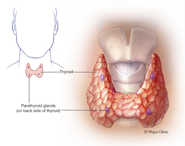
  • Removing all or most of the thyroid (thyroidectomy). In most cases, doctors recommend removing the entire thyroid in order to treat thyroid cancer. Your surgeon makes an incision at the base of your neck to access your thyroid.In most cases, the surgeon leaves small rims of thyroid tissue around the parathyroid glands to reduce the risk of parathyroid damage. Sometimes surgeons refer to this as a near-total thyroidectomy.
  • Removing lymph nodes in the neck. When removing your thyroid, the surgeon may also remove enlarged lymph nodes from your neck and test them for cancer cells.
  • Removing a portion of the thyroid (thyroid lobectomy). In certain situations where the thyroid cancer is very small, your surgeon may recommend removing only one side (lobe) of your thyroid.

Thyroid surgery carries a risk of bleeding and infection. Damage can also occur to your parathyroid glands during surgery, which can lead to low calcium levels in your body. There’s also a risk of accidental damage to the nerves connected to your vocal cords, which can cause vocal cord paralysis, hoarseness, soft voice or difficulty breathing.

Thyroid hormone therapy

After thyroidectomy, you’ll take the thyroid hormone medication levothyroxine (Levoxyl, Synthroid, others) for life.

This medication has two benefits: It supplies the missing hormone your thyroid would normally produce, and it suppresses the production of thyroid-stimulating hormone (TSH) from your pituitary gland. High TSH levels could conceivably stimulate any remaining cancer cells to grow.

You’ll likely have blood tests to check your thyroid hormone levels every few months until your doctor finds the proper dosage for you. Blood tests may continue annually.

Radioactive iodine

Radioactive iodine treatment uses large doses of a form of iodine that’s radioactive.

Radioactive iodine treatment is often used after thyroidectomy to destroy any remaining healthy thyroid tissue, as well as microscopic areas of thyroid cancer that weren’t removed during surgery. Radioactive iodine treatment may also be used to treat thyroid cancer that recurs after treatment or that spreads to other areas of the body.

Radioactive iodine treatment comes as a capsule or liquid that you swallow. The radioactive iodine is taken up primarily by thyroid cells and thyroid cancer cells, so there’s a low risk of harming other cells in your body.

Side effects may include:

  • Nausea
  • Dry mouth
  • Dry eyes
  • Altered sense of taste or smell
  • Fatigue

Most of the radioactive iodine leaves your body in your urine in the first few days after treatment. You’ll be given instructions for precautions you need to take during that time to protect other people from the radiation. For instance, you may be asked to temporarily avoid close contact with other people, especially children and pregnant women.

External radiation therapy

Radiation therapy can also be given externally using a machine that aims high-energy beams, such as X-rays and protons, at precise points on your body (external beam radiation therapy). This treatment is typically administered a few minutes at a time, five days a week, for about five weeks. During treatment, you lie still on a table while a machine moves around you.

External beam radiation therapy may be an option if you can’t undergo surgery and your cancer continues to grow after radioactive iodine treatment. Radiation therapy may also be recommended after surgery if there’s an increased risk that your cancer will recur.

Chemotherapy

Chemotherapy is a drug treatment that uses chemicals to kill cancer cells. Chemotherapy is typically given as an infusion through a vein. The chemicals travel throughout your body, killing quickly growing cells, including cancer cells.

Chemotherapy is not commonly used in the treatment of thyroid cancer, but it may benefit some people who don’t respond to other therapies. For people with anaplastic thyroid cancer, chemotherapy may be combined with radiation therapy.

Injecting alcohol into cancers

Alcohol ablation involves injecting small thyroid cancers with alcohol using imaging such as ultrasound to ensure precise placement of the injection. This treatment is helpful for treating cancer that occurs in areas that aren’t easily accessible during surgery. Your doctor may recommend this treatment if you have recurrent thyroid cancer limited to small areas in your neck.

Targeted drug therapy

Targeted drug therapy uses medications that attack specific vulnerabilities in your cancer cells.

Targeted drugs used to treat thyroid cancer include:

  • Cabozantinib (Cometriq)
  • Sorafenib (Nexavar)
  • Vandetanib (Caprelsa)

These drugs target the signals that tell cancer cells to grow and divide. They’re used in people with advanced thyroid cancer.

Supportive (palliative) care

Palliative care is specialized medical care that focuses on providing relief from pain and other symptoms of a serious illness. Palliative care specialists work with you, your family and your other doctors to provide an extra layer of support that complements your ongoing care. Palliative care can be used while undergoing other aggressive treatments, such as surgery, chemotherapy or radiation therapy.

When palliative care is used along with all of the other appropriate treatments, people with cancer may feel better and live longer.

Palliative care is provided by a team of doctors, nurses and other specially trained professionals. Palliative care teams aim to improve the quality of life for people with cancer and their families. This form of care is offered alongside curative or other treatments you may be receiving.

QUOTE FOR FRIDAY:

“Even though the diagnosis of cancer is terrifying, the prognosis for most patients with papillary and follicular thyroid cancer is usually excellent.”

American Thyroid Association

Part II Thyroid Cancer Month- Dx.

Diagnosing Thyroid Cancer

Thyroid cancer may be diagnosed after a person goes to a doctor because of symptoms, or it might be found during a routine physical exam or other tests. If there is a reason to suspect you might have thyroid cancer, your doctor will use one or more tests to confirm the diagnosis.

This would include:

Family History and Physical Exam

Imaging tests may be done for a number of reasons:

  • To help find suspicious areas that might be cancer
  • To learn how far cancer may have spread
  • To help determine if treatment is working

People who have or may have thyroid cancer will get one or more of these tests.

-Ultrasound

Ultrasound uses sound waves to create images of parts of your body. You are not exposed to radiation during this test.

This test can help determine if a thyroid nodule is solid or filled with fluid. (Solid nodules are more likely to be cancerous.) It can also be used to check the number and size of thyroid nodules as well as help determine if any nearby lymph nodes are enlarged because the thyroid cancer has spread.

For thyroid nodules that are too small to feel, this test can be used to guide a biopsy needle into the nodule to get a sample. Even when a nodule is large enough to feel, most doctors prefer to use ultrasound to guide the needle.

Radioiodine scan

Radioiodine scans can be used to help determine if someone with a lump in the neck might have thyroid cancer. They are also often used in people who have already been diagnosed with differentiated (papillary, follicular, or Hürthle cell) thyroid cancer to help show if it has spread. Because medullary thyroid cancer cells do not absorb iodine, radioiodine scans are not used for this cancer.

For this test, a small amount of radioactive iodine (called I-131) is swallowed (usually as a pill) or injected into a vein. Over time, the iodine is absorbed by the thyroid gland (or thyroid cells anywhere in the body). A special camera is used several hours later to see where the radioactivity is.

For a thyroid scan, the camera is placed in front of your neck to measure the amount of radiation in the gland. Abnormal areas of the thyroid that have less radioactivity than the surrounding tissue are called cold nodules, and areas that take up more radiation are called hot nodules. Hot nodules usually are not cancerous, but cold nodules can be benign or cancerous. Because both benign and cancerous nodules can appear cold, this test by itself can’t diagnose thyroid cancer.

After surgery for thyroid cancer, whole-body radioiodine scans are useful to look for possible spread throughout the body. These scans become even more sensitive if the entire thyroid gland has been removed by surgery because more of the radioactive iodine is picked up by any remaining thyroid cancer cells.

Radioiodine scans work best if patients have high blood levels of thyroid-stimulating hormone (TSH, or thyrotropin). For people whose thyroid has been removed, TSH levels can be increased by stopping thyroid hormone pills for a few weeks before the test. This leads to low thyroid hormone levels (hypothyroidism) and causes the pituitary gland to release more TSH, which in turn stimulates any thyroid cancer cells to take up the radioactive iodine. A downside of this is that it can cause the symptoms of hypothyroidism, including tiredness, depression, weight gain, sleepiness, constipation, muscle aches, and reduced concentration. One way to raise TSH levels without withholding thyroid hormone is to give an injectable form of thyrotropin (Thyrogen®) before the scan.

Because any iodine already in the body can affect this test, people are usually told to avoid foods or medicines that contain iodine for a few days before the scan.

Radioactive iodine can also be used to treat differentiated thyroid cancer, but it is given in much higher doses. This type of treatment is described in Radioactive iodine (radioiodine) therapy.

Chest x-ray

If you have been diagnosed with thyroid cancer (especially follicular thyroid cancer), a plain x-ray of your chest may be done to see if cancer has spread to your lungs.

Computed tomography (CT) scan

The CT scan is an x-ray test that makes detailed cross-sectional images of your body. It can help determine the location and size of thyroid cancers and whether they have spread to nearby areas, although ultrasound is usually the test of choice. A CT scan can also be used to look for spread into distant organs such as the lungs.

One problem using CT scans is that the CT contrast dye contains iodine, which interferes with radioiodine scans. For this reason, many doctors prefer MRI scans for differentiated thyroid cancer.

Magnetic resonance imaging (MRI) scan

MRI scans use magnets instead of radiation to create detailed cross-sectional images of your body. A MRI can be used to look for cancer in the thyroid, or cancer that has spread to nearby or distant parts of the body. But ultrasound is usually the first choice for looking at the thyroid. MRI can provide very detailed images of soft tissues such as the thyroid gland. MRI scans are also very helpful in looking at the brain and spinal cord.

Positron emission tomography (PET) scan

A PET scan can be very useful if your thyroid cancer is one that doesn’t take up radioactive iodine. In this situation, the PET scan may be able to tell whether the cancer has spread.

Biopsy

The actual diagnosis of thyroid cancer is made with a biopsy, in which cells from the suspicious area are removed and looked at in the lab.

Part I Thyroid Cancer Month

The thyroid is a small gland located below the voice box in the front of the neck. It is made up of two lobes and is shaped like a butterfly. As part of your endocrine system, this gland makes hormones that affect your heart rate, temperature, mental function, and metabolism.

If cells in the thyroid gland grow uncontrollably, they form a nodule (tumor). Most (90 percent) of thyroid nodules are benign (noncancerous). But 1 out of 10 are malignant (cancerous).

Approximately 62,500 people are diagnosed with thyroid cancer in the United States each year. The disease usually affects people between the ages of 20 and 55. Women are nearly three times more likely than men to develop thyroid cancer.

Types of Thyroid Cancer

The disease can be hard to catch at first because many thyroid tumors don’t cause symptoms.

Many people with thyroid cancer don’t have any signs or symptoms of the disease.

Some notice small, painless lumps or swellings called nodules in the front of the neck.

Other symptoms can include:

  • hoarseness
  • trouble swallowing
  • breathing problems
  • pain in the throat or neck that doesn’t go away
  • a cough that doesn’t go away

Thyroid Cancer Risk Factors

A risk factor for thyroid cancer is a condition, behavior, or other part of your life that increases the likelihood of developing the disease.

It’s important to understand what risk factors you have for developing thyroid cancer so that your doctors can decide which treatment approach will be most effective for you.

The different types of thyroid cancer have different risk factors.

Risk factors for papillary thyroid cancer include:

  • Radiation exposure. People who were exposed to radiation when they were children have a higher chance of developing papillary thyroid cancer.
  • Inherited conditions. People with a family history of multiple colon growths because of inherited conditions may be at an increased risk for papillary thyroid cancer. An example of this is familial adenomatous polyposis.
  • Family history. For about 5 percent of people who develop papillary thyroid cancer, the disease runs in the family. Researchers are still trying to figure our which gene is to blame.
  • Gender. It’s unclear why, but papillary thyroid cancer occurs about three times more often in women than in men. When it does happen in men, it usually grows and spreads more quickly.

Risk factors for follicular thyroid cancer include:

  • A low-iodine diet. People are more likely to develop follicular carcinoma if they live in a place where iodine isn’t added to salt that’s used in food.
  • Familial conditions. Inherited disorders, such as Werner’s syndrome and Cowden’s syndrome, sometimes include thyroid cancer as part of the disease process.

Risk factors for medullary thyroid cancer (MTC):

  • Family history. Approximately 25 percent of people with MTC have inherited a mutation in a gene called RET.

Information about your health and family medical history can help in figuring out your risk for thyroid cancer. This information is important for building a treatment plan that makes sense for you.