Archive | July 2017

What is a cardiac arrest and what to do if having one?

Above-Normal Sinus Rhythm  Best rhythm to be in.

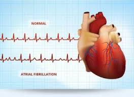
Above-Atrial Fibrillation-rapid rate = Uncontrolled and Controlled is under 100 HB/minute


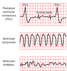
Notice in the PVC and V Tac above the QRS that is wide & in V Fib you see no distinct waves

all fibrillation meaning nothing is working right (NO distinct P wave-you seen none in Ventricular

rhythms to begin with but no QRS or T or U waves) because their are none just fibrillation.

Cardiac arrest is the abrupt loss of heart function in a person who may or may not have diagnosed heart disease. The time and mode of death are unexpected. It occurs instantly or shortly after symptoms appear.

Each year, more than 420,000 emergency medical services-assessed out-of-hospital cardiac arrests occur in the United States.

No. The term “heart attack” is often mistakenly used to describe cardiac arrest. While a heart attack may cause cardiac arrest and sudden death, the terms don’t mean the same thing. Heart attacks are caused by a blockage that stops blood flow to the heart. A heart attack (or myocardial infarction) refers to death of heart muscle tissue due to the loss of blood supply, not necessarily resulting in the death of the heart attack victim.

Cardiac arrest is caused when the heart’s electrical system malfunctions. In cardiac arrest death results when the heart suddenly stops working properly. This may be caused by abnormal, or irregular, heart rhythms (called arrhythmias). A common arrhythmia in cardiac arrest is ventricular fibrillation.

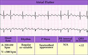
Where in atrial fibrillation the impulse or heart beat intially starts its conduction down the heart to the bottom of the ventricles and with the pulse under 100 = Controlled AFib, you can live a normal life with.  With AFib the atriums are chaotic.

AF occurs if rapid, disorganized electrical signals cause the heart’s two upper chambers—called the atria (AY-tree-uh)—to fibrillate. The term “fibrillate” means to contract very fast and irregularly.

In AF, blood pools in the atria. It isn’t pumped completely into the heart’s two lower chambers, called the ventricles (VEN-trih-kuls). As a result, the heart’s upper and lower chambers don’t work together as they should.

Ventricular Fibrillation doing the same thing but in a lower area of the heart and now in the lower chambers called the Ventricles=much more serious and dangerous if no immediate treatment than death occurs.   This is when the heart’s lower chambers suddenly start beating chaotically and don’t pump blood. Death occurs within minutes after the heart stops. Cardiac arrest may be reversed if CPR (cardiopulmonary resuscitation) is performed and a defibrillator is used to shock the heart and restore a normal heart rhythm within a few minutes.

Heart Attack and Cardiac Arrest

People often use these terms interchangeably, but they are not synonyms. A heart attack is when blood flow to the heart is blocked, and sudden cardiac arrest is when the heart malfunctions and suddenly stops beating unexpectedly. A heart attack is a “circulation” problem and sudden cardiac arrest is an “electrical” problem.

What is a heart attack?


A heart attack occurs when a blocked artery prevents oxygen-rich blood from reaching a section of the heart. If the blocked artery is not reopened quickly, the part of the heart normally nourished by that artery begins to die. The longer a person goes without treatment, the greater the damage. Symptoms of a heart attack may be immediate and intense. More often, though, symptoms start slowly and persist for hours, days or weeks before a heart attack. Unlike with sudden cardiac arrest, the heart usually does not stop beating during a heart attack. The heart attack symptoms in women can be different than men.  A heart attack actually caused scarring to the heart since it causes damaging to the heart muscle tissue.

What is cardiac arrest?


Sudden cardiac arrest occurs suddenly and often without warning. It is triggered by an electrical malfunction in the heart that causes an irregular heartbeat (arrhythmia). With its pumping action disrupted, the heart cannot pump blood to the brain, lungs and other organs. Seconds later, a person loses consciousness and has no pulse. Death occurs within minutes if the victim does not receive treatment.

Fast action can save lives. Find out what to do if someone experiences a heart attack or cardiac arrestFast action can save lives. Find out what to do if someone experiences a heart attack or cardiac arrest.Fast action can save lives. Find out what to do if someone experiences a heart attack or cardiac arrest.

What to do: Heart Attack

Even if you’re not sure it’s a heart attack, don’t wait more than five minutes to call 9-1-1 or your emergency response number. Every minute matters! It’s best to call EMS to get to the emergency room right away. Emergency medical services staff can begin treatment when they arrive — up to an hour sooner than if someone gets to the hospital by car. EMS staff are also trained to revive someone whose heart has stopped. Patients with chest pain who arrive by ambulance usually receive faster treatment at the hospital, too.

What to do: Sudden Cardiac Arrest

Cardiac arrest is reversible in most victims if it’s treated within a few minutes. First, call 9-1-1 for emergency medical services. Then get an automated external defibrillator if one is available and use it as soon as it arrives. Begin CPR immediately and continue until professional emergency medical services arrive. If two people are available to help, one should begin CPR immediately while the other calls 9-1-1 and finds an AED.  Learn CPR  you may just save someone one day being at the right place at the right time.


QUOTE FOR THURSDAY:

“Atrial fibrillation (AFib) is the most common abnormal heart rhythm. In a normal heart, the four chambers of the heart beat in a steady, rhythmic pattern. With AFib, the atria (upper chambers of the heart) fibrillate (quiver or twitch quickly) and create an irregular rhythm.”

Heart Rhythm Society (http://www.hrsonline.org)

QUOTE FOR WEDNESDAY:

“Sinus rhythm refers to the origination of the electrical activity coming from the sinus node — also known as the sinoatrial node, or SA node.  The SA node is the natural pacemaker of the human body.”

American Heart Association

What does a cardiac rhythm actually tell us.

First the engine to the human body is the heart;  our car can’t work without  a good working engine just like the body can’t work without a good working heart.  In the heart we have a natural pacemaker of the heart which is called a Sinus Node.  The sinus node conducts impulses from the top right chamber of the heart to the left chamber in the heart (called Atriums) and follows its way down to the bottom chambers (called ventricles) but for the conduction traveling originally from the right atrium to the left atrium the left side is slightly behind in conduction compared to the right when going down horizontally on each side due to the conduction having to cross over to the left top from the right top of the heart.  Than the conduction continues on the atrio-ventricle valves (called AV valves) causing these valves to open and close completely allowing blood to drop in the ventricles from the atriums when open but without back up in the AV valves if the valve completely seals like any valve operating correctly by closing whether it be pipes, actual engine valves, or our veins/arteries or the actual heart in this case in the human body.  Whatever valves used in our body or inanimate objects all pretty much play the same role.

The Rt. AV valve is the tricuspid valve the Lt. AV valve is the mitral valve.  The conduction continues than to the bottom chambers-ventricles the conduction  goes up and around the entire ventricles sensing up actually the purkinje fibers/papillary muscles  to aid in contracting (depolarization) and relaxation (repolarization) of the ventricles.  This allows the heart to go “Lub Dub” after from the SA node to the end process of conduction (described above) which gives a single beat or pulse.  This allows our red blood cells that are more oxygenated than with carbon dioxide in them on the left side to go out the aorta to the bloodstream giving our tissues oxygen all over where needed when leaving the L side of the heart which after the 02 used up it will be returning to the right side when needing to refill with more oxygen and release the C02 from the red blood cells.   How do we get our red blood cells reoxygenated?  The right side; when the Lt side is doing its function through this conduction process so is the right side.  The Rt side allows the blood on the right side more carbon dioxide red blood cells that are carrying with some oxygen (but very little) so these red blood cells after going through the whole conduction process from the Rt. ventricle enter into the pulmonary artery to the lungs (a much shorter pathway than the Lt. side of the heart sends its RBCs-it goes from the aorta down the body up to the brain and back to the Rt. side of the heart needing more 02).  This side is where the RBCs get reoxygenated than reentry to the left side of the heart going through that side out the aorta when those cells get into the Lt. through through our last process of the conduction system.  This conduction system is so vital in our heart operating properly.   Since the Rt. and Lt. side have 2 completely different functions with our RBCs in dispensing 02 and C02 but both sides need to work correctly and are vital to keep us alive.

On a tele strip a sinus rhythm is made up of a P wave=Atriums contracting (atrial depolarization) than a straight line= atriums relaxing (atrial repolarization) than followed by a QRS wave=Ventricles contracting (ventricular depolarization) followed by a straight line than a T wave (ventricular depolarization) than in some a U wave. A U wave is  on an electrocardiogram that is not always seen. It is typically small, and, by definition, follows the T wave. High probability you will see a U wave in normal sinus rhythm (NSR) or sinus bradycardia (SB).  This is because the HR is slow enough to show a U wave on the rhythm you won’t see them in tachycardias.  U waves are thought to represent repolarization of the papillary muscles or Purkinje fibers.

A regular QRS measures less than 0.12 which is with all atriums rhythms.

In most cases if the sinus node is working properly with the person taking care of their body staying in good shape, eating healthy and getting rest to balance stress and no cardiac disease or in some cases compliance with all above in taking cardiac meds as their M.D. ordered and it shows on the telemetry monitor sinus rhythm heart rate (HR) 80 – 100 but if HR over 100-150 the person has sinus tachycardia (fast pulse beat by the sinus node) or if HR less than 60 it is sinus bradycardia (slow pulse beat by the sinus node) which maybe normal for the person, like an athlete.   All these rhythms derive from the sinus node originally=the natural pacemaker of the heart and have regular rhythms giving you a regular HR not irregular unless you have a premature contraction from the atriums causing a PAC (Premature atrial contraction) or a PVC premature ventricle contraction (deriving from the lower chambers called the Ventricles) that pop up in your sinus regular rhythm causing your beat to be irregular (but remember simple stress or caffeine can cause this premature contraction as well as heart disease of many types).  These premature contractions pop on the telemetry reading with the ingredients that make up a atrial rhythm = p wave + a normal QRS wave + t wave unlikely for a u wave(that follows)  or other rhythms as well which we will get into later.  All these occasional or frequent premature contractions mean is the heart rhythm of the heart is getting irritable and allowing a pulse to pop up from a different area of the heart than from where the actual underlining rhythm is coming from.  If your underlining rhythm through out the recording lets say is coming from the sinus node the underlining rhythm will be sinus rhythm but if occasional or frequent premature contractions are impulsing elsewhere in the heart that causes a premature contraction making your rhythm=pulse irregular slightly if occasional premature contractions but if frequent it makes the rhythm=pulse very irregular.  The more irritable any rhythm gets the higher the probability the rhythm can go into a worse rhythm.  If the heart conduction pulsing elsewhere in than the sinus node causing premature contractions lets say from the upper chambers (the atriums than these are Premature Atrial Contractions called PACs but if from the ventricles they are called PVCs.

PVCs in the rhythm has the features that are the same as a atrial except no p wave since the p wave means a conduction impulse occuring in the upper chambers called atriums.  When an impulse starts in the ventricles this shows the QRS measuring wide because the contraction is in the Ventricle.  For if the impulse started in the atriums the QRS would measure normal.  In the Ventricles where a premature contraction occurs will only have a wide QRS, rather than a regular QRS measurement, because of the chambers the PVC is coming from, being the Ventricles.

PVCs will be discussed with Ventricular Rhythms.

If the sinus node breaks down the heart works by having the next area of conduction take over by compensating having the natural pacing take over in the atriums where we have atrial rhythms which start at a HR over 150 unless controlled atrial fibrillation but we’ll get into that rhythm shortly.  Remember as each area above breaks down in being the pacemaker the lower the start of your rhythm derives from but we don’t find it therapeutic for rhythms to start in the bottom of the heart being in the ventricles but at the top where our natural pacemaker of the heart sits in the right upper atrial chamber of the heart.  The highier the pacemaker site the more therapeutic the rhythm will be as long as it is under a pulse or heart beat of 100 (stable).

The rhythms you see when the atrium is the natural pacemaker of the heart taking over for the SA node that doesn’t work with the heart now compensating with the atrium, they are atrial rhythms such as atrial tachycardia or SVT supraventricular tachycardia, simple meaning above the ventricles.  This is where the rhythm is over 150 to 250 showing a p wave and QRS and usually not a T wave but if the HR is slow enough it might show on the telemetry monitor but usually doesn’t.  If it shows no PACs or PVCs it’s a regular rhythm.

Another atrial rhythm is atrial flutter or A- Flutter)=AFL which shows only QRSs with flutter waves.  A regular rhythm but this rhythm needs to be changed or in time the heart will stress out and lead on to more dangerous rhythms.  This has no p waves but flutter waves with QRS waves.  You can have 2 flutter waves to every QRS wave or 3 to every QRS or 4 flutter waves to every QRS or even 5 but the it can even be a variable ratio of flutter waves to every QRS wave meaning the rhythm is getting more irregular and dangerous.  If left untreated, the side effects of AFL can be potentially life threatening. AFL makes it harder for the heart to pump blood effectively. With the blood moving more slowly, it is more likely to form clots. If the clot is pumped out of the heart, it could travel to the brain and lead to a stroke or heart attack.

The treatment for aflutter is cardioversion using a  defibrillator in sync mode so when the shock is given it lands with the R wave and avoiding the vulnerable T wave section which if the shock landed their could put this rhythm into V Tac or V fib.  The other atrial rhythm is atrial fibrillation (afib) and if under 100 great for if its chronic afib it will be hard to change to NSR but  if a new Dx. of afib higher odds with cardioversion it will shock it the rhythm back into NSR.  Those who are chronic afib or new afib that can’t convert to NSR usually are given Coumadin and ASA aspirin to keep the blood from clotting in the heart and breaking free with the irregular rhythm.  Also possibly used is Beta blockers that slow the conduction of impulses down being a beta blocker it blocks the beta stimulus especially lopressor or Metoprolol that is a selective beta 1 stimulus blocker which is in the heart to slow the HR down.  Than there is  calcium channel blockers possibly used to slow down the HR if needed by blocking cardiac cells sending impulse signals from the top to the bottom of the heart.  Keeping afib under 100 of a pulse rate and more like 80 or less can live a completely normal life if compliant with meds, diet and exercise.

QUOTE FOR TUESDAY:

“Management of heart failure requires a multimodal approach. It involves a combination of lifestyle modifications, medications, and possibly the use of devices or surgery. ”

WOW organization

QUOTE FOR MONDAY:

“The heart works like a pump and beats 100,000 times a day. For the heart to do its function therapeutically it needs another organ to be involved called the lungs or the body could not live”

American Heart Association

How the heart works anatomically!

heart live                                                           heart

The heart works like a pump and beats 100,000 times a day. For the heart to do its function therapeutically it needs another organ to be involved called the lungs or the body could not live. If one of these organs gets damaged in time the other organ gets affected with no treatment the person will die sooner in life (Ex. Heart Failure in time affects the lungs to respiratory failure). A good metaphor for this is the car; if the engine gets damaged with no repair, in time the transmission will get affected; with no repair at all the car will die.

One of the main functions of the red blood cells (RBCs) is to carry oxygen throughout the body to give all tissues our food to stay alive, that would be oxygen. For this to take place this is done through the heart beating nonstop which allows the flow of blood to be running continuous in our blood stream (circulatory system) and at the same time this allows our cells in the blood stream to get to the lungs for oxygen and carbon dioxide exchange. These 3 functions could not take place if the heart wasn’t pumping blood to and from the heart/lungs 24 hrs a day.

This is what takes place when the heart pumps our blood:

The red blood cells carry oxygen and remove the ending result of oxygen used by our tissues, called carbon dioxide=CO2. To get rid of the CO2 (a toxin) in our body the gas is carried from our body by carrying CO2 to the lungs through the RBCs. The high CO2 with low oxygen (O2) concentration RBCs go to one side of the heart (being the right side) to get CO2 removed in exchange for more O2 in a red blood cell (RBC) this is done at the base of the lungs. This is done through breathing.  Then these RBCs get highly oxygenated which then proceed to the left side of the heart. These RBCs get pumped throughout the body where the tissues utilize the oxygen in the RBCs by transferring O2 from the RBCs to the tissue as their food to stay alive. Without oxygen our tissues would starve and we would die. For the oxygen to be transported to the tissues of the body (as their food) it works like this: After the right side of the heart pumps blood to the lungs with high CO2 concentration RBCs, while we inhale to allow more oxygen in the cells at the bottom of the lungs an exchange of gas concentration in the RBCs takes place. The cells release CO2 which is released out of our body through exhaling but also when we inhale more 02 is transported into the RBCs to take another trip around the body releasing the high 02 levels to the tissues that need to utilize it BUT to do this the blood now is sent to the left side of the heart. For all this to take place the heart pumps to transport the oxygen throughout the body giving nutrition to the tissues, without it=cellular starvation but at the same time sends high CO2 RBCs to the right side for a 02/CO2 exchange at the lungs by going through the right side of the heart.  

The heart has two sides, separated by an inner wall called the septum. The septum divides the right from the left side of the heart since the right and left chambers do different functions, as described above. Remember the right side of the heart is sending RBCs with highier CO2 concentration and a low oxygen concentration. You have these RBCs coming from above the heart finally entering the Rt upper chamber (Right Atrium=RA) called the Superior Vena Cava with below the heart the Inferior Vena Cava the meet into each other dumping the high CO2 blood into the RA. Then the blood goes through a valve called the tricuspid valve dumping the blood into the Rt.lower chamber called Rt. Ventricle and through the pulmonary valve going via the pulmonary artery (one of the few arteries with high CO2 concentration, usually arteries high in 02 concentration) dumping the blood at the base of the lungs for 02 and CO2 exchange when we inhale and exhale. After the gas exchange takes place the red blood cells become higher oxygenated in levels and much lower in carbon dioxide levels. Inside a cell is never 100% 02 or CO2. Now this high oxygenated blood goes to the left side of the heart leaving the lungs via the pulmonary vein (one of the few veins high in O2). The blood dumps now in the L upper chamber=Lt. atrium down through the mitral valve to the left ventricle proceeding to the aorta where the high concentrated RBCs go throughout the body dispensing oxygen to our tissues where it is needed.

If you look the right side sends blood from the heart to lungs back to heart=short distance so the muscle on the right side is thin compared to the left side. The reason for this is the left side of the heart receives the oxygen-rich blood from the lungs and pumps it to throughout the body. The left side of the heart has to dispense the high (O2) blood throughout the body so that side of the heart works out more in pumping; so the muscle on the left side is larger than the right side. The muscle of the heart is called myocardium (myo=muscle and cardium=the heart).

Come back tomorrow for the basics of what heart failure is on the the Rt. and Lt. side of the heart.


QUOTE FOR FRIDAY:

“In simple terms Myasthenia Gravis is a disease that causes weakness in the muscles under your control. It happens because of a problem in communication between your nerves and muscles.”

www.myasthenia.org/