Archives

QUOTE FOR TUESDAY:

“Chronic kidney disease (CKD) is a serious condition affecting 37 million people. Often overlooked until symptoms appear, CKD is progressive and can put you at risk for serious health complications including kidney failure. Adopting a healthy lifestyle can help you manage and slow progression of CKD and its complications.”

National Institute of Diabetes and Digestive and Kidney Diseases (NIDDK)

QUOTE FOR MONDAY:

“Acute kidney failure occurs when your kidneys suddenly become unable to filter waste products from your blood.  Acute kidney failure — also called acute renal failure or acute kidney injury — develops rapidly, usually in less than a few days and can be reversible if fairly healthy. ”

MAYO CLINIC

QUOTE FOR THE WEEKEND:

“March is National Kidney Month, a time when communities across the country raise awareness about kidney disease. This year’s focus is on taking charge of your health and the many factors that go into managing your kidney disease.

Chronic kidney disease (CKD) is a serious condition affecting 37 million people. Often overlooked until symptoms appear, “.

National Institute of Diabetes and Digestive and Kidney Diseases (NIDDK)

QUOTE FOR FRIDAY:

“Does the brain always swell? How do you know if the brain is swelling? Doesn’t the CT scan show swelling?
Is it possible that the person’s brain did not swell because of the use of the drug manitol (protocol treatment
in all ICU’s)? Is the chemical released if there is no swelling? If a person didn’t need a shunt, can we assume
there was no swelling?
Pretty much all tissues in the body swell when traumatized. They also require more oxygen to heal. The brain
is unique in that it rests inside a bone case, so when it swells, it experiences more trauma.
The more damage the brain receives, the more it swells. This is caused by leakage from blood vessels. When
the brain swells, because it is housed inside the skull, it has no room to expand. This leads to a rise in
pressure within the brain. This rise in pressure rapidly equals the arterial pressure thereby affecting the blood
flow to the brain. This diffuse pressure which decreases blood flow affects the ability of the cells within the
brain to metabolize properly; the cells are unable to eliminate toxins which then accumulate. ”

Minnesota Brain Injury Alliance

QUOTE FOR THURSDAY:

“Turns out, whether it’s rock ‘n’ roll, jazz, hip-hop or classical, your gray matter prefers the same music you do.  It depends on your personal background.  For a while, researchers believed that classical music increased brain activity and made its listeners smarter, a phenomenon called the Mozart effect. Not necessarily true.  In recent studies, they’ve found that people with dementia respond better to the music they grew up listening to.  If you play someone’s favorite music, different parts of the brain light up.  That means memories associated with music are emotional memories, which never fade out — even in Alzheimer’s patients, depending on its severity.”.

University of Central Florida (https://www.ucf.edu/pegasus/your-brain-on-music) –  neuroscientist Kiminobu Sugaya and world-renowned violinist Ayako Yonetani — have been teaching one of the most popular courses in The Burnett Honors College. “Music and the Brain”.

QUOTE FOR THE WEDNESDAY:

“Playing music has been found to increase the volume and activity in the brain’s corpus callosum — the bridge between the two hemispheres — allowing messages to get across the brain faster and through more diverse routes. This may allow musicians to solve problems more effectively and creatively, in both academic and social settings.”

Brain Pickings (www.brainpickings.org)

QUOTE FOR TUESDAY:

“The idea that music can heal the soul or “soothe the savage breast” is well-known. Music’s healing power over the body has also attracted attention from scientists who aim to test this ancient wisdom. A growing body of research supports the claim that music can alleviate physical pain. Studies have shown music to be an effective pain reliever, both on its own and as an adjuvant in connection with other types of therapy. Long-term studies of music therapy in pain management have shown it to be associated with improved quality of life and reduced consumption of pain relievers.”

Jeanette Bicknell Ph.D.  Psychology Today

QUOTE FOR THE MONDAY:

“Your stroke treatment begins the moment emergency medical services (EMS) arrives to take you to the hospital. Once at the hospital, you may receive emergency care, treatment to prevent another stroke, rehabilitation to treat the side effects of stroke, or all three.”

Centers for Disease Control and Prevention (CDC)

QUOTE FOR THE WEEKEND:

“In the United States, about 795,000 people suffer a stroke each year. Someone has a stroke every 40 seconds, and every 4 minutes someone dies from stroke. There are more than 140,000 deaths each year from stroke. Statistics show that about 40% of stroke deaths occur in males and 60% in females.  Stroke is also the leading cause of serious long-term disability in the United States. There are over 7 million stroke survivors live in United States and two-thirds of them are currently disabled. Around 25% of people who recover their first stroke will have another within 5 years.”

Stroke Awareness Foundation  (www.strokeinfo.org)

Part II Stroke Awareness – Signs/symptoms and diagnostic testing!

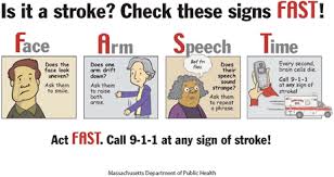
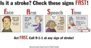
Signs and Symptoms of a stroke happening:

Symptoms of stroke include trouble walking, speaking, and understanding, as well as paralysis or numbness of the face, arm, or leg.

People may experience the following:                                                                      

Muscular: difficulty walking, paralysis with weak muscles, problems with coordination, stiff muscles, overactive reflexes, or paralysis of one side of the body

Visual: blurred vision, double vision, sudden visual loss, or temporary loss of vision in one eye

Whole body: balance disorder, fatigue, or lightheadedness

Speech: difficulty speaking, slurred speech, or speech loss

Sensory: pins and needles or reduced sensation of touch

Facial: muscle weakness or numbness

Limbs: numbness or weakness

Also common: difficulty swallowing, headache, inability to understand, mental confusion, numbness, or rapid involuntary eye movement

What is done for a stroke regarding diagnostic tooling:

To determine the most appropriate treatment for your stroke, your emergency team needs to evaluate the type of stroke you’re having and the areas of your brain affected by the stroke. They also need to rule out other possible causes of your symptoms, such as a brain tumor or a drug reaction. Your doctor may use several tests to determine your risk of stroke, including:

CT scan of brain tissue damaged by stroke

Cerebral angiogram  A cerebral angiogram showing a carotid aneurysm due to a stroke.

Physical examination. Your doctor will ask you or a family member what symptoms you’ve been having, when they started and what you were doing when they began. Your doctor then will evaluate whether these symptoms are still present.

Your doctor will want to know what medications you take and whether you have experienced any head injuries. You’ll be asked about your personal and family history of heart disease, transient ischemic attack or stroke.

Your doctor will check your blood pressure and use a stethoscope to listen to your heart and to listen for a whooshing sound (bruit) over your neck (carotid) arteries, which may indicate atherosclerosis. Your doctor may also use an ophthalmoscope to check for signs of tiny cholesterol crystals or clots in the blood vessels at the back of your eyes.

Blood tests. You may have several blood tests, which tell your care team how fast your blood clots, whether your blood sugar is abnormally high or low, whether critical blood chemicals are out of balance, or whether you may have an infection. Managing your blood’s clotting time and levels of sugar and other key chemicals will be part of your stroke care.

Computerized tomography (CT) scan. A CT scan uses a series of X-rays to create a detailed image of your brain. A CT scan can show a hemorrhage, tumor, stroke and other conditions. Doctors may inject a dye into your bloodstream to view your blood vessels in your neck and brain in greater detail (computerized tomography angiography).

Magnetic resonance imaging (MRI). An MRI uses powerful radio waves and magnets to create a detailed view of your brain. An MRI can detect brain tissue damaged by an ischemic stroke and brain hemorrhages. Your doctor may inject a dye into a blood vessel to view the arteries and veins and highlight blood flow (magnetic resonance angiography, or magnetic resonance venography).

Carotid ultrasound. In this test, sound waves create detailed images of the inside of the carotid arteries in your neck. This test shows buildup of fatty deposits (plaques) and blood flow in your carotid arteries.

Cerebral angiogram. In this test, your doctor inserts a thin, flexible tube (catheter) through a small incision, usually in your groin, and guides it through your major arteries and into your carotid or vertebral artery. Then your doctor injects a dye into your blood vessels to make them visible under X-ray imaging. This procedure gives a detailed view of arteries in your brain and neck.

Echocardiogram. An echocardiogram uses sound waves to create detailed images of your heart. An echocardiogram can find a source of clots in your heart that may have traveled from your heart to your brain and caused your stroke.

You may have a transesophageal echocardiogram. In this test, your doctor inserts a flexible tube with a small device (transducer) attached into your throat and down into the tube that connects the back of your mouth to your stomach (esophagus). Because your esophagus is directly behind your heart, a transesophageal echocardiogram can create clear, detailed ultrasound images of your heart and any blood clots.

In reality going to an ER room if the pt comes suspected of a stroke and has symptoms or not than  nationally in America the hospitals are to do the following:                                                                                 -A neuro assessment should be done in 10 minutes

-A CT SCAN ordered and pt sent off for the test and done within 25 minutes.

-The CT SCAN read and interpreted by the radiologist / neuro doctor.  At this point it tells the MD if the pt has a blockage or a hemmorage in the brain that caused the stroke.  Remember a ischemic stroke and hemmoragic stroke are treated differently.

We’ll get into treatment tomorrow in Part III Treatment of a stroke.