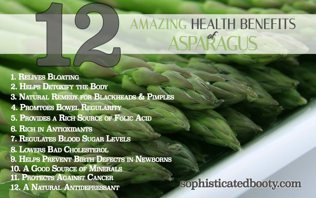
Archives

Part III Spina BIfida Awareness Month – How its diagnosed and treated!


How is Spina Bifida Diagnosed:

In most cases, spina bifida is diagnosed before birth (prenatal). However, some mild cases may go unnoticed until after birth (postnatal). Very mild forms of spinal bifida are found when doing tests for other conditions or may never be detected.

PRENATAL DIAGNOSED:

The most common screening methods used to look for spina bifida during pregnancy are maternal serum alpha fetoprotein (MSAFP) screening and fetal ultrasound.  A doctor can also perform an amniocentesis test.

  • Maternal serum alpha fetoprotein (MSAFP) screen.  At 16 to 18 weeks of pregnancy, a sample of the mother’s blood is taken to measure the level of a protein called alpha-fetoprotein (AFP), which is made naturally by the fetus and placenta.  During pregnancy, a small amount of AFP normally crosses the placenta and enters the mother’s bloodstream.  Abnormally high levels of AFP may indicate that the fetus has spina bifida or other neural tube defect.  This test is not specific for spina bifida and cannot definitively determine that there is a problem with the fetus.  This means that a high AFP level alone is not enough to be sure the fetus has a neural tube defect.  If a high level of AFP is detected, the doctor may request additional testing, such as an ultrasound or amniocentesis.

The second trimester MSAFP screen may be performed alone or as part of a larger, multiple-marker screen.  Multiple-marker screens can look for neural tube defects and other birth defects, including Down syndrome and other chromosomal abnormalities.  First trimester screens for chromosomal abnormalities also exist but signs of spina bifida are not evident until the second trimester when the MSAFP screening is performed.

  • Ultrasound.  A fetal ultrasound uses high-frequency sound waves to create a picture of the developing baby inside the womb.  It is highly accurate in diagnosing some birth defects during pregnancy, including spina bifida.  Fetal ultrasound can be performed during the first trimester (usually between 11-14 weeks) and the second trimester (usually at 18-22 weeks), and diagnosis is more accurate during the second trimester.
  • Amniocentesis.  In this test, a doctor removes a sample of the amniotic fluid that surrounds the fetus and tests it for protein levels that may indicate a neural tube defect and genetic disorders.

POSTNATAL DIAGNOSED:

Closed neural tube defects are often recognized at birth due to an abnormal fatty mass, tuft or clump of hair, or a small dimple or birthmark on the skin at the site of the spinal malformation.  Spina bifida occulta is usually found when x-rays are done for another reason.

In rare cases, myelomeningocele and meningocele are not diagnosed during routine prenatal tests.  The baby will be diagnosed when they are born with a bubble on their back.  Babies with myelomeningocele and closed neural tube defects may have muscle weakness in their feet, hips, and legs that result in joint deformities first noticed at birth.  Mild cases of spina bifida (occulta, closed neural tube defects) not diagnosed during prenatal testing may be detected postnatally using ultrasound or X-ray imaging to look at the spine.

Doctors may use magnetic resonance imaging (MRI) or a computed tomography (CT) scan to get a clearer view of the spinal cord and vertebrae.  To evaluate for hydrocephalus, the doctor will request a head ultrasound, CT or MRI to look for extra cerebrospinal fluid inside the brain.

HOW SPINA BIFIDA CAN BE PREVENTED, THE BEST TREATMENT TO GIVE YOUR BABY; being FOLIC ACID!

Women of childbearing age can reduce their risk of having a child with spina bifida by taking 400 micrograms (mcg) of folic acid every day pre-conception. Because it is water soluble, folic acid does not stay in the body for very long and needs to be taken every day to be effective against neural defects. Since half of all pregnancies in the United States are unplanned, folic acid must be taken whether a woman is planning a pregnancy or not. Research has shown that if all women of childbearing age took a multivitamin with the B-vitamin folic acid, the risk of neural tube defects could be reduced by up to 70%.  If you can prevent this the quality life is sure to be the best!

TREATMENTS FOR SPINA BIFIDA:

Some children with myelomeningocele and closed neural tube defects will need surgery to improve the alignment of their feet, legs, or spine.  Children with myelomeningocele usually have hydrocephalus and may require surgery to help drain fluid in the brain, such as the placement of a shunt or ETV.  Multiple surgeries may be required to replace the shunt, which may become clogged, infected, or disconnected.

Some individuals with myelomeningocele or closed neural tube defects require assistive devices for mobility such as braces, walkers, crutches, or wheelchairs.  The location of the defect on the spine often determines the type of assistive devices needed.  Children with a defect high on the spine will have little movement of the legs and will use a wheelchair for mobility.  Children with a defect lower on the spine usually have more strength in the legs.  They may be able to walk independently, or they may use crutches, leg braces, walkers, and wheelchairs depending on the activity.  Children with myelomeningocele usually have some degree of delayed mobility, so they are referred to physical therapists early on to maximize their strength and function.

Treatment for bladder and bowel dysfunction typically begins soon after birth.  Children with myelomeningocele and some closed neural tube defects have damage to the lowest spinal nerves which control typical bowel and bladder function.  Some children may be able to urinate typically, but most will need to drain their bladders with a catheter or thin tube 4-6 times a day to remain dry in between and to prevent kidney damage.  Kidneys are monitored closely so that medications or surgeries can be performed to prevent renal failure.  To prevent bowel accidents many people with myelomeningocele and closed neural type defects will use rectal medications or large volume enemas to have planned bowel movements.  Close follow-up with a spina bifida specialty clinic is recommended to develop a safe bowel and bladder program.

Treatment for progressive tethering of the spinal cord (called tethered cord syndrome) can be treated with surgery to help prevent further neurological deterioration.


QUOTE FOR TUESDAY:

“The cause of spina bifida is still unknown. Researchers believe it may be a combination of genetics and the environment that causes the condition. There are some things women who are pregnant or could get pregnant can do to help prevent spina bifida, including:

  • Take at least 400 micrograms of folic acid each day. Spina bifida happens early in a pregnancy, often before a woman even knows she is pregnant. All women who are sexually active should take folic acid supplements. While taking folic acid and getting the best prenatal care cannot completely prevent spina bifida, it can reduce the risk.
  • Talk with your primary care physician or pharmacist. If you’re planning to get pregnant or are sexually active, understanding how any prescriptions, over-the-counter medicines or supplements could affect a potential pregnancy can help you make decisions about your care.
  • Work with your provider to monitor and manage your medical conditions. If you have a condition such as Type 1 or Type 2 diabetes or are overweight, you will want to work with your OB/GYN or primary care provider before getting pregnant. Once you are pregnant, your OB/GYN may refer you to a maternal-fetal medicine specialist to get extra care during your pregnancy.
  • Avoid overheating your body (such as in a sauna or hot tub), and treat any fever you may have.”

NORTON Children’s (https://nortonchildrens.com/news/news-what-is-spina-bifida/)

Part II Spina Bifida Awareness-The complications that can arise from Spina Bifida!

  SPINA BIFIDA


TETHERED SPINALCORD (shown above)


FETUS WITH HYDROCEPHALUS (shown above)

  HYDROCEPHALUS

Spina Bifida is one of the causes of Tethered Spinal Cord and Hydrocephalus.

Tethered Spinal Cord is a stretch-induced functional disorder associated with the fixation (tethering) effect of inelastic tissue on the caudal spinal cord, limiting its movement. This abnormal attachment is associated with progressive stretching and increased tension of the spinal cord as a child ages, potentially resulting in a variety of neurological and other symptoms.

Hydrocephalus is caused by either increased production of CSF or impaired circulation and absorption

Spina bifida is a birth defect that affects the spine. Here are the latest national statistics on spina bifida in the United States:

1.)Each year, about 1,400 babies are born with spina bifida, or 1 in every 2,758 births, according to the U.S. Centers for Disease Control and Prevention. The exact cause of spina bifida is unknown. There is no cure but most people with spina bifida lead long and productive lives.

2.)Who it effects the most: Hispanic women have the highest rate of having a child affected by spina bifida, when compared with non-Hispanic white and non-Hispanic black women. Data from 12 state-based birth defects tracking programs were used to estimate the total number of pregnancies affected by spina bifida compared to the total number of live births (also called the prevalence of spina bifida) for each racial/ethnic group:

  • Hispanic: 3.80 per 10,000 live births
  • Non-Hispanic black or African-American: 2.73 per 10,000 live births
  • Non-Hispanic white: 3.09 per 10,000 live births

The symptoms listed below are some of the ways in which tethered spinal cord syndrome may be exhibited in children:

  • Lesion on the lower back
  • Fatty tumor or deep dimple on the lower back
  • Skin discoloration on the lower back
  • Hairy patch on the lower back
  • Back pain, worsened by activity and relieved with rest
  • Leg pain, especially in the back of legs
  • Leg numbness or tingling
  • Changes in leg strength
  • Progressive or repeated muscle contractions
  • Bowel and bladder problems

Spina bifida consequences that may occur, know the type of the spina bifida gives an idea of how many problems the patient will have:

  • Abnormal sensation or paralysis, which mostly occurs with closed neural tube defects and myelomeningocele.  People with these conditions typically have some degree of leg and core muscle weakness and loss of feeling in the groin and feet or legs.  The sensation can be more significant on one side of the body.
  • You may see deterioration of the gait in the patient’s walking.  Typically, the lower in the spine where the condition occurs results in less weakness and loss of feeling.  The strength and feeling do not improve with age due to nerve damage.  People with these types of spina bifida may lose strength and sensation as they grow and mobility can become more difficult with age.  People with these conditions may walk independently or use some combination of leg braces, walkers, crutches, or wheelchairs.  As they age, they may require more of these supports.
  • Chiari II malformation, in which parts of the brain called the brain stem and the cerebellum (hindbrain) protrude downward into the spinal canal or neck area.  It is almost always seen on advanced imagining of the brain in people with myelomeningocele, but it rarely causes symptoms.  When it does, this condition can press on the spinal cord and cause a variety of symptoms including difficulty breathing, swallowing, and arm weakness.  Surgery is sometimes required to reduce pressure in this area.
  • Blockage of cerebrospinal fluid, causing a condition called hydrocephalus.  Hydrocephalus is the abnormal buildup of the fluid that surrounds the brain.  Most people with myelomeningocele have this condition, which is not seen in the other types of spina bifida.  This buildup can put damaging pressure on the brain.  Hydrocephalus is commonly treated by surgically implanting a shunt—a hollow tube—in the brain which allows drainage of the excess fluid into the abdomen where it is absorbed by the body.  The tube is tunneled under the skin and not very noticeable to others.  Another treatment option is an endoscopic third ventriculostomy or ETV, a procedure that creates a new path for the fluid to flow.
  • Meningitis, an infection in the meninges covering the brain.  It can sometimes be associated with shunts.  Meningitis may cause brain injury and can be life-threatening.
  • Tethered cord syndrome can occur with all forms of spina bifida, although it is very rare in individuals with spina bifida occulta.  Usually the spinal cord and nerves float freely.  A tethered cord means that there is some type of tissue attached to and pulling the cord down.  This can cause damage to the nerves and decrease feeling and strength, as well as problems with bowel and bladder control.  It is surgically treated if a person has symptoms.
  • Bowel and bladder incontinence affect most individuals with myelomeningocele and closed neural tube defects.  The nerves at the very bottom of the spine control bowel and bladder function and don’t usually work properly in people with these types of spina bifida.  Most people with myelomeningocele and some types of closed neural tube defects need a regimen or other assistance to drain their bladders periodically or to have regularly scheduled bowel movements.
  • Learning disabilities, including difficulty paying attention, understanding concepts, impaired motor skills, impaired memory, and difficulty with organization and problem solving are commonly seen in children with myelomeningocele.  People with strength lower down in their legs tend to have less difficulty than those with more leg weakness.  Evaluation for an individualized education plan is recommended for all children with myelomeningocele.
  • Other complications such as skin ulcers, low bone mineral density, impaired male fertility, obesity, and kidney failure can be seen in people with myelomeningocele and neural tube defects as they age.  Additionally, people with myelomeningocele are at risk for precocious puberty (when changes to that of an adult occur too soon), sleep apnea, and depression.

The estimated lifetime cost of care for a person with spina bifida, with caregiving costs, is $791,900.

Learn tomorrow how you can prevent Spina Bifida with much more!


QUOTE FOR MONDAY:

“October is Spina Bifida Awareness Month—a time to celebrate the hundreds of thousands of people living with Spina Bifida. Every October, we highlight community stories that challenge us to raise more awareness and support for those living with Spina Bifida.  As a community, we can do this by raising awareness about Spina Bifida either in our own circles or larger networks. It’s also a great time for us to challenge ourselves to find new ways to get involved in raising awareness and advocacy activities.

Spina Bifida is a birth defect that is something you are born with, its not something you develop over time.  There is no cure to Spina Bifida. ”

Spina Bifida Association (https://www.spinabifidaassociation.org/awareness-month/)

Part I Spina Bifida Monthly Awareness-What it is, the risk factors and the 4 different types!

spina3  4 

 spina4

Spina Bifida is the most common permanently disabling birth defect in the United States.

80% of these are located in the lumbar and sacral areas (lower back) of the spine.

Spina Bifida literally means “split spine.”

Spina Bifida happens when a baby is in the womb and the spinal column does not close all of the way. Every day, about 8 babies born in the United States have Spina Bifida or a similar birth defect of the brain and spine.

Spina bifida occurs during the third and fourth weeks of pregnancy when a portion of the fetal spinal cord fails to properly close. As a result, the child is born with a part of the spinal cord exposed on the back.

No one knows for sure the exact cause of spina bifida but have their ideas. Scientists believe that genetic and environmental factors act together to cause the condition.

Spina bifida is a birth defect that mainly affects the spine. Normally in the first month of pregnancy, a special set of cells forms the “neural tube.” The top of the tube becomes the brain and the remainder becomes the spinal cord and structures around it.  In spina bifida, the neural tube doesn’t close all the way and some of the bones of the spine don’t close in the back.

Often, abnormalities of the brain (such as hydrocephalus, described below) accompany abnormalities of the spine because the neural tube closes first in the middle and then closure proceeds both upward and downward—meaning that if something happens that prevents normal formation of the spine, it may also prevent normal formation of the part of the brain that is forming (closing) at the same time.

The term neural tube defect describes a group of conditions, including spina bifida, that occur when the neural tube does not close all the way.

Although doctors and researchers don’t know for sure why spina bifida occurs, they have identified a few risk factors:

  • Race. Spina bifida is more common among whites and Hispanics.
  • Sex. Girls are affected more often.
  • Family history of neural tube defects. Couples who’ve had one child with a neural tube defect have a slightly higher chance of having another baby with the same defect. That risk increases if two previous children have been affected by the condition.
  • In addition, a woman who was born with a neural tube defect, or who has a close relative with one, has a greater chance of giving birth to a child with spina bifida. However, most babies with spina bifida are born to parents with no known family history of the condition.
  • Folate deficiency. Folate (vitamin B-9) is important to the healthy development of a baby. Folate is the natural form of vitamin B-9. The synthetic form, found in supplements and fortified foods, is called folic acid. A folate deficiency increases the risk of spina bifida and other neural tube defects.
  • Some medications. Anti-seizure medications, such as valproic acid (Depakene), seem to cause neural tube defects when taken during pregnancy, perhaps because they interfere with the body’s ability to use folate and folic acid.
  • Diabetes. Women with diabetes who don’t control their blood sugar well have a higher risk of having a baby with spina bifida.
  • Obesity. Pre-pregnancy obesity is associated with an increased risk of neural tube birth defects, including spina bifida.
  • Increased body temperature. Some evidence suggests that increased body temperature (hyperthermia) in the early weeks of pregnancy may increase the risk of spina bifida. Elevating your core body temperature, due to fever or the use of saunas or hot tubs, has been associated with increased risk of spina bifida.
  • If you have known risk factors for spina bifida, talk with your doctor to determine if you need a larger dose or prescription dose of folic acid, even before a pregnancy begins.

There are 4 different types of Spina Bifida:

occulta, closed neural tube defects, meningocele, and myelomeningocele.

1-Occult Spinal Dysraphism (OSD) Infants with this have a dimple in their lower back. Because most babies with dimples do not have OSD, a doctor has to check using special tools and tests to be sure. Other signs are red marks, hyperpigmented patches on the back, tufts of hair or small lumps. In OSD, the spinal cord may not grow the right way and can cause serious problems as a child grows up. Infants who might have OSD should be seen by a doctor, who will recommend tests.

2-Spina Bifida Occulta It is often called “hidden Spina Bifida” because about 15 % of healthy people have it and do not know it. Spina Bifida Occulta usually does not cause harm, & has no visible signs.  Spinal Cord & nerves are usually fine.

Visible indications of spina bifida occulta (SBO) can sometimes be seen on the newborn’s skin above the spinal defect, including:

  • An abnormal tuft of hair
  • A collection of fat
  • A small dimple or birthmark.  Meningocele A meningocele causes part of the spinal cord to come through the spine like a sac that is pushed out. Nerve fluid is in the sac, and there is usually no nerve damage. Individuals with this condition may have minor disabilities.
  • Many people who have spina bifida occulta don’t even know it, unless the condition is discovered during an X-ray or other imaging test done for unrelated reasons. People find out they have it after having an X-ray of their back. It is considered an incidental finding because the X-Ray is normally done for other reasons. However, in a small group of people with SBO, pain and neurological symptoms may occur. Tethered cord can be an insidious complication that requires investigation by a neurosurgeon.

3-Meningomyelocele is a type of spina bifida. Spina bifida is a birth defect in which the spinal canal and the backbone don’t close before the baby is born. This type of birth defect is also called a neural tube defect.  Meningocele occurs when the bones do not close around the spinal cord and the meninges are pushed out through the opening, causing a fluid-filled sac to form. The meninges are three layers of membranes covering the spinal cord, consisting of dura mater, arachnoid mater and pia mater. In most cases, the spinal cord and the nerves themselves are normal or not severely affected. The sac is often covered by skin and may require surgery. This is the rarest type of spina bifida. 

4-Myelomeningocele (Meningomyelocele), also called Spina Bifida Cystica.  Myelomeningocele is the most severe form of spina bifida, occurring nearly once for every 1,000 live births.

Number 4 – This is the most severe form of Spina Bifida.

It happens when parts of the spinal cord and nerves  come through the open part of the spine. It causes nerve damage and other disabilities.  70 to 90% of children with this condition also have too much fluid on their brains HYDROCEPHALUS. This happens because fluid that protects the brain and spinal cord is unable to drain like it should. The fluid builds up, causing pressure and swelling. Without treatment, a person’s head grows too big, and may have brain damage. Children who do not have Spina Bifida can also have this problem, so parents need to check with a doctor. Usually, however, tissues and nerves are exposed, making the baby prone to life-threatening infections.  A portion of the spinal cord or nerves is exposed in a sac through an opening in the spine that may or may not be covered by the meninges. The opening can be closed by surgeons while the baby is in utero or shortly after the baby is born. Myelomeningocele is often called a snowflake condition because no two people with the condition are the same. Typically, the lower in the spine the opening occurs relates to less symptoms in the person. People with myelomeningocele require close follow-up with physicians throughout their childhood and lifespan to maximize their function and prevent complications such as kidney failure.

Neurological impairment is common, there is brain structure changes including:

  • Muscle weakness of the legs, sometimes involving paralysis
  • Bowel and bladder problems
  • Seizures, especially if the child requires a shunt
  • Orthopedic problems — such as deformed feet, uneven hips and a curved spine (scoliosis)Treatment for spina bifida depends on the severity of the condition.

    In general, Spina Bifida Treatment:

  1. Most people with spina bifida occulta require no treatment at all.
  2. Children with meningocele typically require surgical removal of the cyst and survive with little, if any, disability.
  3. Children with myelomeningocele, however take the longest road with treatment.  It require complex and often lifelong treatment and assistance. Almost all of them survive with appropriate treatment starting soon after birth. Their quality of life depends at least partially on the speed, efficiency, and comprehensiveness with which that treatment is provided.  A child born with myelomeningocele requires specialty care.  If the hospital the baby is in does not have newborn neuro surgery than the following is done:
  1. The child should be transferred immediately to a center where newborn surgery can be performed.
  2. Treatment with antibiotics is started as soon as the myelomeningocele is recognized; this prevents infection of the spinal cord, which can be fatal.

The operation involves closing the opening in the spinal cord and covering the cord with muscles and skin taken from either side of the back.

Remember the most common complications are tethered spinal cord and hydrocephalus, which can have very severe consequences.  This is discussed in the next couple of topics!

Updated 10/19/2021

QUOTE FOR THE WEEKEND:

“A pulmonary embolism (PE) is a blood clot that blocks the flow of blood to part of one lung. Many people with this condition have two or more clots in one or both lungs. Symptoms of a pulmonary embolism can vary as subtle to pronounced.  A pulmonary embolism is a serious and potentially life-threatening condition, but there are ways to help prevent it.

Because almost all pulmonary emboli originate from a DVT, and the risk factors and treatment of both are similar, doctors often use the umbrella term venous thromboembolic (VTE) disease to cover both conditions.

Thrombus occurs when a blood clot forms locally in a blood vessel and slows or blocks the flow of blood. There are two types of thrombosis: venous and arterial.

An embolus is any foreign substance that moves in your bloodstream until it blocks a blood vessel. An embolism is often caused when a thrombus or a piece of thrombus breaks off from where it formed and travels to another area of your body.

An embolism is a life-threatening condition and can cause serious complications such as stroke (clot in the brain) and pulmonary embolism (clot or blockage in the lung).

Pulmonary emboli affect about 900,000 people in the U.S. each year. They can happen to anyone at any stage of life, even very healthy people. Depending on the size of the clot and which blood vessels are blocked, a pulmonary embolism can be life-threatening.”

Harvard Health Publishing/Harvard Medical School (Pulmonary embolism: Symptoms, causes, risk factors, and treatment – Harvard Health)

What is a Pulmonary Embolism?

Pulmonary embolism (PULL-mun-ary EM-bo-lizm), or PE, is a sudden blockage in a lung artery. The blockage usually is caused by a blood clot that travels to the lung from a vein in the leg.  The clot gets to the right side of the heart and goes into the upper chamber (atrium) down to the lower chamber (ventricles) to the pulmonary artery bringing the clot into the lungs.

A clot that forms in one part of the body and travels in the bloodstream to another part of the body is called an embolus (EM-bo-lus).

PE is a serious condition that can:

  • Damage part of your lung because of a lack of blood flow to your lung tissue. This damage may lead to pulmonary hypertension (increased pressure in the pulmonary arteries).
  • Cause low oxygen levels in your blood.
  • Damage other organs in your body because of a lack of oxygen.

If a blood clot is large, or if there are many clots, PE can cause death.

Overview

PE most often is a complication of a condition called deep vein thrombosis (DVT). In DVT, blood clots form in the deep veins of the body—most often in the legs. These clots can break free, travel through the bloodstream to the lungs, and block an artery.

Deep vein clots are not like clots in veins close to the skin’s surface. Those clots remain in place and do not cause PE.

Outlook

The exact number of people affected by DVT and PE isn’t known. Estimates suggest these conditions affect 300,000 to 600,000 people in the United States each year.

If left untreated, about 30 percent of patients who have PE will die. Most of those who die do so within the first few hours of the event.

The good news is that a prompt diagnosis and proper treatment can save lives and help prevent the complications of PE.

Other Names for Pulmonary Embolism

  • Venous thromboembolism (VTE). This term is used for both pulmonary embolism and deep vein thrombosis.

What Causes Pulmonary Embolism?

Major Causes

Pulmonary embolism (PE) usually begins as a blood clot in a deep vein of the leg. This condition is called deep vein thrombosis. The clot can break free, travel through the bloodstream to the lungs, and block an artery.

Blood clots can form in the deep veins of the legs if blood flow is restricted and slows down. This can happen if you don’t move around for long periods, such as:

  • After some types of surgery
  • During a long trip in a car or airplane
  • If you must stay in bed for an extended time

Blood clots are more likely to develop in veins damaged from surgery or injured in other ways.

Other Causes

Rarely, an air bubble, part of a tumor, or other tissue travels to the lungs and causes PE. Also, if a large bone in the body (such as the thigh bone) breaks, fat from the bone marrow can travel through the blood. If the fat reaches the lungs, it can cause PE.


Who Is at Risk for Pulmonary Embolism?

Pulmonary embolism (PE) occurs equally in men and women. The risk increases with age. For every 10 years after age 60, the risk of having PE doubles.

Certain inherited conditions, such as factor V Leiden, increase the risk of blood clotting and PE.

Major Risk Factors

Your risk for PE is high if you have deep vein thrombosis (DVT) or a history of DVT. In DVT, blood clots form in the deep veins of the body—most often in the legs. These clots can break free, travel through the bloodstream to the lungs, and block an artery.

Your risk for PE also is high if you’ve had the condition before.

Other Risk Factors

Other factors also can increase the risk for PE, such as:

  • Being bedridden or unable to move around much
  • Having surgery or breaking a bone (the risk goes up in the weeks following the surgery or injury)
  • Having certain diseases or conditions, such as a stroke, paralysis (an inability to move), chronic heart disease, or high blood pressure
  • Smoking

People who have recently been treated for cancer or who have a central venous catheter are more likely to develop DVT, which increases their risk for PE. A central venous catheter is a tube placed in a vein to allow easy access to the bloodstream for medical treatment.  A clot can form at the end of it but usually in preventing this if not heparin bolus (ex.300 U in 5 cc of normal saline) is flushed in the catheter if not just flushing the catheter with 10cc of normal saline the prevent the clot forming every 8 hrs or 12 hrs.

Other risk factors for DVT include sitting for long periods (such as during long car or airplane rides), pregnancy and the 6-week period after pregnancy, and being overweight or obese. Women who take hormone therapy pills or birth control pills also are at increased risk for DVT.

The risk of developing blood clots increases as your number of risk factors increases.


What Are the Signs and Symptoms of Pulmonary Embolism?

Major Signs and Symptoms

Signs and symptoms of pulmonary embolism (PE) include unexplained shortness of breath, problems breathing, chest pain, coughing, or coughing up blood. An arrhythmia (irregular heartbeat) also may suggest that you have PE.

Sometimes the only signs and symptoms are related to deep vein thrombosis (DVT). These include swelling of the leg or along a vein in the leg, pain or tenderness in the leg, a feeling of increased warmth in the area of the leg that’s swollen or tender, and red or discolored skin on the affected leg.

See your doctor right away if you have any signs or symptoms of PE or DVT. It’s also possible to have PE and not have any signs or symptoms.

Other Signs and Symptoms

Some people who have PE have feelings of anxiety or dread, light-headedness or fainting, rapid breathing, sweating, or an increased heart rate.


How Is Pulmonary Embolism Diagnosed?

Pulmonary embolism (PE) is diagnosed based on your medical history, a physical exam, and test results.

Doctors who treat patients in the emergency room often are the ones to diagnose PE with the help of a radiologist. A radiologist is a doctor who deals with x rays and other similar tests.

Medical History and Physical Exam

To diagnose PE, the doctor will ask about your medical history. He or she will want to:

  • Find out your deep vein thrombosis (DVT) and PE risk factors
  • See how likely it is that you could have PE
  • Rule out other possible causes for your symptoms

Your doctor also will do a physical exam. During the exam, he or she will check your legs for signs of DVT. He or she also will check your blood pressure and your heart and lungs.

Diagnostic Tests

Many tests can help diagnose PE. Which tests you have will depend on how you feel when you get to the hospital, your risk factors, available testing options, and other conditions you could possibly have. You may have one or more of the following tests.

Ultrasound

Doctors can use ultrasound to look for blood clots in your legs. Ultrasound uses sound waves to check blood flow in your veins.

For this test, gel is put on the skin of your legs. A hand-held device called a transducer is moved back and forth over the affected areas. The transducer gives off ultrasound waves and detects their echoes as they bounce off the vein walls and blood cells.

A computer turns the echoes into a picture on a computer screen, allowing the doctor to see blood flow in your legs. If the doctor finds blood clots in the deep veins of your legs, he or she will recommend treatment.

DVT and PE both are treated with the same medicines.

Computed Tomography Scans

Doctors can use computed tomography (to-MOG-rah-fee) scans, or CT scans, to look for blood clots in the lungs and legs.

For this test, dye is injected into a vein in your arm. The dye makes the blood vessels in your lungs and legs show up on x-ray images. You’ll lie on a table, and an x-ray tube will rotate around you. The tube will take pictures from many angles.

This test allows doctors to detect most cases of PE. The test only takes a few minutes. Results are available shortly after the scan is done.

Lung Ventilation/Perfusion Scan

A lung ventilation/perfusion scan, or VQ scan, uses a radioactive substance to show how well oxygen and blood are flowing to all areas of your lungs. This test can help detect PE.

Pulmonary Angiography

Pulmonary angiography (an-jee-OG-rah-fee) is another test used to diagnose PE. This test isn’t available at all hospitals, and a trained specialist must do the test.

For this test, a flexible tube called a catheter is threaded through the groin (upper thigh) or arm to the blood vessels in the lungs. Dye is injected into the blood vessels through the catheter.

X-ray pictures are taken to show blood flowing through the blood vessels in the lungs. If a blood clot is found, your doctor may use the catheter to remove it or deliver medicine to dissolve it.

Blood Tests

Certain blood tests may help your doctor find out whether you’re likely to have PE.

A D-dimer test measures a substance in the blood that’s released when a blood clot breaks down. High levels of the substance may mean a clot is present. If your test is normal and you have few risk factors, PE isn’t likely.

Other blood tests check for inherited disorders that cause blood clots. Blood tests also can measure the amount of oxygen and carbon dioxide in your blood. A clot in a blood vessel in your lungs may lower the level of oxygen in your blood.

Other Tests

To rule out other possible causes of your symptoms, your doctor may use one or more of the following tests.

  • Echocardiography (echo). This test uses sound waves to create a moving picture of your heart. Doctors use echo to check heart function and detect blood clots inside the heart.
  • EKG (electrocardiogram). An EKG is a simple, painless test that detects and records the heart’s electrical activity.
  • Chest x ray. This test creates pictures of your lungs, heart, large arteries, ribs, and diaphragm (the muscle below your lungs).
  • Chest MRI (magnetic resonance imaging). This test uses radio waves and magnetic fields to create pictures of organs and structures inside the body. MRI often can provide more information than an x ray.

How Is Pulmonary Embolism Treated?

Pulmonary embolism (PE) is treated with medicines, procedures, and other therapies. The main goals of treating PE are to stop the blood clot from getting bigger and keep new clots from forming.

Treatment may include medicines to thin the blood and slow its ability to clot. If your symptoms are life threatening, your doctor may give you medicine to quickly dissolve the clot. Rarely, your doctor may use surgery or another procedure to remove the clot.

Medicines

Anticoagulants (AN-te-ko-AG-u-lants), or blood thinners, decrease your blood’s ability to clot. They’re used to stop blood clots from getting larger and prevent clots from forming. Blood thinners don’t break up blood clots that have already formed. (The body dissolves most clots with time.)

You can take blood thinners as either a pill, an injection, or through a needle or tube inserted into a vein (called intravenous, or IV, injection). Warfarin is given as a pill. (Coumadin® is a common brand name for warfarin.) Heparin is given as an injection or through an IV tube; usually given as Heparin drip than when Counmadin starts within 24 hrs the drip is discontinued in the hospital and the patient stays on coumadin or some type of blood thinner as the MD prescribes.

At times, your doctor may treat you with both heparin and warfarin at the same time. Heparin acts quickly. Warfarin takes 2 to 3 days before it starts to work. Once warfarin starts to work, heparin usually is stopped.

Pregnant women usually are treated with heparin only, because warfarin is dangerous for the pregnancy.

If you have deep vein thrombosis, treatment with blood thinners usually lasts for 3 to 6 months. If you’ve had blood clots before, you may need a longer period of treatment. If you’re being treated for another illness, such as cancer, you may need to take blood thinners as long as PE risk factors are present.

The most common side effect of blood thinners is bleeding. This can happen if the medicine thins your blood too much. This side effect can be life threatening.

Sometimes the bleeding is internal, which is why people treated with blood thinners usually have routine blood tests. These tests, called PT and PTT tests, measure the blood’s ability to clot. These tests also help your doctor make sure you’re taking the right amount of medicine. Call your doctor right away if you’re bruising or bleeding easily.

Thrombin inhibitors are a newer type of blood-thinning medicine. They’re used to treat some types of blood clots in people who can’t take heparin.

Emergency Treatment

When PE is life threatening, a doctor may use treatments that remove or break up the blood clot. These treatments are given in an emergency room or hospital.

Thrombolytics (THROM-bo-LIT-iks) are medicines that can quickly dissolve a blood clot. They’re used to treat large clots that cause severe symptoms. Because thrombolytics can cause sudden bleeding, they’re used only in life-threatening situations.

Sometimes a doctor may use a catheter (a flexible tube) to reach the blood clot. The catheter is inserted into a vein in the groin (upper thigh) or arm and threaded to the clot in the lung. The doctor may use the catheter to remove the clot or deliver medicine to dissolve it.

Rarely, surgery may be needed to remove the blood clot.

Other Types of Treatment

If you can’t take medicines to thin your blood, or if the medicines don’t work, your doctor may suggest a vena cava filter. This device keeps blood clots from traveling to your lungs.

The filter is inserted inside a large vein called the inferior vena cava. (This vein carries blood from the body back to the heart). The filter catches clots before they travel to the lungs. This type of treatment can prevent PE, but it won’t stop other blood clots from forming.

Graduated compression stockings can reduce the chronic (ongoing) swelling that a blood clot in the leg may cause.

Graduated compression stockings are worn on the legs from the arch of the foot to just above or below the knee. These stockings are tight at the ankle and become looser as they go up the leg. This causes gentle compression (pressure) up the leg. The pressure keeps blood from pooling and clotting.

QUOTE FOR FRIDAY:

“Safety Support Solidarity!

Like safety, there is no one-size-fits-all definition of support for survivors. Often the most important thing you can do to support the survivors in your life is to simply be there for them, without judgement or expectation. If you are an advocate or other helping professional, you likely know this already, as supporting survivors is a huge part of your work. That said, we can all find ways to support the survivors in our lives, regardless of our profession.

What does “safety” look like to you? Freedom from violence? A roof over your head? When we say that survivors deserve safety, we mean that all survivors – and in fact, all people – deserve to live in safety and dignity, however they define it for themselves. This can include, but is not limited to:

  • Physical safety: freedom from violence, safe housing, adequate food, access to medical care
  • Emotional safety: trusting and respectful relationships, being able to express thoughts and feelings without judgement or ridicule, feeling a sense of belonging
  • Financial safety: stability, access to finances, freedom from housing/food insecurity”

VAW net (How can I stand with survivors for Domestic Violence Awareness Month and beyond?)

“October is Domestic Violence Month—and a good time to be reminded that women are not the only victims. Men are victims too.

Domestic violence statistics updated October 2024 by the U.S. Centers for Disease Control and Prevention (CDC):

  • In the United States, nearly every 1 in 2 women and more than 2 in 5 men reported experiencing intimate partner violence at some point in their lifetime.
  • Every minute, 32 people experience intimate partner violence in the United States.
  • 1 in 4 men in the United States has endured severe physical violence from an intimate partner.”

Psychology Today (Male Victims of Intimate Partner Violence | Psychology Today)

Domestic Violence Month Awareness-what makes someone to behave like this & strategies on how to take a stand!


We live in a scary world and for reasons society inflicted on itself; regarding the thought process or how our mind works.  Domestic Violence is violent or aggressive behavior within the home, typically involving the violent abuse of a spouse or partner.

People are often confronted with feelings of disappointment, frustration and anger as they interact with government officials, co-workers, family and even fellow commuters; which is people just in society. Most can control their actions to the extent that relatively few of these interactions end in a radical action like being racist to violence.

What help build a individuals feelings to turn out in a negative result (like bullying someone to protesting to worse rioting to violence or killing) is factors.

Factors being:

  • Feel powerless
  • Suffer from insecurity
  • Need to control others
  • Enjoy the rewards they get from bullying
  • YOUR CHILDHOOD UPBRINGING. Your childhood builds the foundation of how you turn out as an adult. If you have good upbringing where there are good morals, values, ethics with limitations or rules and regulations in what you can and cannot do with mommy and daddy overlooking from a distance in watching the child’s actions/interests/who they play with/what they’re doing on the computer or watching on T.V or even listening to music will help give direction for their child to be effective in society. Including, as the child shows good choices than more independence in getting older with still guidance and direction as needed. Remember your a young adult at 17 and a full fledged adult at 21 years of age to make all decisions in your life.
  1. BEING AN ADULT.   This includes accepting the turn outs of how a situation finally results; before the final result if you did everything you could legally try to reach your hope of a turn out and did reach it great, it makes you a stronger person. Now let’s say you didn’t than acceptance is necessary of what the result turned out as which also makes you a stronger person with being an asset in the community. Than your next step whether it be alone or in society overall move on without being an insult to the community where it effects the society in a negative way (like killing 2 innocent police officers just for wearing the color blue in uniform, prejudice=a radical action).   Being able to allow acceptance in your life which doesn’t always turn out the way you want it to helps you move on in life making you less out to be radical in your behavior. Take the riots (which they call protesting a radical approach from Missouri to New York) and see what their results turned out to be. Stopping people from getting to a destination point who had nothing to do with what the protestors were protesting about, to damage of property of innocent people’s business to the worse DEATH. Like this radical move did anything productive for humans in society. It obviously didn’t.

First let’s look at what turns anger into action?

The answer to this is mostly cognitive control or to use a less technical term, self-control.   University of Michigan professor of social psychology, Richard Nisbett, the world’s greatest authority on intelligence, plainly said that he’d rather have his son being high in self-control than intelligence, one year ago. Self-control is the key to a well-functioning life, because our brain makes us easily [susceptible] to all sorts of influences. Watching a movie showing violent acts predisposes us to act violently. Even just listening to violent rhetoric makes us prone or more inclined to be violent. Ironically, the same mirror neurons that make us empathic make us also very vulnerable to all sorts of influences. This is why control mechanisms are so important. If you think about it, there must be control mechanisms for mirror neurons. Mirror neurons are cells that fire when you grab a cup of coffee (to give you an example) as well as when you see someone else grabbing a cup of coffee. So, how come you don’t imitate all the time? The idea is that there are systems in the brain that help us by imitating only “internally”—they dampen the activity of mirror neurons when we simply watch, so that we can still have the sort of “inner imitation” that allows us to empathize with others, without any overt imitation. The key issue is the balance of power between these control mechanisms that we call top-down—because they are all like executives that control from the top down to the employees—and bottom-up mechanisms, in the opposite direction, like mirror neurons. This is whereby perception—watching somebody making an action—influences decisions—making the same action ourselves.

Neuroscience uncovered why people behave so violently looking into the Virginia Tech Massacre in 2007 with many other like incidents also which were still a small percentage of people. What happens in these individuals is that their cognitive control mechanisms are deranged. Mind you, these individuals are not out-of-control, enraged people. They just use their cognitive control mechanisms in the service of a disturbed goal. There are probably a multitude of factors at play here. The subject is exposed to influences that lead him or her to violent acts—including, unfortunately, not only the violent political rhetoric but also the media coverage of similar acts, as we are doing here. A variety of issues, especially mental health problems that lead to social isolation, lead the subject to a mental state that alters his or her ability to exercise cognitive control in a healthy manner. Again also childhood plays a big role.   The cognitive control capacities of the subject get somewhat redirected—we don’t quite understand how—toward goals and activities that are violent in a very specific way. Not the violent outburst of somebody who has “lost it” in a bar, punching people right and left. The violence is channeled in a very specific plan, with a very specific target—generally fed by the media (like take the protesting that has gone on from Missouri to New York for a month or more with media showing every news flash each day)through some sort of rhetoric, political or otherwise—with very specific tools, in the Giffords case, a 9-millimeter Glock.

Now lets look at what are the signs of a person who is disturbed enough to take some form of action to killing.   The signs are quite visible, although difficult to interpret without a context—and unfortunately they unfold very quickly , and people can rarely witness them before the action is taken (which happened with Brinsley in New York killing officers in Brooklyn on duty just doing their job), . The action itself is a sign, a desperate form of communication from a disturbed individual (Brinsley did put on the internet a warning the day it was going to be done, Sat 12/20/14. Unfortunately, nobody was chatting with the guy when he left his final messages on Internet before getting into action. But I bet that if somebody was communicating with him before the act and saw those signs and read those messages on social network he was using, that person could have done something, could have engaged him in a sort of conversation that might have redirected his deranged plans. Indeed, by connecting with the subject, that person might have redirected some of the activity of mirror neurons toward a truly empathic behavior, rather than in the service of the deranged imitative violence leading to action.

Now we have Ukraine vs Israel War with now Former President Trump having attempted assassinations not once but THREE times because people did not agree to want him as President of the U.S.  What is this world coming to.

My readers I could go on with more examples of people killing but I am sure you listen to the news or read it somehow but I tell you this information not to persecute a person, not even a race or politician but to LEARN HOW THE BRAIN WORKS.   Most importantly to PARENTS bring your children up AS A CHILD not as an adult until they reach adulthood with giving good direction and guidance as their primary mentor. You the parents make our next generation who are now children and even for future parents learn so they will have a more productive working society. For now the society in America works as a   nonproductive unit of people to all races, creeds, genders, sex preferences, & nationalities of all kinds. Especially in being compared to the 1980’s; yes they had their problems but not like today’s with people treating each other with more respect even if things didn’t go their way. Our nation didn’t go off the deep end  where  it was allowing us to have freedom of everything without limitations or better rules/regulations legally in place not followed which we are paying a good price for today for allowing this behaviour and will take a very long time to fix. Remember when someone or now a group of people get hurt you can forgive but healing is like a wound it takes time to heal. Example: Look at Hitler, people who haven’t forgiven him, those that did have not forgotten it and they shouldn’t since he caused such a disaster to the human race. Protesting can be effective where its peaceful, quiet, and not bothering other people in the area who aren’t involved. Look at Missouri and New York City this past 15  years, MUCH DAMAGE due to not thinking first but acting out first.

Take A Stand is a call to action meant to bring attention to the issue of domestic violence for Domestic Violence Awareness Month (DVAM) and throughout the year.  By taking a stand we intend to remind the nation that there are still countless people–victims and survivors, their children and families, their friends and family, their communities–impacted by domestic violence.  We, all of us, should not stop until society has zero tolerance for domestic violence and until all victims and survivors can be heard.

Take A Stand is a call to action meant to bring attention to the issue of domestic violence for Domestic Violence Awareness Month (DVAM) and throughout the year.  By taking a stand we intend to remind the nation that there are still countless people–victims and survivors, their children and families, their friends and family, their communities–impacted by domestic violence.  We, all of us, should not stop until society has zero tolerance for domestic violence and until all victims and survivors can be heard.

Taking a stand against domestic violence means speaking up when we see or hear behaviors that contribute to a culture where violence lives. This could mean trying to stop violence before it starts by promoting healthy relationships and attitudes, supporting survivors of violence when they come forward, or intervening if you see someone acting violently toward another person. Before you intervene in a situation, think about your personal safety, the safety of others, your relationship with the people involved, and what your options are for intervening.

Strategies on how to take a stand:

1) Refocus the Conversation

This is the least direct approach to intervening as a bystander, by refocusing the conversation away from the offensive comment or person.  The goal is to not give an audience to the comment or person by changing the situation.  This approach can work especially well if you are uncomfortable confronting the offender directly for any reason, like someone in a position of power.  You could ask for the time, ask for directions, make a joke, tell a story, or even spill your drink.  If the comment is toward a specific person, you could take a more direct approach and verbally support the target without addressing the offender.

2) Engage Others in Intervention

In this approach, the goal is to use the situation to promote a new positive perspective for all the bystanders. You can do this by asking the group a question that challenges the idea you think is problematic without directly refuting it.  For example, if a friend or family member makes a victim blaming comment about someone returning to a relationship where there is domestic violence, you could say, “I know it’s easy to ask why victims go back when we hear about domestic violence, but maybe a better question would be why someone would think it’s okay to harm or control someone they love.”

3) A Direct Confrontation

In this case, the bystander approaches the person who made the offending comment and explains why what they said is a problem, or how the comment made the bystander feel. You could choose to confront the situation right away or wait for a moment of privacy with this person. It could also be good to seek the support of another friend, especially if you worry that your approach will not be taken seriously.

Your goal is to make whoever you’re approaching consider, even briefly, why their behavior is problematic with the hope of influencing future behavior.

QUOTE THURSDAY:

“An estimated 2.5% of adults worldwide have ADHD and 4.4% of adults in the U.S. have ADHD. More than half of those who have ADHD also have other behavioral health and mood disorder diagnoses like depression or anxiety.

“When adult ADHD is not attended to, with either therapy or medicine, it results in the adult leaving a trail of incompletions in their path, and leaving things incomplete can cause some people to get anxious or depressed,“ says Dr. Manos. “Many times, adults with ADHD can be diagnosed with a depressive disorder or anxiety disorder because of that very thing.”

For this reason, it’s important to get the diagnosis for adult ADHD correct and identify any underlying causes to provide the best course of treatment. When diagnosing ADHD, doctors look at 18 different symptoms — nine inattentive symptoms and nine hyperactivity symptoms.

Diagnosing ADHD is a 3-step process. When diagnosing an adult for ADHD, a doctor looks for:

  • Symptoms of ADHD that are problematic to a person’s life.
  • External factors that could be causing the behavior in question.
  • The presence of another mental health disorder.”

Cleveland Clinic (18 Symptoms That Could Indicate Adult ADHD)