Archive | January 2024

Part I Total Brain Injury (TBI) in regards to Winter Sports.

BRAIN INJURY IMAGE   brain Lobe Regions

concussion 3a concussion 2


We are in winter now with the sports this season that can be very dangerous.  Football with hockey and boxing already started and yet they maybe very exciting they unfortunately can still can be severely dangerous.  One of the reasons when a football quaterback catches after hiked the ball or in a kick off to the other team the player who catches the ball and not running it out starts no longer at the 20 but the 25 yard line, is done all for more safety even though some of us football fanatics think how wimpy.

The NFL now even takes action.  Through Fall and Winter Sports TBI Awareness Month, The Johnny OTM Foundation (Johnny O) is hoping to raise awareness regarding the health risks athletes face when they participate in winter sports, specifically traumatic brain injuries and concussions. September is a great time to put a spotlight on fall/winter sports safety and preventive measures athlete can take to avoid TBIs and concussions.

The mission of Johnny O “is to educate the American public to the growing seriousness of Alzheimer’s, Dementia and Traumatic Brain Injuries in the American population by raising the necessary donations through strategic research initiatives and heightened public awareness to accomplish our objectives.”1 Fall and Winter Sports TBI Awareness Month is one of many initiatives Johnny O is undertaking to not only raise public awareness, but also improve safety and reduce TBIs in Americans of all ages.

 A concussion is a traumatic brain injury that alters the way your brain functions. Effects are usually temporary but can include headaches and problems with concentration, memory, balance and coordination.

Although concussions usually are caused by a blow to the head, they can also occur when the head and upper body are violently shaken. These injuries can cause a loss of consciousness, but most concussions do not. Because of this, some people have concussions and don’t realize it.

Concussions are common, particularly if you play a contact sport, such as football. But every concussion injures your brain to some extent. This injury needs time and rest to heal properly. Most concussive traumatic brain injuries are mild, and people usually recover fully.

Remember the key to a brain concussion fully recovering is not to have impact to the head happening over and over again.  Based on the same concept if you get hit in the same spot over and over again anywhere in the body bruising to actual injury will happen whether it be muscle or bone.  Well get hit in the head over and over again like in sports especially boxing but now the big conversation with football even with a helmet on you will cause a permanent damage to the brain.   A perfect example of this is a boxer that gets hit over an over again to the head in a boxing ring.   The head is just another area of the body and no different than other areas of our body.

What actually happens is the concussion is most often caused by a sudden direct blow or bump to the head.  The brain is made of soft tissue. It’s cushioned by spinal fluid and encased in the protective shell of the skull. When you sustain a concussion, the impact can jolt your brain. Sometimes, it literally causes it to move around in your head. Traumatic brain injuries can cause bruising, damage to the blood vessels, and injury to the nerves.

concussion 5concussion 4

The result? Your brain doesn’t function normally. If you’ve suffered a concussion, vision may be disturbed, you may lose equilibrium, or you may fall unconscious. In short, the brain is confused. That’s why Bugs Bunny often saw stars after getting whacked in the head in his cartoon by some other character.

The new uptake with football is being concerned with players getting concussions from getting hit by their opponent players whether it be defense or offense while playing the game. Concussions have become big business in the football world. With 1,700 players in the NFL, 66,000 in the college game, 1.1 million in high school and 250,000 more in Pop Warner, athletes and families across the country are eager to find ways to cut the risks of brain injury, whose terrifying consequences regularly tear across the sports pages. And a wave of companies offering diagnostic tools and concussion treatments are just as eager to sell them peace of mind.

That’s actually a slogan for one company.   ImPACT, the maker of the world’s most popular concussion evaluation system, offers a 20-minute computerized test that players can take via software or online to measure verbal and visual memory, processing speed, reaction time and impulse control.  The idea behind ImPACT (Immediate Post-Concussion Assessment and Cognitive Testing) and similar batteries is that doctors or athletic trainers can give a baseline test to a healthy athlete, conduct follow-up tests after an injury and then compare the results to help figure out when it’s OK to return the athlete to play. Selling itself as “Valid. Reliable. Safe,” ImPACT dominates the testing market and has spread throughout the sports world: Most NFL clubs use the test, as do all MLB, MLS and NHL clubs, the national associations for boxing, hockey and soccer in the U.S., and nine auto racing circuits.

A total of 87 out of 91 former NFL players have tested positive for the brain disease at the center of the debate over concussions in football, according to new figures from the nation’s largest brain bank focused on the study of traumatic head injury.

Researchers with the Department of Veterans Affairs and Boston University have now identified the degenerative disease known as chronic traumatic encephalopathy, or CTE, in 96 percent of NFL players that they’ve examined and in 79 percent of all football players. The disease is widely believed to stem from repetitive trauma to the head, and can lead to conditions such as memory loss, depression and dementia.

In total, the lab has found CTE in the brain tissue in 131 out of 165 individuals who, before their deaths, played football either professionally, semi-professionally, in college or in high school.

Forty percent of those who tested positive were the offensive and defensive linemen who come into contact with one another on every play of a game, according to numbers shared by the brain bank with FRONTLINE. That finding supports past research suggesting that it’s the repeat, more minor head trauma that occurs regularly in football that may pose the greatest risk to players, as opposed to just the sometimes violent collisions that cause concussions.

But the figures come with several important caveats, as testing for the disease can be an imperfect process. Brain scans have been used to identify signs of CTE in living players, but the disease can only be definitively identified posthumously. As such, many of the players who have donated their brains for testing suspected that they had the disease while still alive, leaving researchers with a skewed population to work with.

 Even with those caveats, the latest numbers are “remarkably consistent” with past research from the center suggesting a link between football and long-term brain disease, said Dr. Ann McKee, the facility’s director and chief of neuropathology at the VA Boston Healthcare System.

“People think that we’re blowing this out of proportion, that this is a very rare disease and that we’re sensationalizing it,” said McKee, who runs the lab as part of a collaboration between the VA and BU. “My response is that where I sit, this is a very real disease. We have had no problem identifying it in hundreds of players.”

In a statement, a spokesman for the NFL said, “We are dedicated to making football safer and continue to take steps to protect players, including rule changes, advanced sideline technology, and expanded medical resources. We continue to make significant investments in independent research through our gifts to Boston University, the [National Institutes of Health] and other efforts to accelerate the science and understanding of these issues.”

The latest update from the brain bank, which in 2010 received a $1 million research grant from the NFL, comes at a time when the league is able to boast measurable progress in reducing head injuries. In its 2015 Health & Safety Report, the NFL said that concussions in regular season games fell 35 percent over the past two seasons, from 173 in 2012 to 112 last season. A separate analysis by FRONTLINE that factors in concussions reported by teams during the preseason and the playoffs shows a smaller decrease of 28 percent.


QUOTE FOR THE WEDNESDAY:

“Esophageal Atresia is a congenital disorder in which a baby’s esophagus does not form properly during pregnancy.  Symptoms include include white, frothy bubbles in the mouth, vomiting, and coughing/choking when feeding.  Treatment includes surgery”.

Yale Medicine (https://www.yalemedicine.org/conditions/tracheoesophageal-fistula-and-esophageal-atresia)

Congenital Defect Esophageal Atresia


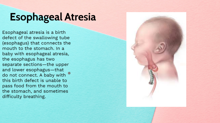
Esophageal atresia is a birth defect in which part of a baby’s esophagus (the tube that connects the mouth to the stomach) does not develop properly.

Esophageal atresia is a birth defect of the swallowing tube (esophagus) that connects the mouth to the stomach. In a baby with esophageal atresia, the esophagus has two separate sections—the upper and lower esophagus—that do not connect. A baby with this birth defect is unable to pass food from the mouth to the stomach, and sometimes difficulty breathing.

Esophageal atresia often occurs with tracheoesophageal fistula, a birth defect in which part of the esophagus is connected to the trachea, or windpipe.

Types of Esophageal Atresia

There are four types of esophageal atresia: Type A, Type B, Type C and Type D.

  • Type A is when the upper and lower parts of the esophagus do not connect and have closed ends. In this type, no parts of the esophagus attach to the trachea.
  • Type B is very rare. In this type the upper part of the esophagus is attached to the trachea, but the lower part of the esophagus has a closed end.
  • Type C is the most common type. In this type the upper part of the esophagus has a closed end and the lower part of the esophagus is attached to the trachea, as is shown in the drawing.
  • Type D is the rarest and most severe. In this type the upper and lower parts of the esophagus are not connected to each other, but each is connected separately to the trachea.

How Many Babies are Born with Esophageal Atresia?

Researchers estimate that about 1 in every 4,100 babies is born with esophageal atresia in the United States.1 This birth defect can occur alone, but often occurs with other birth defects.

Causes

Like many families of children with a birth defect, CDC wants to find out what causes them. Understanding the factors that can increase the chance of having a baby with a birth defect will help us learn more about the causes. CDC funds the Centers for Birth Defects Research and Prevention, which collaborate on large studies such as the National Birth Defects Prevention Study (NBDPS; births 1997-2011) and the Birth Defects Study To Evaluate Pregnancy exposureS (BD-STEPS; began with births in 2014), to understand the causes of and risks for birth defects, including esophageal atresia.

The causes of esophageal atresia in most babies are unknown. Researchers believe that some instances of esophageal atresia may be caused by abnormalities in the baby’s genes. Nearly half of all babies born with esophageal atresia have one or more additional birth defects, such as other problems with the digestive system (intestines and anus), heart, kidneys, or the ribs or spinal column.

Recently, CDC reported on important findings about some factors that increase the risk of having a baby with esophageal atresia:

  • Paternal age – Older age of the father is related to an increased chance of having a baby born with esophageal atresia.
  • Assisted reproductive technology (ART) – Women who used ART to become pregnant have an increased risk of having a baby with esophageal atresia compared to women who did not use ART.

CDC continues to study birth defects, such as esophageal atresia, and how to prevent them. If you are pregnant or thinking about becoming pregnant, talk with your doctor about ways to increase your chances of having a healthy baby.

Diagnosis

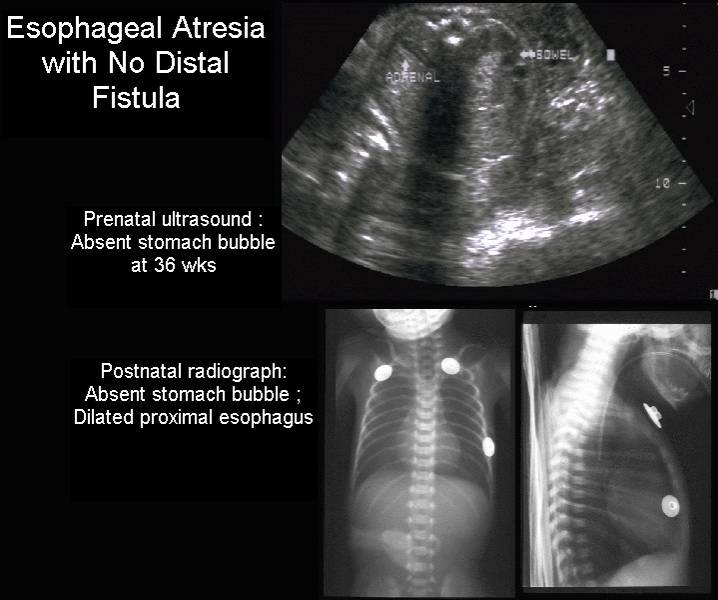
Esophageal atresia is rarely diagnosed during pregnancy. Esophageal atresia is most commonly detected after birth when the baby first tries to feed and has choking or vomiting, or when a tube inserted in the baby’s nose or mouth cannot pass down into the stomach. An x-ray can confirm that the tube stops in the upper esophagus.

Treatment

Once a diagnosis has been made, surgery is needed to reconnect the two ends of the esophagus so that the baby can breathe and feed properly. Multiple surgeries and other procedures or medications may be needed, particularly if the baby’s repaired esophagus becomes too narrow for food to pass through it; if the muscles of the esophagus don’t work well enough to move food into the stomach; or if digested food in the stomach consistently moves back up into the esophagus.

QUOTE FOR TUESDAY:

“Radon is a naturally occurring radioactive gas emitted from the ground. Radon is odorless, tasteless and colorless, and can enter a home through cracks in floors, basement walls, foundations and other openings. Radon can be present at high levels inside homes, schools and other buildings. Radon in homes is more common than you think. Pennsylvania has one of the nation’s most serious radon problems. High levels of radioactive radon gas have been found in every county but most places in the Commonwealth remain undertested, so this isn’t something that should be taken lightly.”

American Lung Association (https://www.lung.org/media/press-releases/pa-radon-2024)

Part II January National Radon Month

National Radon Month3  National Radon Month5

Four Things You Can Do During National Radon Action Month:

1-Test your home –

EPA and the U.S. Surgeon General recommend that all homes in the U.S. be tested for radon.  Testing is easy and inexpensive.

Learn more about testing your home, including how to obtain an easy-to-use test kit.

2-Attend a National Radon Action Month event in your area –

Look for radon events in your community.

3-Spread the word –

Spend time during National Radon Action Month encouraging others to learn about radon and test their homes.

Tell your family and friends about the health risk of radon.  Encourage them to test their homes.

Plan an activity in your community to help raise awareness.

Write an op-ed or letter to the editor using samples from the event planning resources.

Attract media attention by working with a local official to get a radon proclamation.

View or order EPA’s free radon publications.

4-Buy a radon-resistant home –

– If you are considering buying a new home, look for builders who use radon-resistant new construction. Read more about radon-resistant new construction, “Building Radon Out: A Step-by-Step Guide to Build Radon-Resistant Homes“.

Build Green: It’s Easy to Build New Homes Radon-Resistant …”The good news is you can build your customers a safer, healthier, radon-resistant home. The techniques to prevent radon from entering a home are practical and straightforward for any builder. It’s an inexpensive way to offer families a benefit that could reduce their risk of lung cancer. And it’s a smart way to build trust between you and your customer.”  Fuad Reveiz, Member of the National Association of Home Builders.

5 WAYS YOU CAN PARTICIPATE:

 Promote radon risk reduction during National Radon Action Month and throughout the year.

Set a bold goal for your community to reduce radon risk.  Whether you are conducting a single National Radon Action Month event, or developing an entire radon risk reduction program for your community, it is important to set goals to define what you will achieve.

Collaborate with local radon partners.  Coordinate with your state radon program and local radon professionals to hold events and measure results.

Plan a National Radon Action Month activity or event to kick off your campaign.  Conduct activities and events designed to raise awareness, recognize successful efforts and share best practices. Set goals and establish ways to measure results from your activities and events.

An excellent way to get your entire community involved is the National Radon Poster Contest. Students ages 9-14 are encouraged to create posters that raise radon awareness. Entries will be accepted from March through October from students in all U.S. states, tribal nations and U.S. territories.

Visit to Kansas State University National Radon Services for more information about this year’s contest and how you can get involved.

Promote your activity or event on EPA’s Web site and elsewhere. Use tools such as blogs and the Share Your Story feature on Radonleaders.org to promote your activities and share outreach ideas with the broader radon community.

Work with the media to spread your message.  Work with your local media outlets to run Public Service Announcements (PSAs) and generate media coverage of radon topics, events and activities in your community.

QUOTE FOR MONDAY:

“You can’t see, smell or taste radon, but it could be present at a dangerous level in your home. Radon is the leading cause of lung cancer deaths among nonsmokers in America and claims the lives of about 21,000 Americans each year. In fact, the EPA and the U.S. Surgeon General urge all Americans to protect their health by testing their homes, schools and other buildings for radon.

Exposure to radon is a preventable health risk and testing radon levels in your home can help prevent unnecessary exposure. If a high radon level is detected in your home, you can take steps to fix the problem to protect yourself and your family.”

United States Environmental Protection – EPA

(https://www.epa.gov/radon/national-radon-action-month-information)

Part I January National Radon Month Awareness

 National Radon Month   National Radon Month4

Radon is a naturally occurring gas that seeps out of rocks and soil. Radon comes from uranium that has been in the ground since the time the earth was formed, and the rate of radon seepage is very variable, partly because the amounts of uranium in the soil vary considerably. Radon flows from the soil into outdoor air and also into the air in homes from the movement of gases in the soil beneath homes. Outside air typically contains very low levels of radon, but it builds up to higher concentrations indoors when it is unable to disperse. Some underground mines, especially uranium mines, contain much higher levels of radon.

Radon is present outdoors and indoors. It is normally found at very low levels in outdoor air and in drinking water from rivers and lakes. It can be found at higher levels in the air in houses and other buildings, as well as in water from underground sources, such as well water.

Radon breaks down into solid radioactive elements called radon progeny (such as polonium-218, polonium-214, and lead-214). Radon progeny can attach to dust and other particles and can be breathed into the lungs. As radon and radon progeny in the air break down, they give off radiation that can damage the DNA inside the body’s cells.

 How are people exposed to radon?

1-At home and in other buildings.

For both adults and children, most exposure to radon comes from being indoors in homes, offices, schools, and other buildings. The levels of radon in homes and other buildings depend on the characteristics of the rock and soil in the area. As a result, radon levels vary greatly in different parts of the United States, sometimes even within neighborhoods. Elevated radon levels have been found in every state.

Radon gas given off by soil or rock can enter buildings through cracks in floors or walls; construction joints; or gaps in foundations around pipes, wires, or pumps. Radon levels are usually highest in the basement or crawl space. This level is closest to the soil or rock that is the source of the radon. Therefore, people who spend much of their time in basement rooms at home or at work have a greater risk for being exposed.

Small amounts of radon can also be released from the water supply into the air. As the radon moves from the water to air, it can be inhaled. Water that comes from deep, underground wells in rock may have higher levels of radon, whereas surface water (from lakes or rivers) usually has very low radon levels. For the most part, water does not contribute much to overall exposure to radon.

Radon exposure can also occur from some building materials if they are made from radon-containing substances. Almost any building material made from natural substances, including concrete and wallboard, may give off some level of radon. In most cases these levels are very low, but in a few instances these materials may contribute significantly to radon exposure.

Some granite countertops may expose people to different levels of radon. Most health and radiation experts agree that while a small portion of granite countertops might give off increased levels of radon, most countertops give off extremely low levels.

According to the US Environmental Protection Agency (EPA), it’s very unlikely that a granite countertop in a home would increase the radiation level above the normal, natural background level that comes from nearby soil and rocks. Still, people concerned about radon from countertops and from other household sources can test these levels using home detection kits or can hire a professional to do the testing (see the section “How can I avoid exposure to radon?”).

According to the EPA, the average indoor radon level is about 1.3 picocuries per liter (pCi/L). People should take action to lower radon levels in the home if the level is 4.0 pCi/L or higher. The EPA estimates that nearly 1 out of every 15 homes in the United States has elevated radon levels.

Outdoors, radon generally disperses and does not reach high levels. Average levels of radon outdoors, according to the EPA, are about 0.4 pCi/L.

2-At certain jobs

 In the workplace, people working underground, such as some types of miners, are among the most likely to be exposed to high levels of radon. High death rates from lung problems among miners in some parts of the world were first noted hundreds of years ago, long before people knew what radon was. Studies of radon-exposed miners during the 1950s and 1960s confirmed the link between radon exposure and lung cancer.

Higher levels of radon exposure are also more likely for people who work in uranium processing factories or who come in contact with phosphate fertilizers, which may have high levels of radium (an element that can break down into radon).

Does radon cause cancer?

 Being exposed to radon for a long period of time can lead to lung cancer. Radon gas in the air breaks down into tiny radioactive elements (radon progeny) that can lodge in the lining of the lungs, where they can give off radiation. This radiation can damage lung cells and eventually lead to lung cancer.

Cigarette smoking is by far the most common cause of lung cancer in the United States, but radon is the second leading cause. Scientists estimate that about 20,000 lung cancer deaths per year are related to radon.

Exposure to the combination of radon gas and cigarette smoke creates a greater risk for lung cancer than either factor alone. Most radon-related lung cancers develop in smokers. However, radon is also thought to cause a significant number of lung cancer deaths among non-smokers in the United States each year.

Although radon is chemically inert and electrically uncharged, it is radioactive, which means that radon atoms in the air can spontaneously decay, or change to other atoms. When the resulting atoms, called radon progeny, are formed, they are electrically charged and can attach themselves to tiny dust particles in indoor air. These dust particles can easily be inhaled into the lung and can adhere to the lining of the lung. The deposited atoms decay, or change, by emitting a type of radiation called alpha radiation, which has the potential to damage cells in the lung. Alpha radiations can disrupt DNA of these lung cells. This DNA damage has the potential to be one step in a chain of events that can lead to cancer. Alpha radiations travel only extremely short distances in the body. Thus, alpha radiations from decay of radon progeny in the lungs cannot reach cells in any other organs, so it is likely that lung cancer is the only potentially important cancer hazard posed by radon.

For centuries, it has been known that some underground miners suffered from higher rates of lung cancer than the general population. In recent decades, a growing body of evidence has causally linked their lung cancers to exposure to high levels of radon and also to cigarette smoking. The connection between radon and lung cancer in miners has raised concern that radon in homes might be causing lung cancer in the general population, although the radon levels in most homes are much lower than in most mines. The National Research Council study, which has been carried out by the sixth Committee on Biological Effects of Ionizing Radiation (BEIR) VI, has used the most recent information available to estimate the risks posed by exposure to radon in homes.

You can’t see, smell or taste radon, but it could be present at a dangerous level in your home. Radon is the leading cause of lung cancer deaths among nonsmokers in America and claims the lives of about 21,000 Americans each year. In fact, the EPA and the U.S. Surgeon General urge all Americans to protect their health by testing their homes, schools and other buildings for radon.

Exposure to radon is a preventable health risk and testing radon levels in your home can help prevent unnecessary exposure. If a high radon level is detected in your home, you can take steps to fix the problem to protect yourself and your family.

Radon, being naturally occurring, cannot be entirely eliminated from our homes. Of the deaths that attributes to radon (both independently and through joint action with smoking), perhaps one-third could be avoided by reducing radon in homes where it is above the “action guideline level” of 148 Bqm-3 (4 pCiL-1) to below the action levels recommended by the Environmental Protection Agency.4

The risk of lung cancer caused by smoking is much higher than the risk of lung cancer caused by indoor radon. Most of the radon-related deaths among smokers would not have occurred if the victims had not smoked. Furthermore, there is evidence for a synergistic interaction between smoking and radon.

QUOTE FOR THE WEEKEND:

“A few precautions that are recommended for all pregnant women include the following:

  • Limit your exposure to mercury by not eating bigeye tuna, king mackerel, marlin, orange roughy, shark, swordfish, or tilefish. Limit eating white (albacore) tuna to 6 ounces a week. You do not have to avoid all fish during pregnancy. In fact, fish and shellfish are nutritious foods with vital for a pregnant woman and her fetus. Be sure to eat at least 8–12 ounces of low-mercury fish and shellfish per week.
  • Avoid exposure to lead. Lead can be found in old paint, construction materials, alternative medicines, and items made in foreign countries, such as jewelry and pottery.
  • Avoid taking high levels of vitamin A. Very high levels of vitamin A have been linked to severe birth defects. You should consume no more than 10,000 international units of vitamin A a day.
  • Women who are obese (defined as having a of 30 or greater) when they get pregnant have an increased risk of having babies with certain birth defects than women who are a normal weight. Among the most common obesity-related birth defects are NTDs, heart defects, and . If you are planning a pregnancy, the best way to prevent problems caused by obesity is to be at a normal weight before you get pregnant.”

American College of Obstetricians and Gynecologists ACOG

(https://www.acog.org/womens-health/faqs/reducing-risks-of-birth-defects)

What to know in Prevention for Congenital Defects.

Though not all birth defects can be avoided, prenatal care and awareness of past or current conditions can help with prevention.

  • Prenatal care. Taking a daily prenatal vitamin that includes at least 400 micrograms of folic acid can help prevent a variety of birth defects. You should be taking prenatal vitamins if you’re of reproductive age, when you’re actively trying to conceive and/or as soon as you find out you are pregnant. In addition to taking your prenatal vitamin, avoiding alcohol, tobacco and illegal drugs can significantly help prevent birth defects and pregnancy complications.
  • Awareness of past or current conditions. If you previously had a pregnancy with a birth defect, it’s important to find out the most likely causes because it can help your physician plan preventive measures for your next pregnancy. For example, spina bifida is caused by a deficiency in folate, so if your previous pregnancy had spina bifida, you can take a high dose of folate to help prevent future spina bifida diagnoses.

Are all birth defects discovered before a baby is born?

It’s not always possible to detect all birth defects in utero. However, high-resolution ultrasounds done by certified prenatal ultrasound groups make it possible to diagnose defects that will cause a significant impact before birth.

Baschat says: “At the Center for Fetal Therapy, we recommend that pregnant women have the first-trimester nuchal translucency scan between 11 and 14 weeks and the anatomy scan between 18 and 20 weeks. These two ultrasounds provide us with the best opportunity for detecting birth defects.”

In recent years, some pregnant women have turned down the first-trimester ultrasound because a maternal screening blood test now exists for Down syndrome. The ultrasound is still strongly recommended, though, because there are many other serious birth defects that can be detected this early in pregnancy.

Is it possible to treat birth defects while a baby is in utero?

Absolutely. While there are many different types of birth defects, it’s extremely important to try to correct those that damage vital organs before the baby is born. The Center for Fetal Therapy specializes in treating several of these defects in utero, including:

  • Congenital diaphragmatic hernia. This condition, in which a hole in the diaphragm allows abdominal content to enter the chest and restrict lung development, can be significantly helped in utero through fetoscopic endotracheal occlusion, a surgery that improves lung function and significantly increases survival rates.
  • Lower urinary tract obstruction. This occurs when the flow of urine is blocked from exiting the fetus’ body, leading to permanent kidney damage. Relieving this obstruction before birth protects the kidneys.

Fetal treatments also exist for conditions that make the baby unhealthy, even if they are not considered to be birth defects. For example, if a fetus has an irregular heartbeat, you can give the mother medication that will cross the placenta and treat the fetus.

How successful are in utero treatments for fetal conditions?

By treating fetal conditions in utero rather than waiting until after birth, fetuses are given significantly better chances of survival and a reduced need for major surgery after birth. For example, with a condition like twin-to-twin transfusion syndrome, in which identical twins develop a blood volume imbalance, both babies could die without any intervention. By performing laser surgery in utero, there is approximately a 95 percent chance that at least one baby will survive.

Explains Baschat, “Success rates vary depending on the condition, available treatments and the individual patient, but overall, where fetal interventions are available, we see a much higher rate of survival for affected fetuses.”

If a baby has fetal surgery, will he/she need different care after birth?

This will depend on the individual condition and type of surgery performed. For all fetal surgeries, your baby needs to be delivered at a hospital where pediatric subspecialties are in-house so the baby’s care can be managed after birth. Baschat says: “Many of the treatments we perform require patients to deliver at the highest-level multispecialty hospital, like The Johns Hopkins Hospital. This way, all prenatal and postnatal care is available to you in one location.”

If you have one child affected by a birth defect, will all of your future children have that same condition?

All future children will not definitively be affected by the same birth defect, but it will depend on what the cause was. If the birth defect was caused by a genetic mutation, there may be a higher likelihood of recurrence, but if you seek care from a specialized center, maternal-fetal medicine specialists and genetic counselors can work with you to assess future risk.

The risk of some conditions can be determined before you become pregnant through genetic testing. If that doesn’t exist for the condition in question, maternal-fetal medicine specialists and genetic counselors can genetically test your fetus during pregnancy to see if she or he exhibits the mutation that affected your previous child.

Are there communities in which you can speak with other families who have had children affected by birth defects?

There are many forums online, whether on the web or social media platforms, in which parents come together. “At our center, we reach out to previous patients to see if they are interested in communicating with new patients affected by the same condition,” Baschat explains. “They can empathize about the specific birth defect, but they can also provide guidance and advice about the experience of working with our center throughout the pregnancy and after the baby is born.”

In the future, what advances will take place in treating disease before birth?

Through prenatal diagnosis and understanding fetal disease better, physicians are learning more about what harms the fetus and what may be of benefit before birth. Additionally, new uses for minimally invasive surgical equipment are regularly being discovered.

There are also stem cell therapies, genomic medicine and a whole host of related treatments that are currently used for children and adults. One day, these may also apply to the fetus. “While it’s hard to predict the future, we’re currently doing things we couldn’t have foreseen 10 years ago, so we’re very hopeful for continued advancements in fetal care,” says Baschat.

QUOTE FOR FRIDAY:

“The reported incidence of PDA in term neonates is only 1 in 2,000 births, accounting for 5%–10% of all congenital heart disease. The incidence of PDA in preterm neonates is far greater, with reports ranging from 20%–60% (depending on population and diagnostic criteria). The increased incidence of PDA in the preterm infant is attributable to the lack of normal closure mechanisms due to immaturity. Gestational age and weight are intimately linked to PDA in preterm neonates. Specifically, PDA is present in 80% of infants weighing less than 1,200 g at birth, compared to 40% of infants weighing less than 2,000 g at birth., Furthermore, symptomatic PDA is present in 48% of infants with a birth weight of less than 1,000 g.”

National Library of Medicine (https://www.ncbi.nlm.nih.gov/pmc/articles/PMC3462096/)