Archive | August 2023

QUOTE FOR THURSDAY:

“Fibrodysplasia ossificans progressiva is a disorder in which muscle tissue and connective tissue such as tendons and ligaments are gradually replaced by bone (ossified), forming bone outside the skeleton (extra-skeletal or heterotopic bone) that limits movement. This process generally becomes noticeable in early childhood, starting with the neck and shoulders and proceeding down the body and into the limbs.”

Fibrodysplasia Ossificans Progressiva/Stone Man Syndrome

Fibrodysplasia ossificans progressiva (FOP) 

It is a disorder in which skeletal muscle and connective tissue, such as tendons and ligaments, are gradually replaced by bone (ossified). This condition leads to bone formation outside the skeleton (extra-skeletal or heterotopic bone) that restricts movement.

This process generally becomes noticeable in early childhood, starting with the neck and shoulders and moving down the body and into the limbs. People with FOP are born with abnormal big toes (hallux valgus) which can be helpful in making the diagnosis. Trauma, such as a fall or invasive medical procedure, or a viral illness may trigger episodes of muscle swelling and inflammation (myositis). These flareups lasts for several days to months and often result in permanent bone growth in the injured area.

FOP is almost always caused by a mutation at the same place in the ACVR1 gene (The ACVR1 gene provides instructions for making the activin receptor type-1 (ACVR1) protein, which is a member of a protein family called bone morphogenetic protein (BMP) type I receptors.)  and is inherited in an autosomal dominant manner. This condition occurs in about 1 in 1,600,000 newborns and about 800 people worldwide are known to have FOP.

SIGNS AND SYMPTOMS

-Fibrodysplasia ossificans progressiva (FOP) is characterized by the gradual replacement of muscle tissue and connective tissue (such as tendons and ligaments) by bone, restricting movement. This process generally becomes noticeable in early childhood, starting with the neck and shoulders and proceeding down the body and into the limbs.

-The formation of extra-skeletal bone causes progressive loss of mobility as the joints become affected. Speaking and eating may also become difficult as the mouth becomes affected. Over time, people with FOP may become malnourished because of the inability to eat. They may also develop breathing difficulties as a result of extra bone formation around the rib cage that restricts expansion of the lungs.

-Any trauma to the muscles of an individual with FOP (a fall or an invasive medical procedure) may trigger episodes of muscle swelling and inflammation followed by more rapid ossification in the injured area. Flare-ups may also be caused by viral illnesses such as the flu.

-Affected individuals may also have short thumbs and other skeletal abnormalities.

Inheritance

-Fibrodysplasia ossificans progressiva is inherited in an autosomal dominant pattern, which means one copy of the altered gene in each cell is sufficient to cause the disorder.

-Most cases of fibrodysplasia ossificans progressiva result from new mutations in the gene. These cases occur in people with no history of the disorder in their family. In only a small number of cases, an affected person has inherited the mutation from one affected parent.

How this disease is diagnosed:

-Making a diagnosis for a genetic or rare disease can often be challenging. Healthcare professionals typically look at a person’s medical history, symptoms, physical exam, and laboratory test results in order to make a diagnosis. The following resources provide information relating to diagnosis and testing for this condition. If you have questions about getting a diagnosis, you should contact a healthcare professional.

-The Genetic Testing Registry (GTR) provides information about the genetic tests for this condition. The intended audience for the GTR is health care providers and researchers. Patients and consumers with specific questions about a genetic test should contact a health care provider or a genetics professional.

TREATMENT:

There is currently no definitive treatment.  However, a brief course of high-dose corticosteroids, such as Prednisone, started within the first 24 hours of a flare-up, may help reduce the intense inflammation and tissue swelling seen in the early stages of fibrodysplasia ossificans progressiva.  Other medications, such as muscle relaxants, mast cell inhibitors, and aminobisphosphonates, if appropriate, should be closely monitored by a physician.  Surgery to remove heterotopic and extra-skeletal bone is risky and can potentially cause painful new bone growth.

References:


  • Fibrodysplasia ossificans progressiva. Genetics Home Reference (GHR). August 2007; http://ghr.nlm.nih.gov/condition/fibrodysplasia-ossificans-progressiva.
  • FOP Fact Sheet. International Fibrodysplasia Ossificans Progressiva Association. http://www.ifopa.org/what-is-fop/overview.html. Accessed 6/5/2014.
  • Pignolo R, Kaplan F. Pediatric Fibrodysplasia Ossificans Progressiva. E-medicine. July 30, 2009; http://emedicine.medscape.com/article/1007104-overview. Accessed 3/17/2011


QUOTE FOR WEDNESDAY:

“An accurate diagnosis is necessary to treating gastroparesis, since the treatment depends on the cause. If your doctor diagnosed an underlying disease or condition that is causing the gastroparesis, the treatment will focus on correcting or reversing that condition; if there is no underlying cause or if it is not possible to treat it, then the goal of treatment is to promote gastric emptying and relieve your symptoms.

The first step is to stop taking medications that inhibit or delay gastric emptying.”

John Hopkins Medicine (https://www.hopkinsmedicine.org/health/conditions-and-diseases/gastroparesis/gastroparesis-treatment)

Part II Gastroparesis August Awareness Month

How is Gastroparesis Treated?
The treatment for gastroparesis in an individual depends on the severity of symptoms. Treatments are aimed at managing symptoms over a long-term.

Treatment approaches may involve one or a combination of:

  • dietary and lifestyle measures,
  • medications, and/or
  • procedures that may include surgery, such as
  • enteral nutrition,
  • parenteral nutrition,
  • gastric electrical stimulation (Enterra), or
  • other surgical procedures

Some people with gastroparesis have mild symptoms that come and go, which can be managed with dietary and lifestyle measures.

Others have moderate to more severe symptoms that additionally may be treated with medications to stimulate motility and/or reduce nausea and vomiting.

Some people have severe symptoms that are difficult to treat or do not respond to initial treatment approaches. They may require additional procedures to maintain nutrition and/or reduce symptoms.

Goals of Treatment
The goals of treatment are to manage and reduce symptoms, maintain quality of daily living, and minimize related problems such as:

  • Severe dehydration due to persistent vomiting
  • Bezoars (solid collections of food, fiber, or other material), which can cause nausea, vomiting, obstruction, or interfere with absorption of some medications in pill form
  • Difficulty managing blood glucose levels in people with diabetes
  • Malnutrition due to poor absorption of nutrients or a low calorie intake

Manage Risk and Benefit
No single treatment helps all persons with gastroparesis. All drugs and procedures have inherent risks, some more than others. Some of the risks are unavoidable, while others can be avoided and managed. For patients and families it is important to talk to the doctor or health care team about both benefit and risk.

As a patient, in the context of your personal illness status, consider:

  • How severe is your own condition – what effect is it having on your life
  • What is the possible benefit from the treatment suggested or prescribed to you
  • What are the chances that you will receive benefit from the treatment
  • How much benefit should you reasonably expect
  • What possible side effects or complications might there be from the treatment
  • What are the chances that you will experience a side effect or serious adverse event from the treatment
  • What can you do to reduce the chances of side effects or complications
  • How will you know when a side effect occurs
  • Exactly what should you do if a side effect or complication occurs

How to live with Gastroparesis:

Gastroparesis is a long-term condition that can impair quality of life and well-being. Living with gastroparesis affects not only those who suffer but also many others, especially family members and friends. It also touches on relationships in the classroom, in the workplace, or in social interactions.

It takes skills and strengths to deal with a challenging digestive condition like gastroparesis. It means being a kind of active researcher, always looking for what does and does not help, hurt, and work best.

It is important to understand the condition and to advocate for better health. If you or a friend or loved one has gastroparesis, it is also important to understand that you are not alone with this diagnosis.

QUOTE FOR TUESDAY:

“Gastroparesis is a condition that affects the normal spontaneous movement of the muscles (motility) in your stomach. Ordinarily, strong muscular contractions propel food through your digestive tract. But if you have gastroparesis, your stomach’s motility is slowed down or doesn’t work at all, preventing your stomach from emptying properly.”

MAYO CLINIC (https://www.mayoclinic.org/diseases-conditions/gastroparesis/symptoms-causes/)

Part I Gastroparesis August Awareness Month


Should focus attention on important health messages about gastroparesis diagnosis, treatment, and quality of life issues. The goals include improving understanding of gastroparesis to help patients and families manage the condition, and encouraging preventive strategies.

The number of people with gastroparesis appears to be rising. Yet gastroparesis is poorly understood. More community awareness is needed about the condition.

The more awareness for gastroparesis, the greater the ability to impact positive outcomes, such as additional research and improved patient care for the functional GI and motility disorders.

Gastroparesis is also called delayed gastric emptying. The term “gastric” refers to the stomach.

Normally, the stomach empties its contents in a controlled manner into the small intestines. In gastroparesis, the muscle contractions (motility) that move food along the digestive tract do not work properly and the stomach empties too slowly.

Gastroparesis is characterized by the presence of certain long-term symptoms together with delayed stomach emptying in the absence of any observable obstruction or blockage. The delayed stomach emptying is confirmed by a test.

Signs and Symptoms:

The signs and symptoms of gastroparesis may differ among persons with the condition. Symptoms usually occur during and after eating a meal.

Symptoms that are characteristic of gastroparesis include:

  • Nausea and/or vomiting
  • Retching (dry heaves)
  • Stomach fullness after a normal sized meal
  • Early fullness (satiety) – the inability to finish a meal

Diagnosing Gastroparesis:

The symptoms of gastroparesis are similar to those that occur in a number of other illnesses. When symptoms persist over time or keep coming back, it’s time to see a doctor to diagnose the problem. An accurate diagnosis is the starting point for effective treatment.

Diagnosis of gastroparesis begins with a doctor asking about symptoms and past medical and health experiences (history), and then performing a physical exam. Any medications that are being taken need to be disclosed.

Tests will likely be performed as part of the examination. These help to identify or rule out other conditions that might be causing symptoms. Tests also check for anything that may be blocking or obstructing stomach emptying. Examples of these tests include:

  • a blood test,
  • an upper endoscopy, which uses a flexible scope to look into the stomach,
  • an upper GI series that looks at the stomach on an x-ray, or
  • an ultrasound, which uses sound waves that create images to look for disease in the pancreas or gallbladder that may be causing symptoms.

If – after review of the symptoms, history, and examination – the doctor suspects gastroparesis, a test to measure how fast the stomach empties is required to confirm the diagnosis.

Slow gastric emptying alone does not correlate directly with a diagnosis of gastroparesis.

There are several different ways to measure the time it takes for food to empty from the stomach into the small intestine. These include scintigraphy, wireless motility capsule, or breath test. Your doctor will provide details of the one chosen.

Gastric Emptying Study (Scintigraphy)
The diagnostic test of choice for gastroparesis is a gastric emptying study (scintigraphy). The test is done in a hospital or specialty center.

It involves eating a bland meal of solid food that contains a small amount of radioative material so that it can be tracked inside the body. The abdomen is scanned over the next few hours to see how quickly the meal passes out of the stomach. A radiologist will interpret the study at periodic intervals after the meal.

A diagnosis of gastroparesis is confirmed when 10% or more of the meal is still in the stomach after 4 hours.

Other methods for measuring gastric emptying include a wireless motility capsule and a breath test.

Wireless Motility Capsule
The ingestible wireless motility capsule (SmartPill) is swallowed and transmits data to a small receiver that the patient carries. The data collected is interpreted by a radiologist. While taking the test, people can go about their daily routine. After a day or two, the disposable capsule is excreted naturally from the body.

Breath Test
The breath test involves eating a meal that contains a nonradioactive component that can be tracked and measured in the breath over a period of hours. The results can then be calculated to determine how quickly the stomach empties.

Stay tune for Part II tomorrow!


QUOTE FOR MONDAY:

“It all comes down to the pathophysiology.  Sepsis ultimately results from a complex interaction of pro-inflammatory, anti-inflammatory, activated complement system, and coagulation mediators that in association with detector and signaling markers, trigger a host response. Initiators (microbes, trauma, hypoxia, ischemia, toxins) cause local tissue damage, which release local pro- and anti-inflammatory markers. Proinflammatory signalers include TNF, IL-1, and IL-6, while anti-inflammatory markers include IL-4, IL-10, IL-11 and soluble TNF receptors. These are designed to function and contain at a local level. If the initiators overwhelm the local response, the mediators affect multiple systems in the body: dermal, cardiovascular, gastrointestinal, renal, neurologic, hematologic/coagulopathic, pulmonary, and endocrine.3,4 All of these mimics have a similar endgame: triggering a systemic reaction that looks just like sepsis.”

emDOCS (http://www.emdocs.net/mimics-of-sepsis/)

Part II Sepsis and SIRS (systemic inflammatory response syndrome)-Still a major problem in hospitals!

Patty Duke and sepsissepsismultihit

               sepsis inflammatory process

Part II talks to you about the multi-hit theory of SIRS with Inflammatory Cascade of SIRS and lastly the coagulation process in SIRS.   It also tells you an extensive amount of infectious and non-infectious causes of SIRS. Lastly the key antidote to SIRS.

Multi-hit theory

A multi hit theory behind the progression of SIRS to organ dysfunction and possibly multiple organ dysfunction syndrome (MODS). In this theory, the event that initiates the SIRS cascade primes the pump. With each additional event, an altered or exaggerated response occurs, leading to progressive illness. The key to preventing the multiple hits is adequate identification of the ETIOLOGY or CAUSE of SIRS and appropriate resuscitation and therapy.

Inflammatory cascade

Trauma, inflammation, or infection leads to the activation of the inflammatory cascade. Initially, a pro-inflammatory activation occurs, but almost immediately thereafter a reactive suppressing anti-inflammatory response occurs. This SIRS usually manifests itself as increased systemic expression of both pro-inflammatory and anti-inflammatory species. When SIRS is mediated by an infectious insult, the inflammatory cascade is often initiated by endotoxin or exotoxin. Tissue macrophages, monocytes, mast cells, platelets, and endothelial cells are able to produce a multitude of cytokines. The cytokines tissue necrosis factor–alpha (TNF-α) and interleukin-1 (IL-1) are released first and initiate several cascades.

The release of certain factors without getting into medical specific terms they ending line induces the production of other pro-inflammatory cytokines, worsening the condition.

Some of these factors are the primary pro-inflammatory mediators. In research it suggests that glucocorticoids may function by inhibit-ing certain factors that have been shown to be released in large quantities within 1 hour of an insult and have both local and systemic effects. In studies they have shown that certain cytokines given individually produce no significant hemodynamic response but that they cause severe lung injury and hypotension. Others responsible for fever and the release of stress hormones (norepinephrine, vasopressin, activation of the renin-angiotensin-aldosterone system).

Other cytokines, stimulate the release of acute-phase reactants such as C-reactive protein (CRP) and pro-calcitonin.

The pro-inflammatory interleukins either function directly on tissue or work via secondary mediators to activate the coagulation cascade and the complement cascade and the release of nitric oxide, platelet-activating factor, prostaglandins, and leukotrienes.

High mobility group box 1 (HMGB1) is a protein present in the cytoplasm and nuclei in a majority of cell types. In response to infection or injury, as is seen with SIRS, HMGB1 is secreted by innate immune cells and/or released passively by damaged cells. Thus, elevated serum and tissue levels of HMGB1 would result from many of the causes of SIRS.

HMGB1 acts as a potent pro-inflammatory cytokine and is involved in delayed endotoxin lethality and sepsis.

Numerous pro-inflammatory polypeptides are found within the complement cascade. It is thought they are felt to contribute directly to the release of additional cytokines and to cause vasodilatation and increasing vascular permeability. Prostaglandins and leukotrienes incite endothelial damage, leading to multi-organ failure.

Polymorphonuclear cells (PMNs) from critically ill patients with SIRS have been shown to be more resistant to activation than PMNs from healthy donors, but, when stimulated, demonstrate an exaggerated micro-bicidal response (agents that kill microbes). This may represent an auto-protective mechanism in which the PMNs in the already inflamed host may avoid excessive inflammation, thus reducing the risk of further host cell injury and death.[4]

Coagulation

The correlation between inflammation and coagulation is critical to understanding the potential progression of SIRS. IL-1 and TNF-α directly affect endothelial surfaces, leading to the expression of tissue factor. Tissue factor initiates the production of thrombin, thereby promoting coagulation, and is a proinflammatory mediator itself. Fibrinolysis is impaired by IL-1 and TNF-α via production of plasminogen activator inhibitor-1. Pro-inflammatory cytokines also disrupt the naturally occurring anti-inflammatory mediators anti-thrombin and activated protein-C (APC).

If unchecked, this coagulation cascade leads to complications of micro-vascular thrombosis, including organ dysfunction. The complement system also plays a role in the coagulation cascade. Infection-related pro-coagulant activity is generally more severe than that produced by trauma.

What the causes of SIRS can be:

The etiology of systemic inflammatory response syndrome (SIRS) is broad and includes infectious and noninfectious conditions, surgical procedures, trauma, medications, and therapies.

The following is partial list of the infectious causes of SIRS:

  • Bacterial sepsis
  • Burn wound infections
  • Candidiasis
  • Cellulitis
  • Cholecystitis
  • Community-acquired pneumonia
  • Diabetic foot infection
  • Erysipelas
  • Infective endocarditis
  • Influenza
  • Intra-abdominal infections (eg, diverticulitis, appendicitis)
  • Gas gangrene
  • Meningitis
  • Nosocomial pneumonia
  • Pseudomembranous colitis
  • Pyelonephritis
  • Septic arthritis
  • Toxic shock syndrome
  • Urinary tract infections (male and female)
  • *The following is a partial list of the noninfectious causes of SIRS:
  • Acute mesenteric ischemia
  • Adrenal insufficiency
  • Autoimmune disorders
  • Burns
  • Chemical aspiration
  • Cirrhosis
  • Cutaneous vasculitis
  • Dehydration
  • Drug reaction
  • Electrical injuries
  • Erythema multiforme
  • Hemorrhagic shock
  • Hematologic malignancy
  • Intestinal perforation
  • Medication side effect (eg, from theophylline)
  • Myocardial infarction
  • Pancreatitis
  • Seizure
  • Substance abuse – Stimulants such as cocaine and amphetamines
  • Surgical procedures
  • Toxic epidermal necrolysis
  • Transfusion reactions
  • Upper gastrointestinal bleeding
  • VasculitisThe treatment is don’t get it since it is hard to get rid of especially for people over 65 and in hospitals.  There is no one Rx for it.  If you’re unfortunate enough to be diagnosed with SIRS the sooner you get diagnosed with it including being in stage one as opposed to three the higher the odds the turn out will be for you.  Again the key is prevention; don’t get it. There is no one antidote to this SIRS

PREVENTION IS THE KEY ANTIDOTE!   So stay healthy and out of  hospitals!

QUOTE FOR THE WEEKEND:

“Sepsis is a clinical syndrome that complicates severe infection and is characterized by the systemic inflammatory response syndrome (SIRS), immune dysregulation, microcirculatory derangements, and end-organ dysfunction. In this syndrome, tissues remote from the original insult display the cardinal signs of inflammation, including vasodilation, increased microvascular permeability, and leukocyte accumulation.

Although inflammation is an essential host response, the onset and progression of sepsis center upon a “dysregulation” of the normal response, usually with an increase in both proinflammatory and antiinflammatory mediators, initiating a chain of events that leads to widespread tissue injury.”

Uptodate.com (https://www.uptodate.com/contents/systemic-inflammatory-response-syndrome-sirs-and-sepsis-in-children-definitions-epidemiology-clinical-manifestations-and-diagnosis)

Part I Sepsis & SIRS (systemic inflammatory response syndrome)-Not new in hospitals.

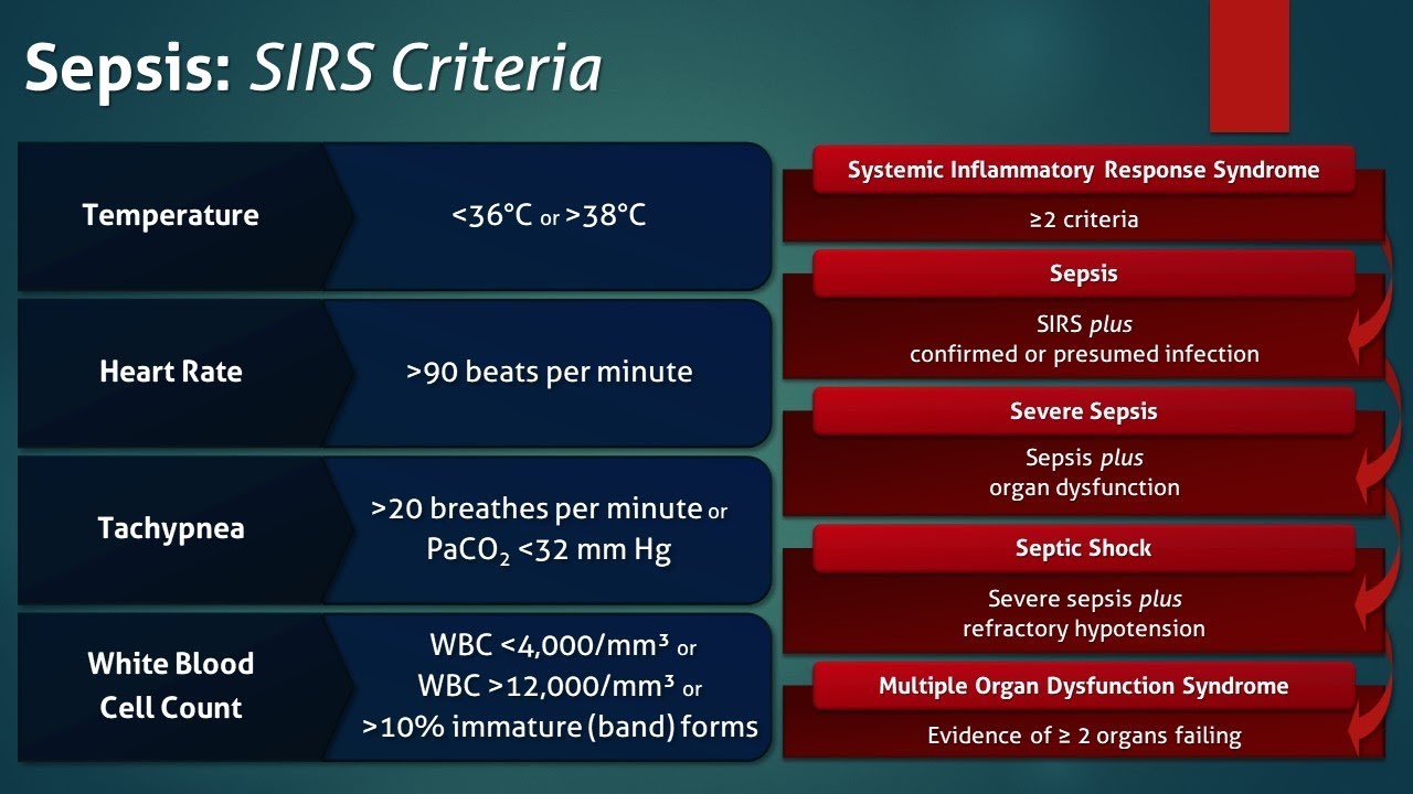
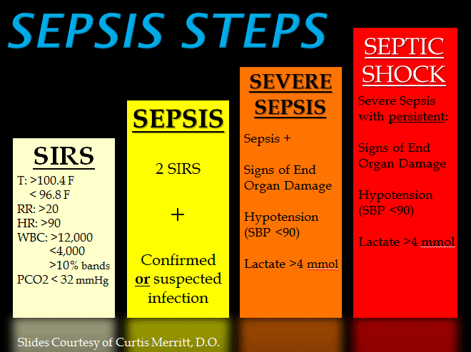
SIRS was first described by Dr. William R. Nelson, of the University of Toronto, in a presentation to the Nordic Micro Circulation meeting in Geilo, Norway-February 1983.  In 1992, the American College of Chest Physicians (ACCP) and the Society of Critical Care Medicine (SCCM) introduced definitions for systemic inflammatory response syndrome (SIRS), sepsis, severe sepsis, septic shock, and multiple organ dysfunction syndrome (MODS), they are interrelated with each other in SIRS.  The idea behind defining SIRS was to define a clinical response to a nonspecific insult of either infectious or noninfectious origin. SIRS is defined as 2 or more of the following variables:

  • Fever of more than 38°C (100.4°F) or less than 36°C (96.8°F)
  • Heart rate of more than 90 beats per minute
  • Respiratory rate of more than 20 breaths per minute or arterial carbon dioxide tension (PaCO 2) of less than 32 mm Hg, which is normally in our body at 35-45 mm Hg whereas the oxygen= PaO2 in our body greater than 80mm Hg for the norm.
  • Abnormal white blood cell count (>12,000/µL or < 4,000/µL or >10% immature [band] forms)

SIRS is nonspecific and can be caused by ischemia, inflammation, trauma, infection, or several insults combined. Thus, SIRS is not always related to infection but commonly is.  SIRS is an inflammatory state affecting the whole body, frequently a response of the immune system to infection, but not always.  It is frequently related to sepsis, a condition in which individuals meet criteria for SIRS and have a known infection.

It is the body’s response to an infectious or noninfectious insult to it. Although the definition of SIRS refers to it as an “inflammatory” response, it actually has pro- and anti-inflammatory components.  SIRS describes the host response to a critical illness of infectious or noninfectious cause, such as burns, trauma, and pancreatitis. More specific definitions are as follows: Sepsis is SIRS resulting from a presumed or known site of infection. Severe sepsis is sepsis with an acute associated multiple organ failure.

What causes sepsis?

Bacterial infections are the most common cause of sepsis. Sepsis can also be caused by fungal, parasitic, or viral infections. The source of the infection can be any of a number of places throughout the body. Common sites and types of infection that can lead to sepsis include:

  • The abdomen—An inflammation of the appendix (appendicitis), bowel problems, infection of the abdominal cavity (peritonitis), and gallbladder or liver infections
  • The central nervous system—Inflammation or infections of the brain or the spinal cord
  • The lungs—Infections such as pneumonia
  • The skin—Bacteria can enter skin through wounds or skin inflammations, or through the openings made with intravenous (IV) catheters (tubes inserted into the body to administer or drain fluids). Conditions such as cellulitis (inflammation of the skin’s connective tissue) can cause sepsis.
  • The urinary tract (kidneys or bladder)—Urinary tract infections are especially likely if the patient has a urinary catheter to drain urine

Who is at risk for sepsis?

Sepsis can strike anyone, but those at particular risk include:

  • People with weakened immune systems
  • Patients who are in the hospital
  • People with pre-existing infections or medical conditions
  • People with severe injuries, such as large burns or bullet wounds
  • People with a genetic tendency for sepsis
  • The very old or very young

What are the symptoms of sepsis?

Because of the many sites on the body from which sepsis can originate, there is a wide variety of symptoms. The most prominent are:

  • Decreased urine output
  • Fast heart rate
  • Fever
  • Or the opposite Hypothermia (very low body temperature)
  • Shaking
  • Chills
  • Warm skin or a skin rash
  • Confusion or delirium
  • Hyperventilation (rapid breathing)

How is sepsis diagnosed?

A person may have sepsis if he or she has:

  • A high or low white blood cell count
  • A low platelet count
  • Acidosis (too much acid in the blood); in the hospital what is checked is lactic acid blood level.
  • A blood culture that is positive for bacteria
  • Abnormal kidney or liver function

How is sepsis treated?

The most important intervention in sepsis is quick diagnosis and prompt treatment. Patients diagnosed with severe sepsis are usually placed in the intensive care unit (ICU) of the hospital for special treatment. The doctor will first try to identify the source and the type of infection, and then administer antibiotics to treat the infection. (Note: antibiotics are ineffective against infections caused by viruses; if anything what is used is antiviral medications.)

The doctor also administers IV fluids to prevent blood pressure from dropping too low. In some cases, vasopressor medications (which constrict blood vessels) are needed to achieve an adequate blood pressure. Some patients are given new drug therapies, such as activated protein C (APC). And finally, if organ failures occur, appropriate supportive care is provided (for example, dialysis for kidney failure, mechanical ventilation for respiratory failure, etc.).  Commonly what is used when initially sepsis is diagnosed is Vancomycin with other antibiotics like Imipenum, Cefepime, and others depending on what the blood culture shows as the microorganism if SIRS is caused by a bacterial infection (many times it is).