Archive | August 2020

QUOTE FOR THURSDAY:

“August is National and World Breastfeeding Awareness Month, and experts and mothers from more than 170 countries agree: Breast milk is best when it comes to feeding a baby in the first year of life. Like trees, teeth contain growth rings that can be counted to estimate age. Teeth rings also incorporate dietary minerals as they grow. Breast milk contains barium, which accumulates steadily in an infant’s teeth and then drops off after weaning.”

NIH – National Institute of Health


QUOTE FOR WEDNESDAY:

“It it estimated for lung cancer in the United States for 2020 are: About 228,820 new cases of lung cancer (116,300 in men and 112,520 in women) About 135,720 deaths from lung cancer (72,500 in men and 63,220 in women). Jan 8, 2020″

American Cancer Society

Part III Lung Cancer

For many people, the first sign that they may have lung cancer is the appearance of a suspicious spot on a chest x-ray or a CT scan. But an image alone is not enough to tell you whether you have cancer and, if so, what type of cancer it is.

Most people who come to us for a lung cancer diagnosis first meet with a surgeon. He or she will work with pathologists, radiologists, and other lung cancer specialists to determine the specific type of lung cancer you have and how advanced it is. These findings help your disease management team develop the most successful treatment plan for you.

The first step is for your doctor to get a tissue sample using one of several biopsy methods. Then a pathologist — a type of doctor who specializes in diagnosing disease —who focuses on lung cancer studies the tissue under a microscope to determine whether you have lung cancer and, if so, what type. He or she will be able to tell this by looking closely at the cancer cells’ shape and other features.

Knowing which type of lung cancer you have will help your doctors to stage the tumor accurately and to begin identifying the best treatment approach. Understanding what type of cancer you have is also important because each type responds differently to certain chemotherapy drugs.

Testing healthy people for lung cancer

Several organizations recommend people with an increased risk of lung cancer consider annual computerized tomography (CT) scans to look for lung cancer. If you’re 55 or older and smoke or used to smoke, talk with your doctor about the benefits and risks of lung cancer screening.

 Some studies show lung cancer screening saves lives by finding cancer earlier, when it may be treated more successfully. But other studies find that lung cancer screening often reveals more benign conditions that may require invasive testing and expose people to unnecessary risks and worry.

Tests to diagnose lung cancer

If there’s reason to think that you may have lung cancer, your doctor can order a number of tests to look for cancerous cells and to rule out other conditions. In order to diagnose lung cancer, your doctor may recommend:

  • Imaging tests. An X-ray image of your lungs may reveal an abnormal mass or nodule. A CT scan can reveal small lesions in your lungs that might not be detected on an X-ray.
  • Sputum cytology. If you have a cough and are producing sputum, looking at the sputum under the microscope can sometimes reveal the presence of lung cancer cells.
  • Tissue sample (biopsy). A sample of abnormal cells may be removed in a procedure called a biopsy.Your doctor can perform a biopsy in a number of ways, including bronchoscopy, in which your doctor examines abnormal areas of your lungs using a lighted tube that’s passed down your throat and into your lungs; mediastinoscopy, in which an incision is made at the base of your neck and surgical tools are inserted behind your breastbone to take tissue samples from lymph nodes; and needle biopsy, in which your doctor uses X-ray or CT images to guide a needle through your chest wall and into the lung tissue to collect suspicious cells.A biopsy sample may also be taken from lymph nodes or other areas where cancer has spread, such as your liver.

QUOTE FOR TUESDAY:

“Lung cancer staging is way of describing where the lung cancer is located, if or where it has spread, and whether it is affecting other parts of the body. Treatment options are available for all stages of lung cancer, and knowing the stage helps your healthcare team:

• Understand how advanced your lung cancer is
• Recommend those treatment options that are likely to be most effective for you
• Evaluate your response to treatment”.

LUNGEVITY (Lungevity.org)

Part II Lung Cancer

What is staging and why is it important?

Understanding if and where lung cancer has spread (the stage) is important to determining what options are available for treatment. Imaging tests, biopsies and laboratory tests help to determine staging.

Non-Small Cell Lung Cancer

Non-small cell lung cancer is one of several cancers staged using the TNM system. The cancer is staged according to the size of the tumor (T), the extent to which the cancer has spread to the lymph nodes (N), and the extent to which the cancer has spread beyond the lymph nodes, or metastasis (M).

How Does The TNM Staging System Work?

The TNM staging system:

  • Was created by merging the staging systems of the American Joint Committee on Cancer (AJCC) http://www.cancerstaging.org/ and the International Union Against Cancer (UICC) http://www.uicc.org/ in 1987
  • Is one of the most commonly used cancer staging systems
  • Standardizes cancer staging internationally

T is for Tumor

How big is the tumor? Where is it located? Has it spread to nearby tissue?

TX The primary tumor cannot be assessed, or the presence of a tumor was only proven by the finding of cancer cells in sputum or bronchial washings but not seen in imaging tests or bronchoscopy.
T0 No evidence of a primary tumor.
Tis “In situ” – cancer is only in the area where the tumor started and has not spread to nearby tissues.
T1 The tumor is less than 3 cm (just slightly over 1 inch), has not spread to the membranes that surround the lungs (visceral pleura), and does not affect the air tunes (bronchi) that brand out on either side from the windpipe (trachea).
T1a The tumor is less than 2 cm.
T1b The tumor is larger than 2 cm but less than 3 cm.
T2 The tumor is larger than 3 cm but less than 7 cm or involves the main air tubes (bronchus) that brand out from the windpipe (trachea) or the membranes that surround the lungs (visceral pleura). The tumor may partially block the airways but has not caused the entire lung to collapse (atelectasis) or to develop pneumonia).
T2a The tumor is larger than 3 cm but less than or equal to 5 cm.
T2b The tumor is larger than 5 cm but less than or equal to 7 cm.
T3 The tumor is more than 7 cm or touches an area near the lung (such as the chest wall or diaphragm, or sac surrounding the heart (pericardium) or has grown into the main air tubes (bronchus) that brand out from the windpipe (trachea) but not the area where the windpipe divides or has caused one lunch to collapse (atelectasis) or pneumonia in an entire lung or there is a separate tumor(s) in the same lobe.
T4 The tumor is of any size and has spread to the area between the lungs (mediastinum), heart, trachea, esophagus, backbone or the place where the windpipe (trachea) branches or there is a separate tumor(s) in a different lobe of the same lung.

N is for Lymph Node

Has the cancer spread to the lymph nodes in and around the lungs? For more information on the lymph system and lymph nodes, see Lymph System

NX Regional lymph nodes cannot be assessed.
N0 No cancer found in lymph nodes.
N1 Cancer has spread to lymph nodes within the lung or to the area where the air pipes (bronchus) that branch out from the windpipe enter the lung, but only on the same side of the lung as the tumor (ipsilateral).
N2 Cancer has spread to lymph nodes near where the windpipe (trachea) branches into the left and right air tubes (bronchi) or near the area in the center of the lung (mediastinum) but only on the same side of the lung as the tumor.
N3 Cancer has spread to lymph nodes found on the opposite side of the lung as the tumor (contralateral) or lymph nodes in the neck.

M is for Metastasis

Has the cancer spread to other parts of the body?

MX Cancer spread cannot be assessed
M0 Cancer has not spread.
M1 Cancer has spread.
M1a Cancer has spread: separate tumor(s) in a lobe in the opposite lung from the primary tumor (contralateral), or malignant nodules in the membrane that surround the lung (pleura) or malignant excess fluid (effusion) in the pleura or membrane that surround the hear (pericardium).
M1b Cancer has spread to distant part of the body such as brain, kidney, bone.

Stages

After the Tumor (T), Lymph Nodes (N) and Metastasis (M) have been determined, the cancer is then staged accordingly:

Overall Stage T N M
Stage 0 Tis (in situ) N0 M0
Stage IA T1a, b N0 M0
Stage IB T2a N0 M0
Stage IIA T1a, b
T2a
T2b
N1
N1
N0
M0
M0
M0
Stage IIB T2b
T3
N1
N0
M0
M0
Stage IIIA T1, T2
T3
T4
N2, N1
N2, N0
N1
M0
M0
M0
Stage IIIB T4
Any T
N2
N3
M0
M0
Stage IV Any T Any N M1a, b

Small Cell Lung Cancer

Small cell lung cancer is most often staged as either limited-stage or extensive-stage.

Limited-Stage

Indicates that the cancer has not spread beyond one lung and the lymph nodes near that lung.

Extensive-Stage

The cancer is in both lungs or has spread to other areas of the body.

Source:

International Association for the Study of Lung Cancer. Goldstraw P, ed. Staging Handbook in Thoracic Oncology. Orange Park: Editorial Rx Press; 2009.

QUOTE FOR MONDAY:

“Lung cancer is a type of cancer that begins in the lungs. Your lungs are two spongy organs in your chest that take in oxygen when you inhale and release carbon dioxide when you exhale.  Lung cancer is the leading cause of cancer deaths worldwide. People who smoke have the greatest risk of lung cancer, though lung cancer can also occur in people who have never smoked.”

MAYO CLINIC

Part I Lung Cancer

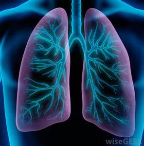
When you breathe in, air enters through your mouth and nose and goes into your lungs through the trachea (windpipe). The trachea divides into tubes called the bronchi (singular, bronchus), which enter the lungs and divide into smaller branches called the bronchioles. At the end of the bronchioles are tiny air sacs known as alveoli.

Many tiny blood vessels run through the alveoli. They absorb oxygen from the inhaled air into your bloodstream and pass carbon dioxide (a waste product from the body) into the alveoli. This is expelled from the body when you exhale. Taking in oxygen and getting rid of carbon dioxide are your lungs’ main functions.

A thin lining called the pleura surrounds the lungs. The pleura protects your lungs and helps them slide back and forth as they expand and contract during breathing. The space inside the chest that contains the lungs is called the pleural space (or pleural cavity).

Below the lungs, a thin, dome-shaped muscle called the diaphragm separates the chest from the abdomen. When you breathe, the diaphragm moves up and down, forcing air in and out of the lungs.
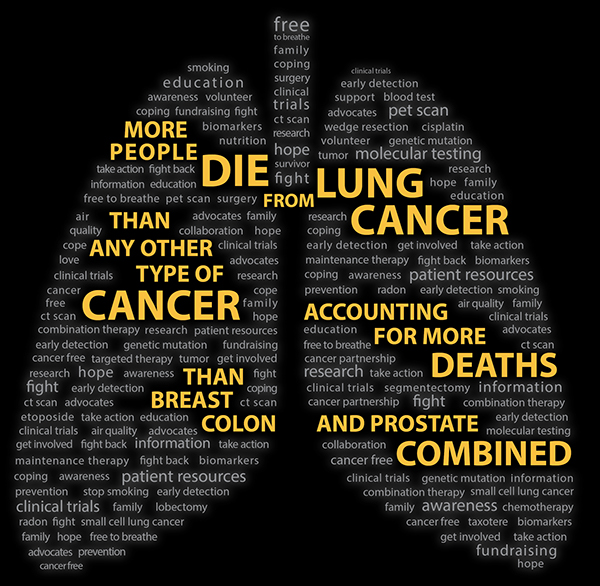
LUNG CANCER

There are 3 types of lungs cancer.  The two most common types of lung cancer that exist are 1 non-small cell lung cancer (NSCLC), which is the most common, and 2 small cell lung cancer (SCLC), an aggressive cancer that occurs in just over 10 percent of all lung cancer cases.

The third group is 3 lung carcinoid tumors (also known as lung carcinoids) are a type of lung cancer, which is a cancer that starts in the lungs. Cancer starts when cells begin to grow out of control. Cells in nearly any part of the body can become cancer, and can spread to other areas of the body.

Lung carcinoid tumors are uncommon and tend to grow slower than other types of lung cancers. They are made up of special kinds of cells called neuroendocrine cells.

Lung Cancer Symptoms

Both major types of lung cancer have similar symptoms. These symptoms often include a cough that doesn’t go away and shortness of breath.

Sometimes lung cancer does not cause any signs or symptoms. It may be found during a chest X-ray done for another condition. Signs and symptoms may be caused by lung cancer or by other conditions. Check with your doctor if you have any of the following:

  • Chest discomfort or pain
  • A cough that doesn’t go away or gets worse over time
  • Trouble breathing
  • Wheezing
  • Blood in sputum (mucus coughed up from the lungs)
  • Hoarseness
  • Loss of appetite
  • Weight loss for no known reason
  • Tiredness/lethargy
  • Trouble swallowing
  • Swelling in the face and/or veins in the neck

For both conditions, early detection through a low-dose computed topography (CT) scan is especially critical. Identifying lung cancer in its earliest stages even before you have symptoms can reduce the risk of death by 20 percent, according to recent studies.

Non-small cell lung Cancer (NSCLC)

Non-small cell lung cancer (NSCLC) is the most common type of cancer in lung tissues. Your risk of developing this disease increases if you are a longtime or former smoker, have been exposed to passive smoke, or have had environmental or occupational exposure to radon, asbestos, uranium, and other substances. The primary types of NSCLC are named for the type of cells found in the cancer:

  • Squamous-cell carcinoma (also called epidermoid carcinoma)
  • Adenocarcinoma
  • Large-cell carcinoma
  • Adenosquamous carcinoma
  • Undifferentiatiated carcinoma

Small Cell Lung Cancer (SCLC)

In small cell lung cancer (SCLC), small cancerous cells arise in the airway, usually in a central location. This is an aggressive cancer that spreads quickly throughout the body through the blood and lymphatic (node) systems. Typically occurring in people who smoke or who used to smoke, SCLC accounts for just over 10 percent of all lung cancers.

QUOTE FOR WEEKEND:

“Most people with spinal muscular atrophy are missing a piece of the SMN1 gene, which impairs SMN protein production. A shortage of SMN protein leads to motor neuron death, and as a result, signals are not transmitted between the brain and muscles. Muscles cannot contract without receiving signals from the brain, so many skeletal muscles become weak and waste away, leading to the signs and symptoms of spinal muscular atrophy.”

U.S. Library of Medicine (NIH)

QUOTE FOR TUESDAY:

“Muscular dystrophy is a group of inherited diseases characterized by weakness and wasting away of muscle tissue, with or without the breakdown of nerve tissue. There are 9 types of muscular dystrophy, with each type involving an eventual loss of strength, increasing disability, and possible deformity.

The most well known of the muscular dystrophies is Duchenne muscular dystrophy (DMD), followed by Becker muscular dystrophy (BMD).”

John Hopkins Medicine

QUOTE FOR MONDAY:

“Gastroparesis is rare.  Fewer than 200,000 cases a year.   Gastroparesis can affect digestion. The cause might be damage to a nerve that controls stomach muscles.  Symptoms include nausea and a full feeling after little food is eaten.  Diet changes and medications may offer relief.”

MAYO CLINIC