Archives

QUOTE FOR TUESDAY:

“The word BSE is short but it stands for a disease with a long name, bovine spongiform encephalopathy. “Bovine” means that the disease affects cows, “spongiform” refers to the way the brain from a sick cow looks spongy under a microscope, and “encephalopathy” meaning that it is a disease of the brain. BSE is commonly called “mad cow disease.

Cattle affected by BSE experience progressive degeneration of the nervous system. Signs usually don’t appear until about 3–6 years after initial infection.

Here’s what to look for:

  • Changes in temperament (nervousness or aggression)
  • Abnormal posture
  • Coordination problems and difficulty in rising
  • Weight loss
  • Decreased milk production
  • Loss of condition without noticeable loss of appetite

After signs appear, the animal’s condition deteriorates until it dies. This usually takes anywhere from 2 weeks to 6 months.”

U.S.D.A. US Dept or Agriculture (https://www.aphis.usda.gov/livestock-poultry-disease/cattle/bse)


Part I MAD COW DISEASE=bovine spongiform encephalopathy (BSE) & in humans called Creutzfeldt-Jakob disease (vCJD).

Mad Cow Disease (Spongiform Encephalopathy or BSE)

Mad cow disease, or bovine spongiform encephalopathy (BSE), is a disease that was first found in cattle. It’s related to a disease in humans called variant Creutzfeldt-Jakob disease (vCJD). Both disorders are universally fatal brain diseases caused by a prion. A prion is a protein particle that lacks DNA (nucleic acid). It’s believed to be the cause of various infectious diseases of the nervous system. Eating infected cattle products, including beef, can cause a human to develop mad cow disease.

What is mad cow disease?

Mad cow disease is a progressive, fatal neurological disorder of cattle resulting from infection by a prion. It appears to be caused by contaminated cattle feed that contains the prion agent. Most mad cow disease has happened in cattle in the United Kingdom (U.K.), a few cases were found in cattle in the U.S. between 2003 and 2006. There were 4 more reported up to 2018.  Feed regulations were then tightened.

In addition to the cases of mad cow reported in the U.K. (78% of all cases were reported there) and the U.S., cases have also been reported in other countries, including France, Spain, Netherlands, Portugal, Ireland, Italy, Japan, Saudi Arabia, and Canada. Public health control measures have been implemented in many of the countries to prevent potentially infected tissues from entering the human food chain. These preventative measures appear to have been effective. For instance, Canada believes its prevention measures will wipe out the disease from its cattle population by 2017.

What is variant Creutzfeldt-Jakob Disease (vCJD)?

Creutzfeldt-Jakob Disease (CJD) is a rare, fatal brain disorder. It causes a rapid, progressive dementia (deterioration of mental functions), as well as associated neuromuscular disturbances. The disease, which in some ways resembles mad cow disease, traditionally has affected men and women between the ages of 50 and 75. The variant form, however, affects younger people (the average age of onset is 28) and has observed features that are not typical as compared with CJD. About 230 people with vCJD have been identified since 1996. Most are from the U.K. and other countries in Europe. It is rare in the U.S., with only 4 reported cases since 1996 until May of 2023 in Chicago found in baby cow.

What is the current risk of acquiring vCJD from eating beef and beef products produced from cattle in Europe?

Currently this risk appears to be very small, perhaps fewer than 1 case per 10 billion servings–if the risk exists at all. Travelers to Europe who are concerned about reducing any risk of exposure can avoid beef and beef products altogether, or can select beef or beef products, such as solid pieces of muscle meat, as opposed to ground beef and sausages. Solid pieces of beef are less likely to be contaminated with tissues that may hide the mad cow agent. Milk and milk products are not believed to transmit the mad cow agent. You can’t get vCJD or CJD by direct contact with a person who has the disease. Three cases acquired during transfusion of blood from an infected donor have been reported in the U.K. Most human Creutzfeldt-Jakob disease is not vCJD and is not related to beef consumption but is also likely due to prion proteins

The Risk of getting Mad Cow Disease in the US, based on CDC-Centers for Disease Prevention and Control show the following statistics:

On December 23, 2003, the U.S. Department of Agriculture (USDA) announced a presumptive diagnosis of the first known case of BSE in the United States. It was in an adult Holstein cow from Washington State. This diagnosis was confirmed by an international reference laboratory in Weybridge, England, on December 25. Trace-back based on an ear-tag identification number and subsequent genetic testing confirmed that the BSE-infected cow was imported into the United States from Canada in August 2001.

Because the animal was non-ambulatory (a “downer cow”) at slaughter, brain tissue samples were taken by USDA’s Animal and Plant Health Inspection Service as part of its targeted surveillance for BSE. However the animal’s condition was attributed to complications from calving. After the animal was examined by a USDA Food Safety and Inspection Service (FSIS) veterinary medical officer both before and after slaughter, the carcass was released for use as food for human consumption. During slaughter, the tissues considered to be at high risk for the transmission of the BSE agent were removed.

On December 24, 2003, FSIS recalled beef from cattle slaughtered in the same plant on the same day as the BSE positive cow.

On June 24, 2005, the USDA announced receipt of final results from The Veterinary Laboratories Agency in Weybridge, England, confirming BSE in a cow that had conflicting test results in 2004. This cow was from Texas, died at approximately 12 years of age, and represented the first endemic case of BSE by a cow in the United States.

On March 15, 2006, the USDA announced the confirmation of BSE in a cow in Alabama. The case was identified in a non-ambulatory (downer) cow on a farm in Alabama. The animal was euthanized by a local veterinarian and buried on the farm. The age of the cow was estimated by examination of the dentition as 10 years old.

It had no ear tags or distinctive marks; the herd of origin could not be identified despite an intense investigation.

In August 2008, several ARS investigators reported that a rare, genetic abnormality that may persist within the cattle population “is considered to have caused” BSE in this atypical (H-type) BSE animal from Alabama.

On April 24, 2012, the USDA confirmed a BSE case in a dairy cow in California. This cow was tested as part of the USDA targeted BSE surveillance at rendering facilities in the United States. The cow was 10 years and 7 months old and was classified as having the L-type BSE strain.

On July 18, 2017, the U.S. Department of Agriculture (USDA) announced the confirmation of the fifth case of bovine spongiform encephalopathy (BSE) in an 11-year-old cow in Alabama. The cow was found through USDA’s routine surveillance. The cow was found to be positive for an atypical (L-type) strain of BSE. Atypical BSE usually occurs in older cattle and seems to arise spontaneously in cattle populations.

On August 29, 2018 the U.S. Department of Agriculture (USDA) announced a confirmed atypical, H-type case of bovine spongiform encephalopathy (BSE) in a six year old mixed-breed beef cow in Florida. USDA reported that this animal never entered the food supply and at no time presented a risk to human health.

How does the cow even get Mad Cow Disease?

The parts of a cow that are not eaten by people are cooked, dried, and ground into a powder. The powder is then used for a variety of purposes, including as an ingredient in animal feed. A cow gets BSE by eating feed contaminated with parts that came from another cow that was sick with BSE. The contaminated feed contains the abnormal prion, and a cow becomes infected with the abnormal prion when it eats the feed. If a cow gets BSE, it most likely ate the contaminated feed during its first year of life. Remember, if a cow becomes infected with the abnormal prion when it is one-year-old, it usually will not show signs of BSE until it is five-years-old or possibly older.

Learn more tomorrow in Part II on Mad Cow Disease.


QUOTE FOR MONDAY:

“Meningitis is an inflammation of the membranes that cover the brain and spinal cord. It can be caused by a number of infectious agents including viruses and bacteria. The type of meningitis and its cause can only be determined by conducting laboratory tests.

Viral meningitis (also called aseptic meningitis) is the most common type of meningitis and is less severe than bacterial meningitis. In Illinois, an average of 600 cases of aseptic meningitis is reported annually, with most occurring in late summer and early autumn. The majority of cases of aseptic meningitis are due to viruses called enteroviruses that can infect the stomach and small intestine. A small number of cases are caused by different viruses, which can be transmitted by infected mosquitoes; these are called arboviruses. Fatal cases of viral meningitis are rare and complete recovery is the rule.

Bacterial meningitis is often more severe than aseptic meningitis, particularly in infants and the elderly. Before antibiotics were widely used, 70 percent or more of bacterial meningitis cases were fatal; with antibiotic treatment, the fatality rate has dropped to 15 percent or less. Bacterial meningitis is most common in the winter and spring. Three bacteria cause the majority of cases: Haemophilus influenzae, Neisseria meningitidis or Streptococcus pneumoniae.”

Illinois Dept. Public Health-IDPH (https://dph.illinois.gov/topics-services/diseases-and-conditions/diseases-a-z-list/meningitis.html)


Meningococcal Meningitis


Meningococcal meningitis is a form of meningitis caused by a specific bacterium known as Neisseria meningitidis. Meningitis is characterized by inflammation of the membranes (meninges) around the brain or spinal cord. This inflammation can begin suddenly (acute) or develop gradually (subacute).

Meningococcal meningitis is a form of meningitis caused by a specific bacterium known as Neisseria meningitidis. Meningitis is characterized by inflammation of the membranes (meninges) around the brain or spinal cord. This inflammation can begin suddenly (acute) or develop gradually (subacute). Symptoms may include fever, headache, and a stiff neck, sometimes with aching muscles. Nausea, vomiting and other symptoms may also occur. Skin rashes occur in about half of all individuals with meningococcal meningitis.

Meningococcal meningitis is still associated with a high mortality rate and persistent neurological defects, particularly among infants and young children. Meningococcal meningitis without antibiotic therapy is uniformly fatal.

Meningococcal meningitis evolves when the bacteria, Neisseria meningitidis (N.meningitidis) progresses from initial adherence to the nasopharyngeal (nose and throat) mucosa to invasion of the deeper mucosal layers (the submucosa). These bacteria rapidly multiply, and can lead to a mild (subclinical) infection. However, in approximately 10-20% of cases, the N.meningitidis enters the bloodstream (meningococcemia). This systemic form of the disease, meningococcemia, usually precedes the development of meningococcal meningitis by 24-48 hours.

Key facts

  • Meningitis is a devastating disease with a high case fatality rate and leading to serious long-term complications (sequelae).
  • Meningitis remains a major global public-health challenge.
  • Epidemics of meningitis are seen across the world, particularly in sub-Saharan Africa.
  • Many organisms can cause meningitis including bacteria, viruses, fungi, and parasites.
  • Bacterial meningitis is of particular concern. Around 1 in 10 people who get this type of meningitis die and 1 in 5 have severe complications.
  • Safe affordable vaccines are the most effective way to deliver long-lasting protection

4 main causes of acute bacterial meningitis:

  • Neisseria meningitidis (meningococcus)
  • Streptococcus pneumoniae (pneumococcus)
  • Haemophilus influenzae
  • Streptococcus agalactiae (group B streptococcus)

The WHO World Health Organization states that these bacteria are responsible for more than half of the deaths from meningitis globally and they cause other severe diseases like sepsis and pneumonia; including other bacteria e.g., Mycobacterium tuberculosis, Salmonella, Listeria, Streptococcus and Staphylococcus, viruses such as enteroviruses and mumps, fungi especially Cryptococcus, and parasites like Amoeba are also important causes of meningitis.

Those at Risk:

Although meningitis affects all ages, young children are most at risk. Newborn babies are at most risk from Group B streptococcus, young children are at higher risk from meningococcus, pneumococcus and Haemophilus influenzae. Adolescents and young adults are at particular risk of meningococcal disease while the elderly are at particular risk of pneumococcal disease.

People all over the world are at risk of meningitis. The highest burden of disease is seen in a region of sub-Saharan Africa, known as the African Meningitis Belt, especially recognised to be at high risk of epidemics of meningococcal but also pneumococcal meningitis.

Higher risk is seen when people are living in close proximity, for example at mass gatherings, in refugee camps, in overcrowded households or in student, military and other occupational settings. Immune deficiencies such as HIV infection or complement deficiency, immunosuppression, and active or passive smoking can also raise the risk of different types of meningitis.


QUOTE FOR THE WEEKEND:

“With the onset of spring in the Northern Hemisphere, animals that hibernate are waking up from a long-period of deep sleep. They spent the winter hibernating to conserve energy when food was scarce. Animals that hibernate include bats, black bears, Arctic ground squirrels, and common poorwill birds. Many other species such as raccoons and skunks go into a state of torpor during the cold weather, which is a type of light hibernation. Most hibernators wake up during the months of March and April, but some do so as late as May.”

Earth Sky (https://earthsky.org/earth/spring-means-these-4-hibernators-are-waking-up/)

Those creatures waking up from hibernating since last year!

For animals that hibernate, making it to spring is no small feat. Torpor — the state of reduced bodily activity that occurs during hibernation — is not restful. By the time they emerge, hibernating animals are often sleep-deprived: Most expend huge bursts of energy to arouse themselves occasionally in the winter so their body temperatures don’t dip too low. This back-and-forth is exhausting, and hibernators do it with little to no food and water. By winter’s end, some have shed more than half their body weight.

But just because it’s spring doesn’t mean it’s time to celebrate. Spring means getting ready for the full speed of summer — and

With the onset of spring in the Northern Hemisphere, animals that hibernate are waking up from a long-period of deep sleep. They spent the winter hibernating to conserve energy when food was scarce. Animals that hibernate include bats, black bears, Arctic ground squirrels, and common poorwill birds. Many other species such as raccoons and skunks go into a state of torpor during the cold weather, which is a type of light hibernation. Most hibernators wake up during the months of March and April, but some do so as late as May.

This would include the following creatures:

1.) Bats  

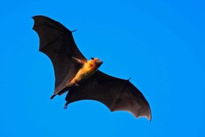
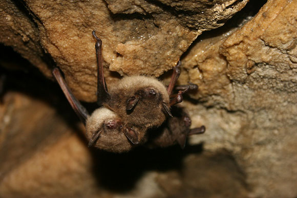
Many types of bats hibernate through the long, cold winter in caves. Bats that hibernate include the little brown bat, the big brown bat, and the northern long-eared bat. During hibernation, their body temperature, heart rate, breathing rate, and metabolism drop to very low levels. This allows them to get by without food or water and stay in a dormant state for long periods of time.

Fertilization happens a few days after females emerge from hibernation. After leaving their winter caves, they move to a large tree or another cave. “They want a warm, stable environment where they can develop their young,” said Joy M. O’Keefe, a bat expert and assistant professor at Indiana State University.

Bats often return to the same maternity spot year after year, sometimes traveling hundreds of miles to get there. Dozens of mothers will congregate at these sites, cuddling to keep warm. When their pups are born, 50 to 60 days later, mothers may help each other by taking turns foraging for insects and roosting with the group.  With no parenting responsibilities, and perhaps to avoid competing with the females, males will stay in torpor for longer — making their hibernation spaces real man caves in the spring.

As spring arrives, so do bats! Many naturalists state during this season looking for migrating salamanders and blossoming bloodroot.   They never thought much about what bats are doing this time of year.

It turns out these flying mammals, who retreated into hibernation back in the fall, are emerging from April through May, as the weather grows consistently warmer and insects again fill the air.

2.) Bears

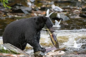
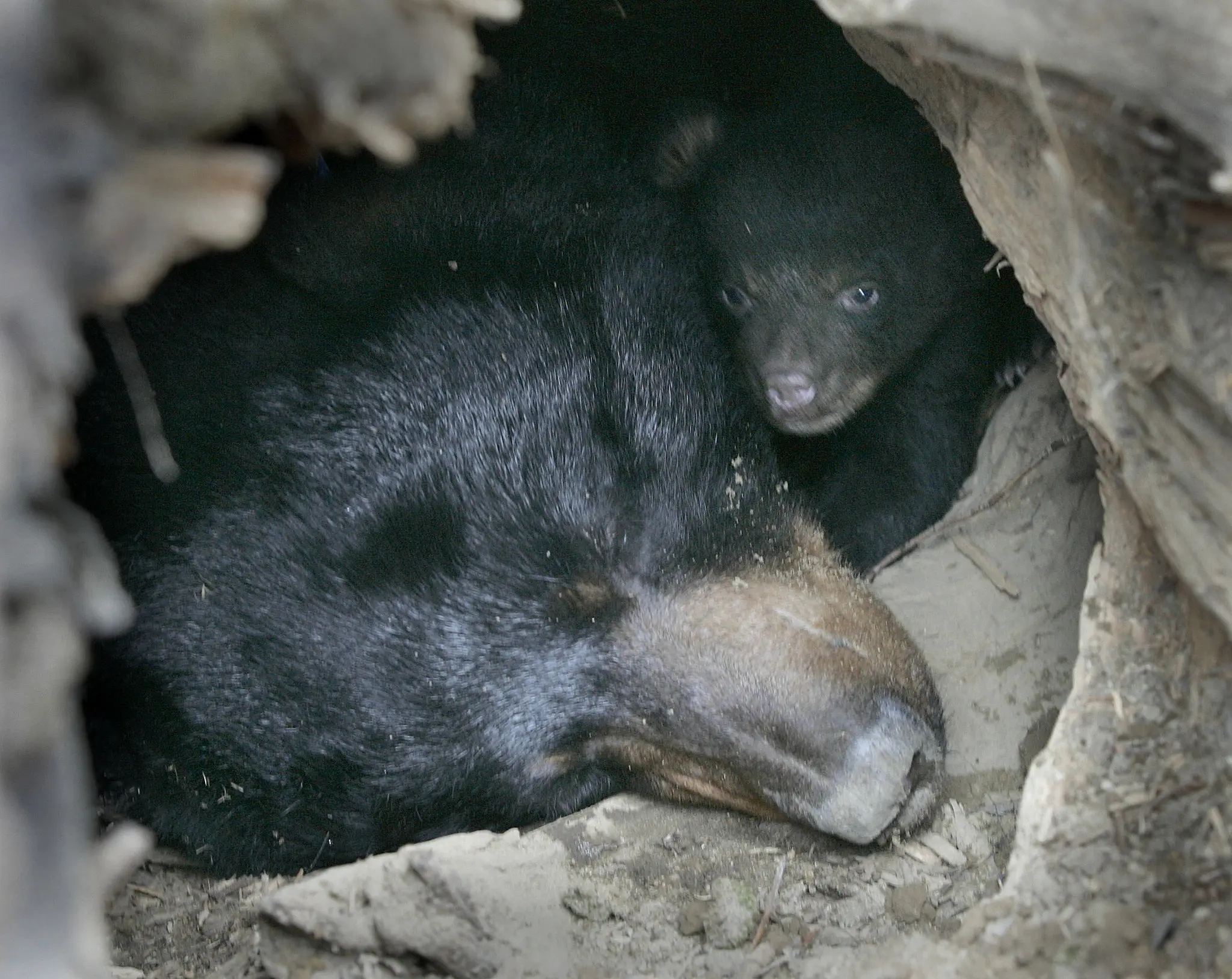
When spring arrives and the snow begins to melt, bears start to wake up after months of hibernation. It is an exciting time of the year for bears and park visitors. When bears emerge from their dens, understandably hungry, they immediately begin to search for food. And there is plenty to eat.  Receding snow reveals vegetation rich in nutrients. Winter kill – deer, elk, moose or anything else that may fancy a bear’s taste buds, are easy pickings. It’s an important time of the year for a bear as it begins the process of nourishing itself, continually gorging on food throughout the year in preparation for hibernation in the fall.  For visitors beginning their spring and summer vacations, the emergence of bears means a chance to see a bruin in its natural habitat, its home. But it also means that another food source presents itself to bears – the food you may accidentally (or intentionally) leave behind or provide. Storing your food and disposing of garbage properly can mean life or death to a bear. Be sure to always properly store food in bear country. 

One of the many reasons people visit national parks with bears is to experience a wild place capable of supporting healthy populations of black and grizzly bears. When visitors become careless and do not properly store their food, bears are undoubtedly going to find it; their sense of smell is amazing. When visitors feed bears, it’s a recipe for trouble. If bears become used to approaching people and eating human food (we call that habituation), the bear no longer seeks the natural food it is supposed to be foraging for. This creates a management and safety problem for park visitors and bears. While park staff work to manage bears and visitors, sometimes there is a need to remove a bear from a park. Imagine what that does to the ecosystem and your experience as a visitor coming to see a bear. For many, it means the park experience is diminished, and the ecosystem isn’t as intact.

When we visit a park with bears, we are entering their home. As guests, proper behavior and etiquette on our part can contribute to a safe and enjoyable visit for us as our hosts.

3.) Arctic Squirrels: 

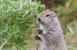
Arctic ground squirrels are the largest of the North American ground squirrel species, ranging from 524 up to 1,500 grams in weight, and 332 to 495 mm in length. They undergo seasonal changes in body mass and lose weight during hibernation. They exhibit sexual dimorphism, with males being larger than females. Body mass drastically varies seasonally, between summer foraging bouts and winter hibernation. They have tawny brown coloration with white flecks on the dorsal side of the pelage and a light tan or beige coloration on their undersides. Their undersides lighten during winter months.

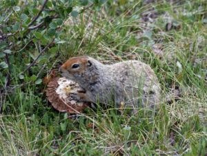
During the onset of cold weather, Arctic ground squirrels dig deep burrows in the ground and hibernate. One scientist attached temperatures sensors to their abdomens and recorded body temperatures in hibernating squirrels as low as -2.9 degrees Celsius (26.8 degrees Fahrenheit), which is below the temperature that water freezes! The squirrel’s blood, however, does not freeze in part because it is salty and also because they have some sort of “super cool” supercooling mechanism that protects them. Scientists are actively researching the brain activity of hibernating Arctic ground squirrels for insights into how to protect people from neurodegenerative diseases like Alzheimer’s and to help them recover from brain injuries. Specifically, the brains of Arctic ground squirrels show a remarkable ability to bounce back after months of dormancy that degrades neuronal connections.

Arctic ground squirrels generally begin hibernation in the beginning of August and wake up in early April, when the males dig their way out from underground.
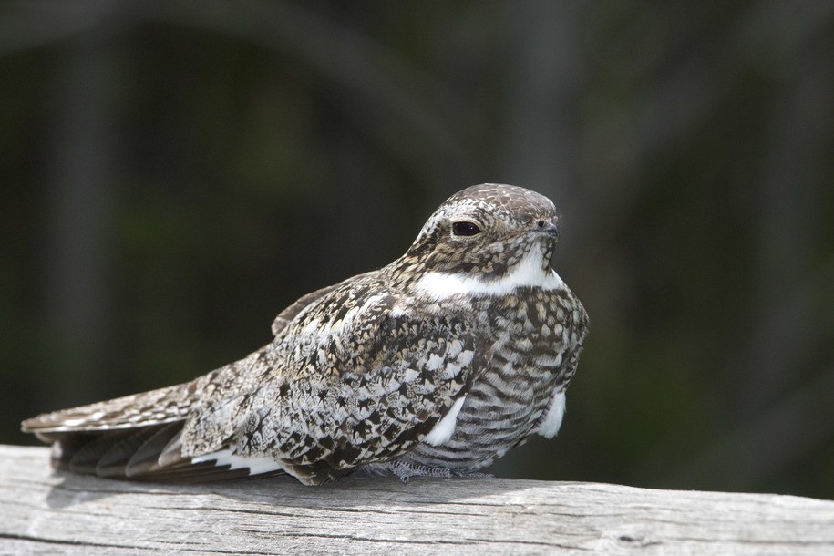
4.) Common poorwill  

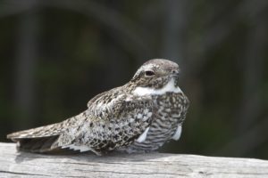
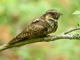
Most birds migrate south when the weather turns cold, but the common poorwill stays put and hibernates. Poorwills are the only bird species known to hibernate. They can be found in the western United States and Canada. Native Americans often referred to this bird as “the sleeping one.”

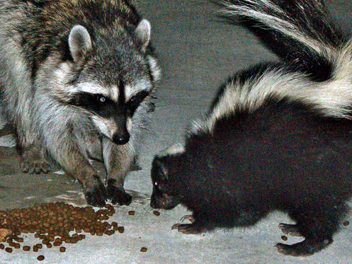
5.) Torpor = Racco0n and Skunk

Torpor is a state of light hibernation that many animals enter into to survive the winter. Animals that use torpor as a survival strategy include raccoons and skunks. 

While there is no bright line that separates animals that hibernate from those that use torpor, it generally comes down to the length of time that an animal spends in dormancy and the extent to which its body temperature and metabolic rate are depressed. Torpor is associated with brief periods of dormancy, sometimes for only a few hours, and small physiological changes, whereas hibernation is associated with lengthy periods of dormancy and large physiological changes.
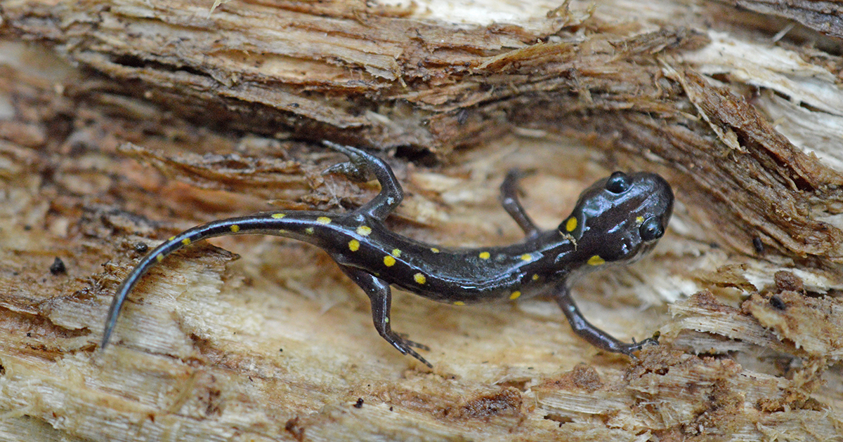
6.) Reptiles:

Scientists use the term brumation to refer to hibernating-like states in reptiles,

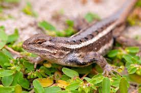
which are not warm-blooded animals so the physiological responses are a bit different from those in mammals and birds. Insects enter cold-induced dormant periods too, and this is referred to by the term diapause. Often on the internet, the term hibernation will be used as a catch-all phrase for all of these types of dormant states.

The exact triggers that cause an animal to enter into and emerge from hibernation aren’t well known, but combinations of factors such as changes in temperature, daylight, and food availability are thought to play an important role. Especially critical is an animal’s internal biological clock, which will initiate hormone changes when it is time for the animal to wake up.

Bottom line: Hibernation is a survival strategy that animals use during the winter to conserve energy when food is scarce. Animals that hibernate include bats, black bears, Arctic ground squirrels, and common poorwill birds.

QUOTE FOR FRIDAY:

“Prevent Blindness has declared April as Women’s Eye Health and Safety Month in an effort to educate the public on the increased risk for women and vision health issues, as well as steps that can be taken to prevent vision loss.

Two out of every three people living with blindness or vision problems are women according to the National Eye Institute. Women are at greater risk of developing a number of eye conditions, including age-related macular degeneration (AMD), cataracts and glaucoma.

At Texas Retina, we know that women often manage their family’s health and take care of everyone else first. However, it is important that women make their own eye health a priority as early detection and consistent treatment can significantly reduce the risk of vision loss. Here are a few tips:

  • Women 40 and older should have an annual dilated eye exam.
  • They should also see their optometrist or ophthalmologist any time they experience a change in vision.
  • Eat a healthy diet rich in vitamin C, vitamin E, beta carotene, lutein, zeaxanthin, zinc and omega-3 fatty acids. This should include foods such as salmon, broccoli and dark leafy greens.
  • Stay active as regular exercise is known to reduce the risk of eye disease.
  • Protect your eyes from the sun by wearing proper sunglasses that block ultraviolet radiation.
  • Avoid smoking.

Texas Retina (https://www.texasretina.com/2024/04/02/april-is-womens-eye-health-and-safety-month)

Women’s eye health and safety month 2024

Two out of every three people living with blindness or vision problems are women, according to the National Eye Institute. And, data from The Future of Vision: Forecasting the Prevalence and Costs of Vision Problems study shows that women are at higher risk for certain eye diseases and conditions. Prevent Blindness has declared April as Women’s Eye Health and Safety Month in an effort to educate the public on the increased risk for women and vision health issues, as well as steps that can be taken to prevent vision loss.

Women have a higher prevalence of major vision problems, including:

  • Age-related Macular Degeneration
  • Autoimmune Diseases (such as Lupus or Sjögren’s Syndrome)
  • Cataract
  • Dry Eye
  • Glaucoma
  • Low Vision
  • Thyroid Eye Disease
  • Refractive Error

According to the World Health Organization’s World Report on Vision, women, on average, live longer than men, and are thus at greater risk of developing eye conditions associated with ageing. However, even after controlling for age, global estimates suggest that women with moderate and severe presenting distance vision impairment outnumber men by approximately 7 percent.

Gender and financial disparities can also create barriers to eyecare access for women.  A recent study published in JAMA Ophthalmology found that although women were more likely to use eye care, they are also more likely to report difficulty affording eyeglasses than men.

In addition to eye disease and conditions, women have unique health issues related to pregnancy and menopause due to fluctuating hormone levels. Women may notice changes in their ability to see clearly during pregnancy. Women with pre-existing conditions, like glaucoma, high blood pressure or diabetes, need to alert their eye doctor that they are pregnant (or planning to become pregnant). Additionally, dry eye leading to a clinical diagnosis or severe symptoms affects more than 3.2 million American women middle-aged and older.

As ophthalmologists and optometrists re-open their practices temporary closing of the office due to the COVID-19 pandemic, it is important that women make their vision health a priority to help detect vision issues early. Early and consistent treatment for most eye diseases can significantly reduce the risk of vision loss.

QUOTE FOR THURSDAY:

“Head and neck cancers usually begin in the squamous cells that line the mucosal surfaces inside the mouth, nose, throat, and salivary glands. They are often referred to as squamous cell carcinomas of the head and neck.

Head and neck cancers account for nearly 4 percent of all cancers in the United States, according to the National Cancer Institute (NCI).

Counting cancers of the oral cavity, pharynx, and larynx, the NCI estimates that about 66,920 cases occurred in the U.S. in 2023. About 15,400 people died from these diseases, according to the NCI’s Surveillance, Epidemiology and End Results (SEER) program.

Head and neck cancer is more common among men than women. These cancers are also diagnosed more often among people over age 50 than among younger people.

Symptoms of head and neck cancers may include a lump or a sore that doesn’t heal. In addition, symptoms can include sore throat that doesn’t go away, difficulty in swallowing, and a hoarse voice.

Alcohol and tobacco use are major risk factors for head and neck cancers. Also, infection with cancer-causing types of human papillomavirus (HPV) increases a person’s risk of certain head and neck cancers.”

American Association for Cancer Research – AACR (https://www.aacr.org/patients-caregivers/awareness-months/head-and-neck-cancer-awareness-month/)

Oral, Head, and Neck Cancer Month Awareness!


  ORAL CANCER RECONCONSTRUCTION SURVIVORS

KNOW THE SYMPTOMS

Most oral, head, and neck cancers begin in the cells that make up the moist tissues (mucosal surfaces) of the head and neck, including in the mouth, nose, and throat. Like other cancers, oral, head, and neck cancers occur when abnormal cells begin to grow and divide uncontrollably and form a mass, called a malignant tumor.

The National Cancer Institute estimates that 85 percent of oral, head, and neck cancers are linked to tobacco use. Men (age 50 or older) who use both tobacco and alcohol are at the highest risk.

Cancers of the brain, eye, thyroid, as well as the skin, bones, muscles and nerves of the head and neck are not included under the heading of “oral, head and neck” cancer.

Doctors identify oral, head, and neck cancers by the area in which the cancer begins:

Lip and oral cavity cancer occurs on the lips, the front two-thirds of the tongue, the gums, the lining of the cheeks and lips, the floor of the mouth under the tongue, the palate, and the area behind the wisdom teeth.

Where does oral cancer affect:

Oral cancer (mouth cancer) is the broad term for cancer that affects the inside of your mouth. Oral cancer can look like a common problem with your lips or in your mouth, like white patches or sores that bleed. Oral cancer (mouth cancer) is the most common form of head and neck cancer. It typically affects people age 60 and older. Oral cancer affects your lips and the first parts of your tongue, mouth roof and floor. It also affects your oropharynx — the last part of your tongue and roof of your mouth, your tonsils, the sides and back of your throat that can include your sinuses, and pharynx (throat). The difference between a common problem and potential cancer is these changes don’t go away. Left untreated, oral cancer can spread throughout your mouth and throat to other areas of your head and neck. Approximately 63% of people with oral cavity cancer are alive five years after diagnosis.

 It can be life-threatening if not diagnosed and treated early.

When it is caught early, like other cancers is much easier for doctors to treat when caught early. Yet most people get a diagnosis when their condition is too advanced to treat effectively. If you see your dentist or doctor regularly and learn how to spot suspicious changes, you’ll have a much better shot at an early diagnosis.

Overall, about 11 people in 100,000 will develop oral cancer during their lifetime. Men are more likely than women to develop oral cancer. People who are white are more likely to develop oral cancer than people who are Black.

Risk factors in developing oral cancer include:

  • Smoking. Cigarette, cigar, or pipe smokers are six times more likely than nonsmokers to develop oral cancers.
  • Smokeless tobacco use. Users of dip, snuff, or chewing tobacco products are 50 times more likely to develop cancers of the cheek, gums, and lining of the lips.
  • Excessive consumption of alcohol. Oral cancers are about six times more common in drinkers than in nondrinkers.  Using alcohol and tobacco together increases your chances even more.
  • Family history of cancer.
  • Excessive sun exposure, especially at a young age. Ultraviolet radiation from the sun can cause lip cancers.
  • Human papillomavirus (HPV). Certain HPV strains are etiologic risk factors for Oropharyngeal Squamous Cell Carcinoma (OSCC). Almost everyone who’s sexually active will get HPV at some point in life. A specific type of this virus is causing a growing number of otherwise healthy men under 50 to get cancers in the back of their mouths and throats from oral sex. The more people you and your partners have sex with, the bigger your risk.
  • Age. Oral cancers can take years to grow. Most people find they have it after age 55. But more younger men are getting cancers linked to HPV.
  • Gender. Men are at least twice as likely as women to get oral cancer. It could be because men drink and smoke more than women do.
  • Poor diet. Studies have found a link between oral cancer and not eating enough vegetables and fruits.

It is important to note that over 25% of all oral cancers occur in people who do not smoke and who only drink alcohol occasionally.

The overall 5-year survival rate for patients with an early diagnosis of oral cavity and pharynx cancers is 84%. If the cancer has spread to nearby tissues, organs, or lymph nodes, the 5-year survival rate drops to 65%.

How it is diagnosed?

As part of your routine dental exam, your dentist will conduct an oral cancer screening exam.  Your dentist knows what a healthy mouth should look like and probably has the best chance of spotting any cancer. Experts recommend getting checked every year starting at age 18, and sooner if you start smoking or having sex.

More specifically, your dentist will feel for any lumps or irregular tissue changes in your neck, head, face, and oral cavity. When examining your mouth, your dentist will look for any sores or discolored tissue as well as check for any signs and symptoms mentioned above.

A biopsy may be needed to determine the makeup of a suspicious-looking area. There are different types of biopsies and your doctor can determine which one is best.

Treatments:

It varies from person to person depending on the stage of oral cancer the person has and how advanced the cancer is.

Whether a patient has surgery, radiation, and surgery, or radiation, surgery, and chemotherapy, you need to remember it depends on the stage of cancer development. Each case is individual.

Patients with cancers treated in their early stages may have little in the way of post-treatment disfigurement. For those whose cancer is caught at a later stage, the results of surgical removal of the disease may require reconstruction of portions of their oral cavity or facial features. There may be needed adjunctive therapy to assist in speech, chewing of foods, the problems associated with the lack of salivary function, and the fabrication of dental or facial prostheses.